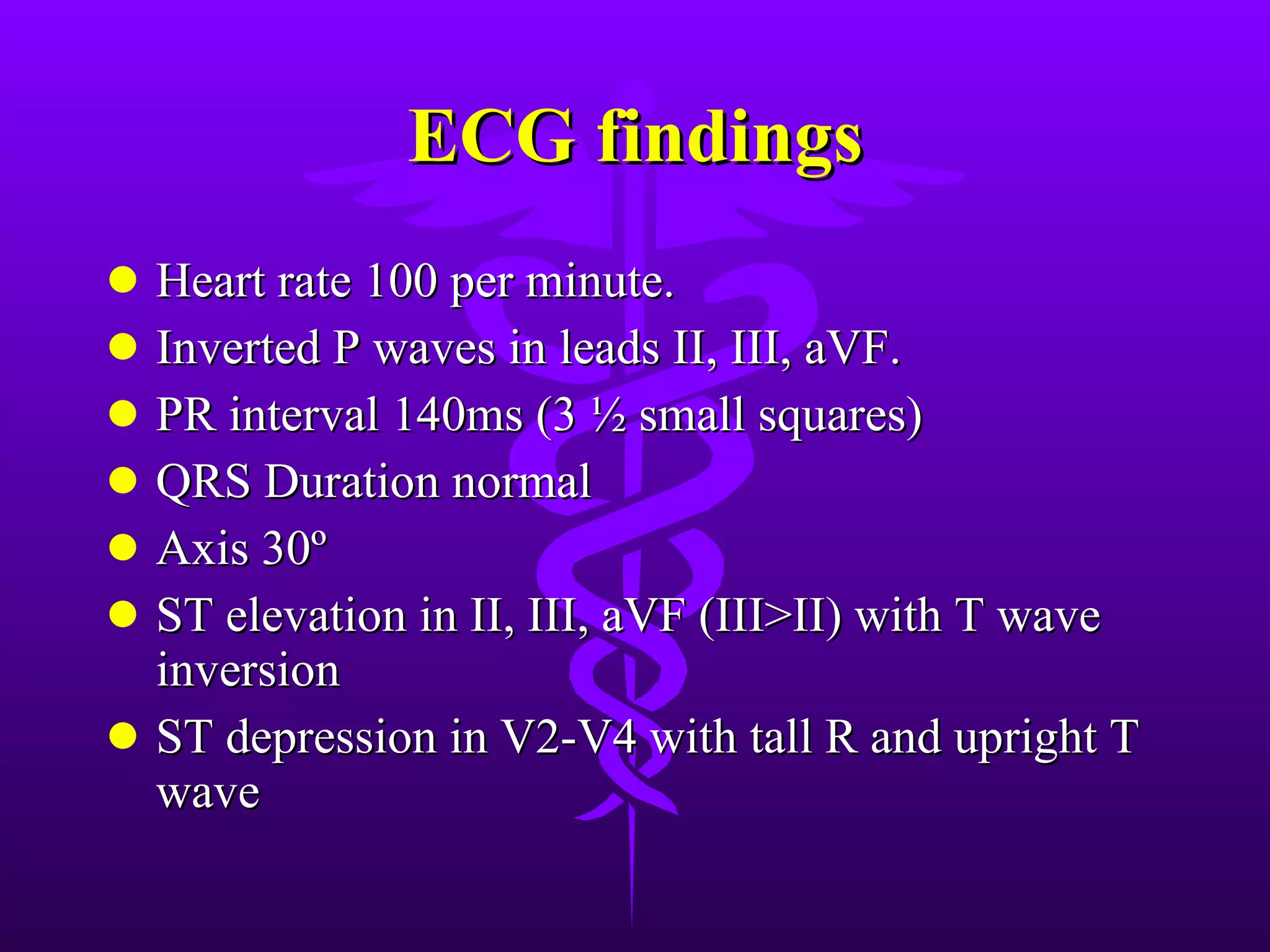
ECG of the week By Dr. C. R. Rajakumar M5 unit  Prof. P. Vijayaraghavan’s unit
Patient presented with c/o Chest pain for the past 3 hrs He was a known hypertensive on drugs for the past 5yrs. ECG was taken
 
ECG findings Heart rate 100 per minute. Inverted P waves in leads II, III, aVF. PR interval 140ms (3 ½ small squares) QRS Duration normal Axis 30º ST elevation in II, III, aVF (III>II) with T wave inversion ST depression in V2-V4 with tall R and upright T wave
Diagnosis  Coronary sinus rhythm with Infero posterior wall MI
Atrial Rhythms Atrial rhythm resembles sinus rhythm, but origins from a different atrial focus.  It can be recognised by the abnormal configuration of the p-wave
Characteristics of atrial rhythm Atrial Rhythms Atrial frequency 50 – 100 bpm Ventricular frequency 1:1 Regularity  Regular Origin  Atrium P wave Present but differs form sinus rhythm Effect of adenosine Slows down
FREQUENCY OF ECTOPIC SITE
Frequency of atrial ectopic sites
Algorithm to identify site of atrial rhythm
Coronary sinus rhythm; inverted p waves in lead 2 ,3 aVF  and  PR interval  normal
Which chamber? Right atrial A negative   or biphasic (positive, then negative) P-wave in lead V 1  was   associated with a 100% specificity for a tachycardia   arising from the RA. Left atrial A positive or biphasic (negative, then   positive) P-wave in lead V 1  was associated with a 100% sensitivity for a tachycardia originating in the LA.
Superior or inferior location of ectopic impulse? In general, the polarity   of leads II, III, aVF is deeply negative for an inferiorly located ectopic impulse. Low amplitude, positive, or biphasic for a superiorly   located ectopic impulse.
 

ECG: Coronary Sinus Rhythm

  • 1.
    ECG of theweek By Dr. C. R. Rajakumar M5 unit Prof. P. Vijayaraghavan’s unit
  • 2.
    Patient presented withc/o Chest pain for the past 3 hrs He was a known hypertensive on drugs for the past 5yrs. ECG was taken
  • 3.
  • 4.
    ECG findings Heartrate 100 per minute. Inverted P waves in leads II, III, aVF. PR interval 140ms (3 ½ small squares) QRS Duration normal Axis 30º ST elevation in II, III, aVF (III>II) with T wave inversion ST depression in V2-V4 with tall R and upright T wave
  • 5.
    Diagnosis Coronarysinus rhythm with Infero posterior wall MI
  • 6.
    Atrial Rhythms Atrialrhythm resembles sinus rhythm, but origins from a different atrial focus. It can be recognised by the abnormal configuration of the p-wave
  • 7.
    Characteristics of atrialrhythm Atrial Rhythms Atrial frequency 50 – 100 bpm Ventricular frequency 1:1 Regularity Regular Origin Atrium P wave Present but differs form sinus rhythm Effect of adenosine Slows down
  • 8.
  • 9.
    Frequency of atrialectopic sites
  • 10.
    Algorithm to identifysite of atrial rhythm
  • 11.
    Coronary sinus rhythm;inverted p waves in lead 2 ,3 aVF and PR interval normal
  • 12.
    Which chamber? Rightatrial A negative or biphasic (positive, then negative) P-wave in lead V 1 was associated with a 100% specificity for a tachycardia arising from the RA. Left atrial A positive or biphasic (negative, then positive) P-wave in lead V 1 was associated with a 100% sensitivity for a tachycardia originating in the LA.
  • 13.
    Superior or inferiorlocation of ectopic impulse? In general, the polarity of leads II, III, aVF is deeply negative for an inferiorly located ectopic impulse. Low amplitude, positive, or biphasic for a superiorly located ectopic impulse.
  • 14.