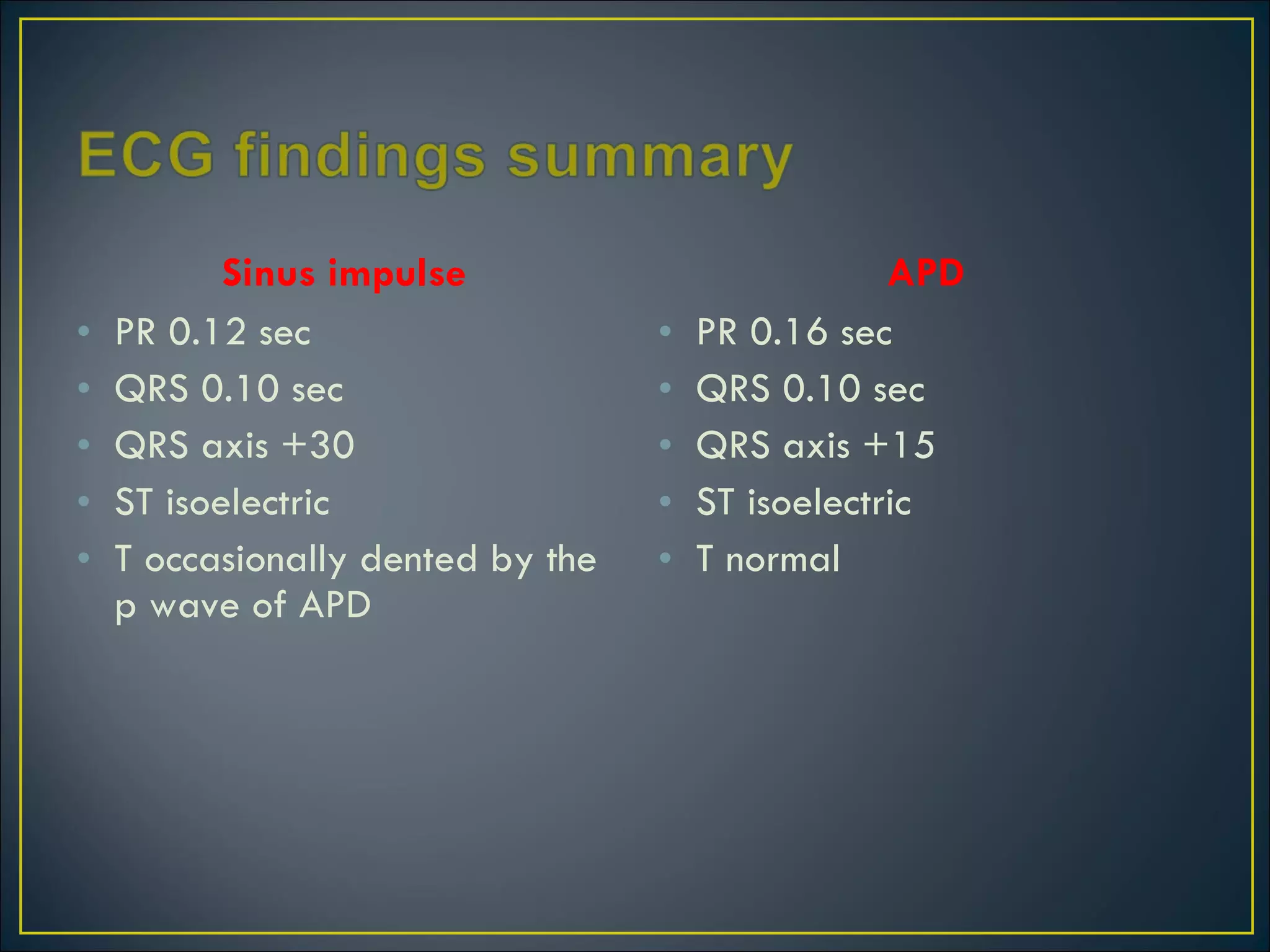
Prof P. Vijayaraghavan’s Unit by Dr J. Stalin Roy
Munirathinam 70 yr old male, a known Hypertensive came for routine check up. His pulse was regularly irregular, an ECG was ordered.
 
 
Two foci of origin of atrial depolarisation as evidenced by two different p wave morphologies  (the sinus p wave is + followed by – ie right atrial followed by left atrial; the ectopic p is – followed by + ie left atrial followed by right atrial) The sinus impulse is followed by an Atrial Premature Depolarisation (APD) which is coupled to the sinus impulse by a constant interval (0.48sec) The APD is followed by a normal ventricular depolarisation as evidenced by normal QRS This rhythm is known as Atrio-Ventricular Bigeminy or simply Atrial Bigeminy
Sinus impulse PR 0.12 sec QRS 0.10 sec QRS axis +30 ST isoelectric T occasionally dented by the p wave of APD APD PR 0.16 sec QRS 0.10 sec QRS axis +15 ST isoelectric T normal
 
A P-wave algorithm constructed on the basis of findings from 130 atrial tachycardias correctly localized the focus in 93%
Right atrial A negative   or biphasic (positive, then negative) P-wave in lead V 1  was   associated with a 100% specificity and PPV for a tachycardia   arising from the RA. Left atrial A positive or biphasic (negative, then   positive) P-wave in ECG lead V 1  was associated with a 100% sensitivity   and NPV for tachycardia originating in the LA.
Lead V1 is oriented towards the right atrium, aVL towards the left atrium, so localization is mainly based on morphologies of p waves in these leads. Lead V1 is always positive when the impulse originates near the pulmonary veins (left atrial); and negative when the origin is near the tricuspid annulus (right atrial). In general, the polarity   of leads II, III, aVF is deeply  negative for an inferoanterior   location, and low amplitude, positive, or biphasic for a superior   location. Perinodal (near the AV node) and right septal foci are associated with an  isoelectric V1.
Rare rhythm disorder characterized by an APD occurring after each sinus impulse followed by a non-compensatory pause (characteristic of APD) The APD usually arises from a  single  irritable focus within the atria. An atrial or junctional focus becomes irritable due to: Excess adrenaline due to increased sympathetic stimuli Caffeine amphetamines, cocaine and other  β 1 receptor stimulants Excess digitalis, certain toxins, occasionally ethanol Hyperthyroidism  Stretch  Hypoxia
Usually benign condition  But it may rarely precipitate supraventricular and ventricular arrhythmias most commonly Atrial Fibrillation Management includes correction of predisposing conditions
 

ECG: Atrial Bigeminy

  • 1.
    Prof P. Vijayaraghavan’sUnit by Dr J. Stalin Roy
  • 2.
    Munirathinam 70 yrold male, a known Hypertensive came for routine check up. His pulse was regularly irregular, an ECG was ordered.
  • 3.
  • 4.
  • 5.
    Two foci oforigin of atrial depolarisation as evidenced by two different p wave morphologies (the sinus p wave is + followed by – ie right atrial followed by left atrial; the ectopic p is – followed by + ie left atrial followed by right atrial) The sinus impulse is followed by an Atrial Premature Depolarisation (APD) which is coupled to the sinus impulse by a constant interval (0.48sec) The APD is followed by a normal ventricular depolarisation as evidenced by normal QRS This rhythm is known as Atrio-Ventricular Bigeminy or simply Atrial Bigeminy
  • 6.
    Sinus impulse PR0.12 sec QRS 0.10 sec QRS axis +30 ST isoelectric T occasionally dented by the p wave of APD APD PR 0.16 sec QRS 0.10 sec QRS axis +15 ST isoelectric T normal
  • 7.
  • 8.
    A P-wave algorithmconstructed on the basis of findings from 130 atrial tachycardias correctly localized the focus in 93%
  • 9.
    Right atrial Anegative or biphasic (positive, then negative) P-wave in lead V 1 was associated with a 100% specificity and PPV for a tachycardia arising from the RA. Left atrial A positive or biphasic (negative, then positive) P-wave in ECG lead V 1 was associated with a 100% sensitivity and NPV for tachycardia originating in the LA.
  • 10.
    Lead V1 isoriented towards the right atrium, aVL towards the left atrium, so localization is mainly based on morphologies of p waves in these leads. Lead V1 is always positive when the impulse originates near the pulmonary veins (left atrial); and negative when the origin is near the tricuspid annulus (right atrial). In general, the polarity of leads II, III, aVF is deeply negative for an inferoanterior location, and low amplitude, positive, or biphasic for a superior location. Perinodal (near the AV node) and right septal foci are associated with an isoelectric V1.
  • 11.
    Rare rhythm disordercharacterized by an APD occurring after each sinus impulse followed by a non-compensatory pause (characteristic of APD) The APD usually arises from a single irritable focus within the atria. An atrial or junctional focus becomes irritable due to: Excess adrenaline due to increased sympathetic stimuli Caffeine amphetamines, cocaine and other β 1 receptor stimulants Excess digitalis, certain toxins, occasionally ethanol Hyperthyroidism Stretch Hypoxia
  • 12.
    Usually benign condition But it may rarely precipitate supraventricular and ventricular arrhythmias most commonly Atrial Fibrillation Management includes correction of predisposing conditions
  • 13.