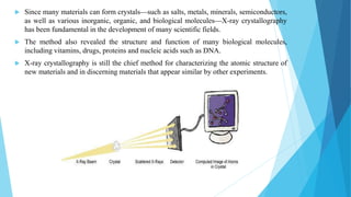
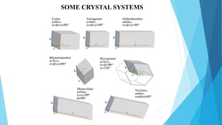
Downloaded 115 times

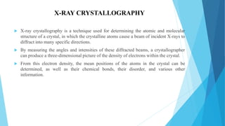
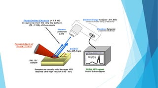
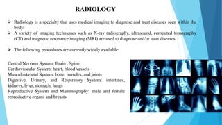
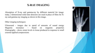
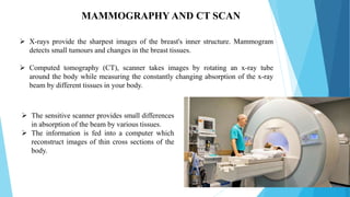
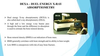
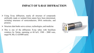
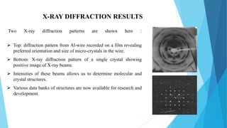
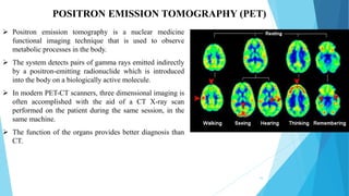
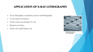


The document is a presentation by Himanshu Dixit on the various applications of X-ray technology across different fields, covering techniques such as X-ray crystallography, photoelectron spectroscopy, and medical imaging. It details how X-rays are utilized in diagnostics, treatment, and material analysis, emphasizing their significance in biology and materials science. The presentation also discusses X-ray lithography as a method for creating high-resolution microstructures.


