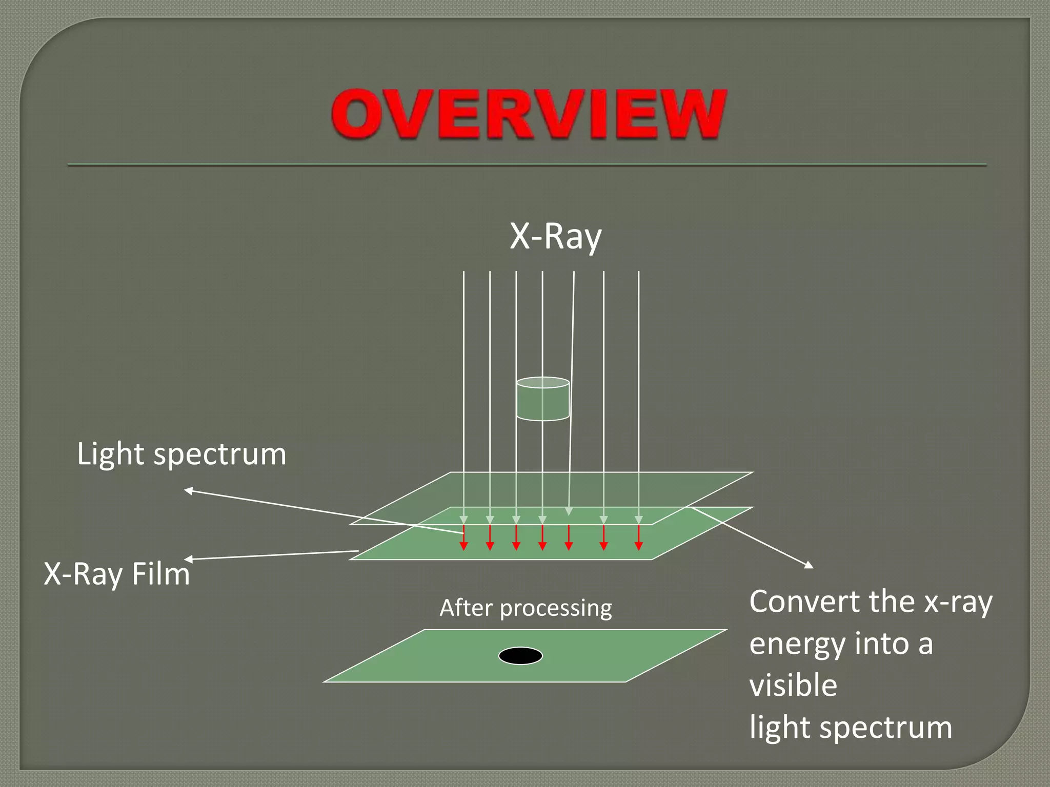
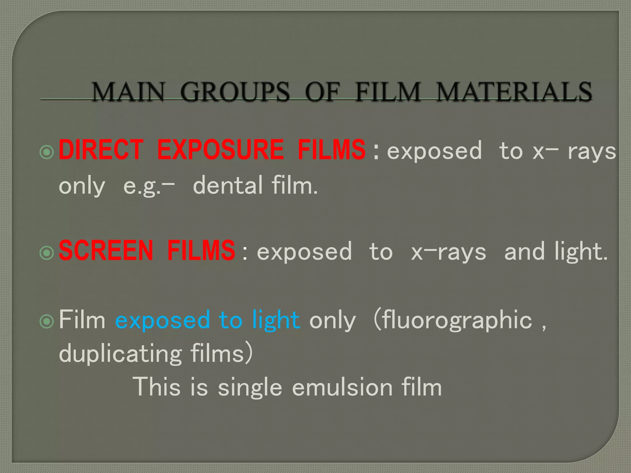
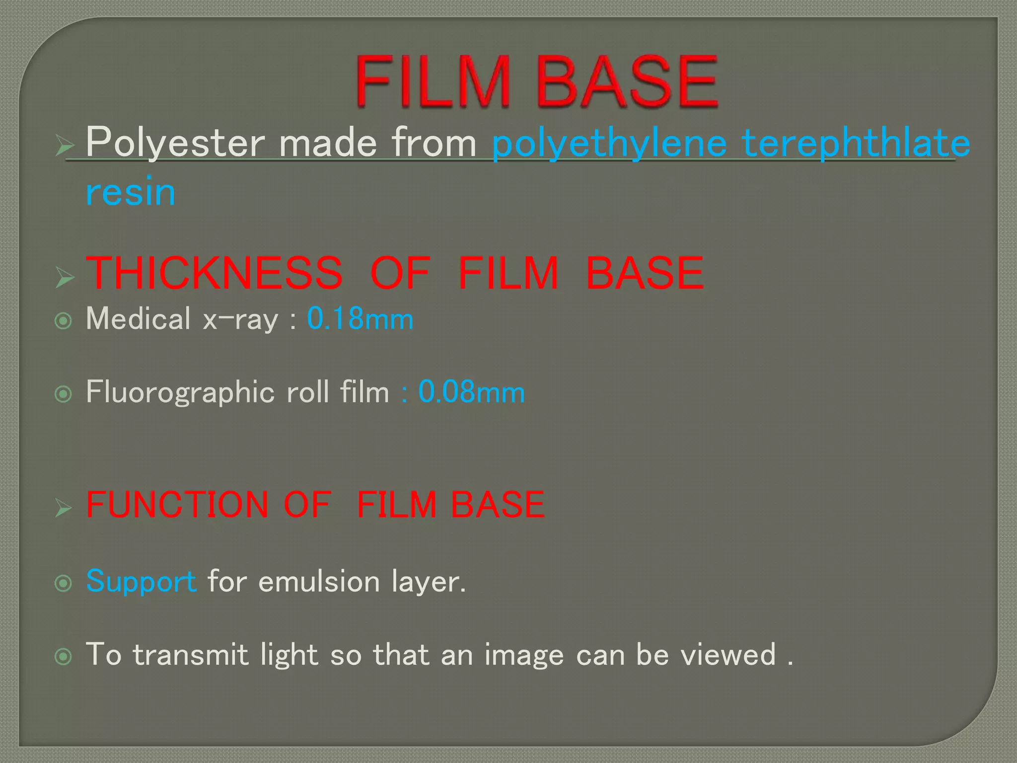
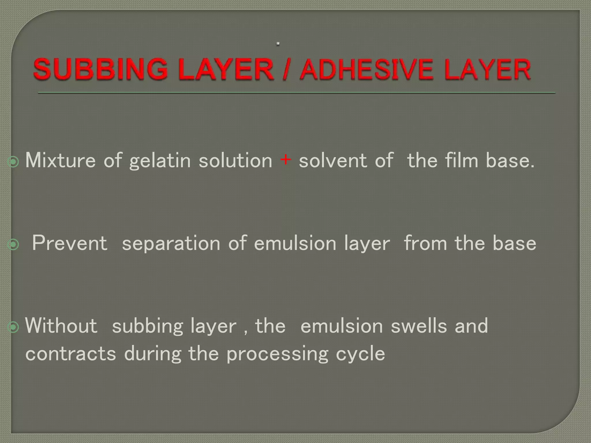
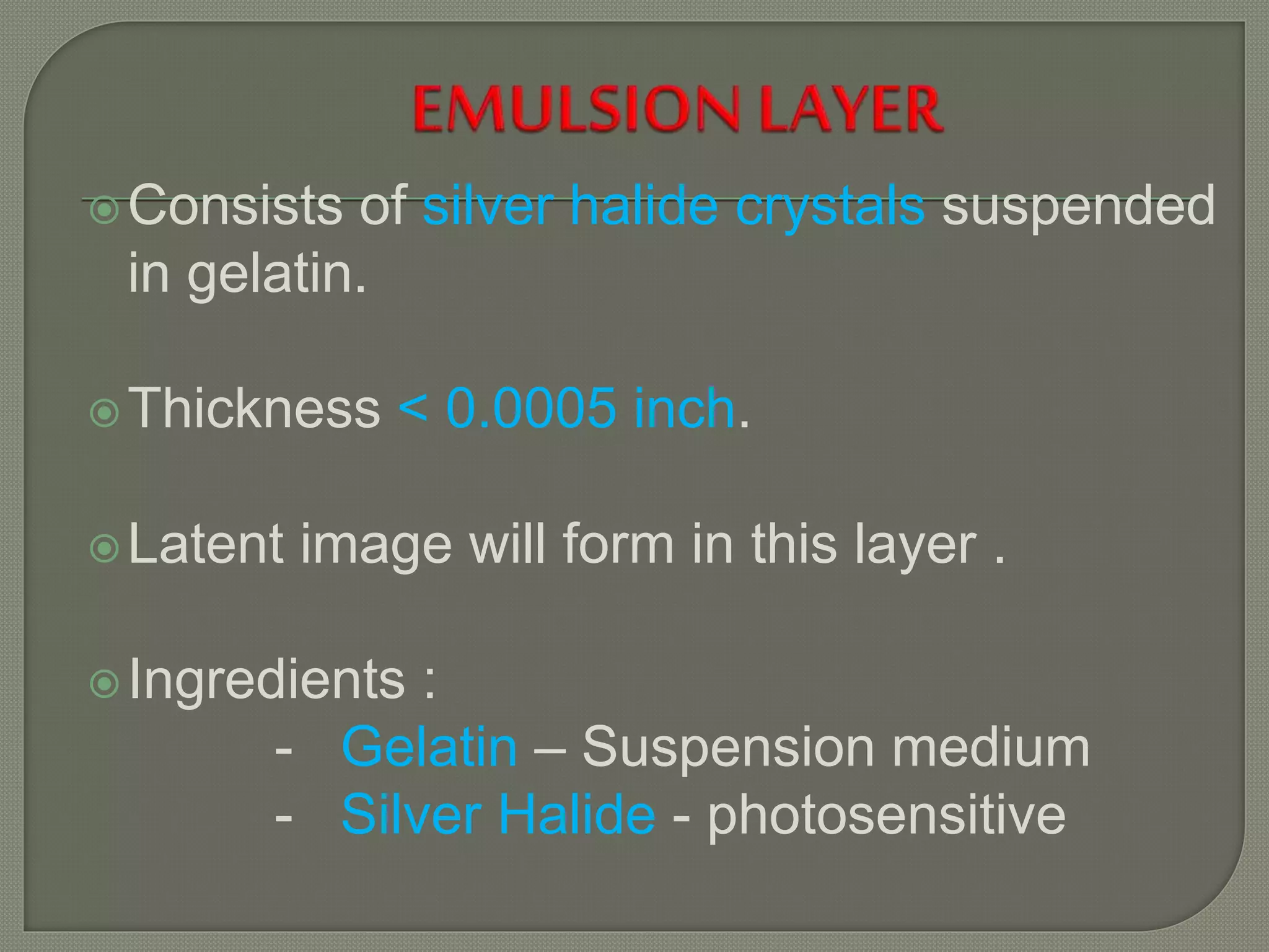
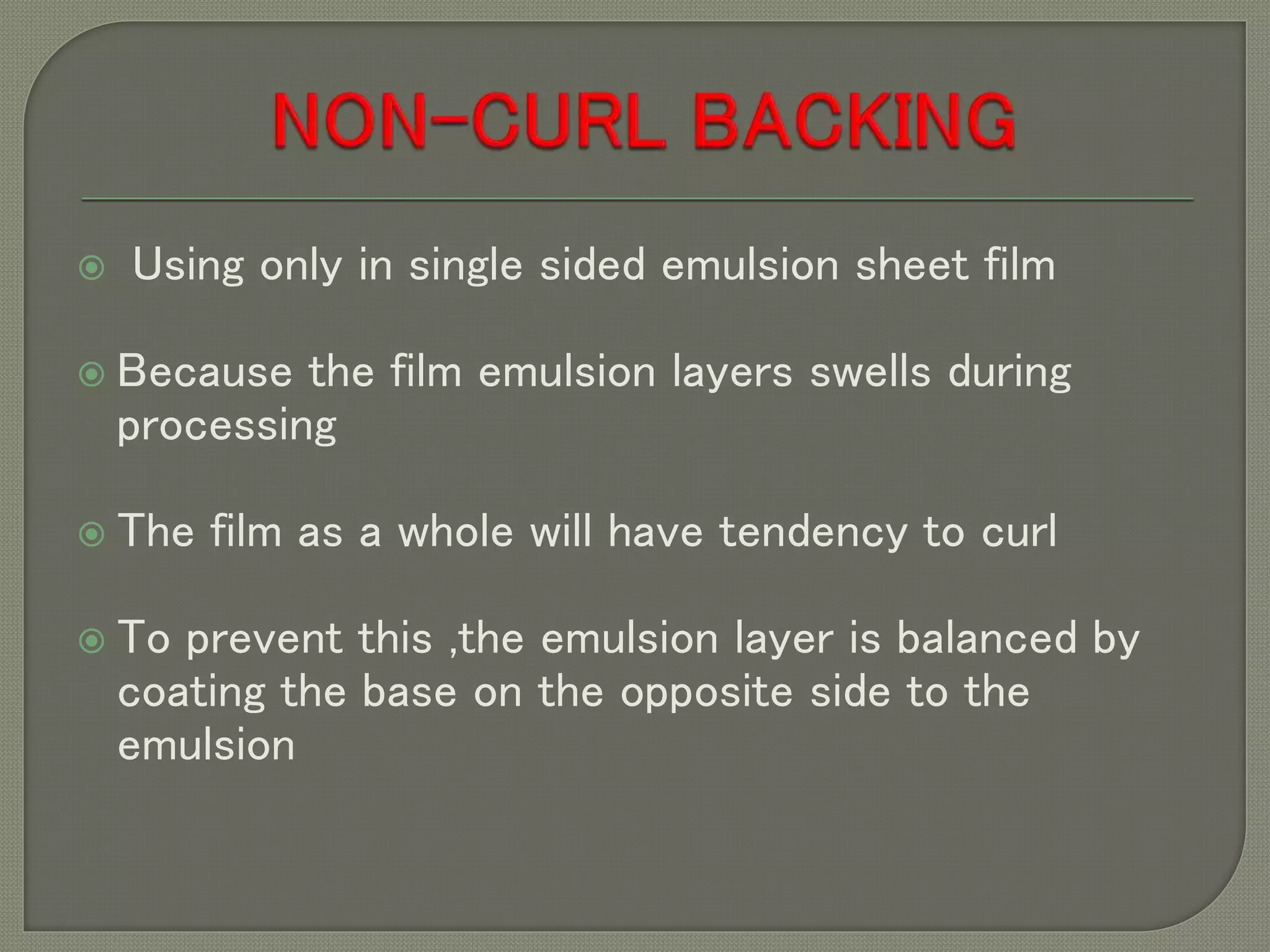
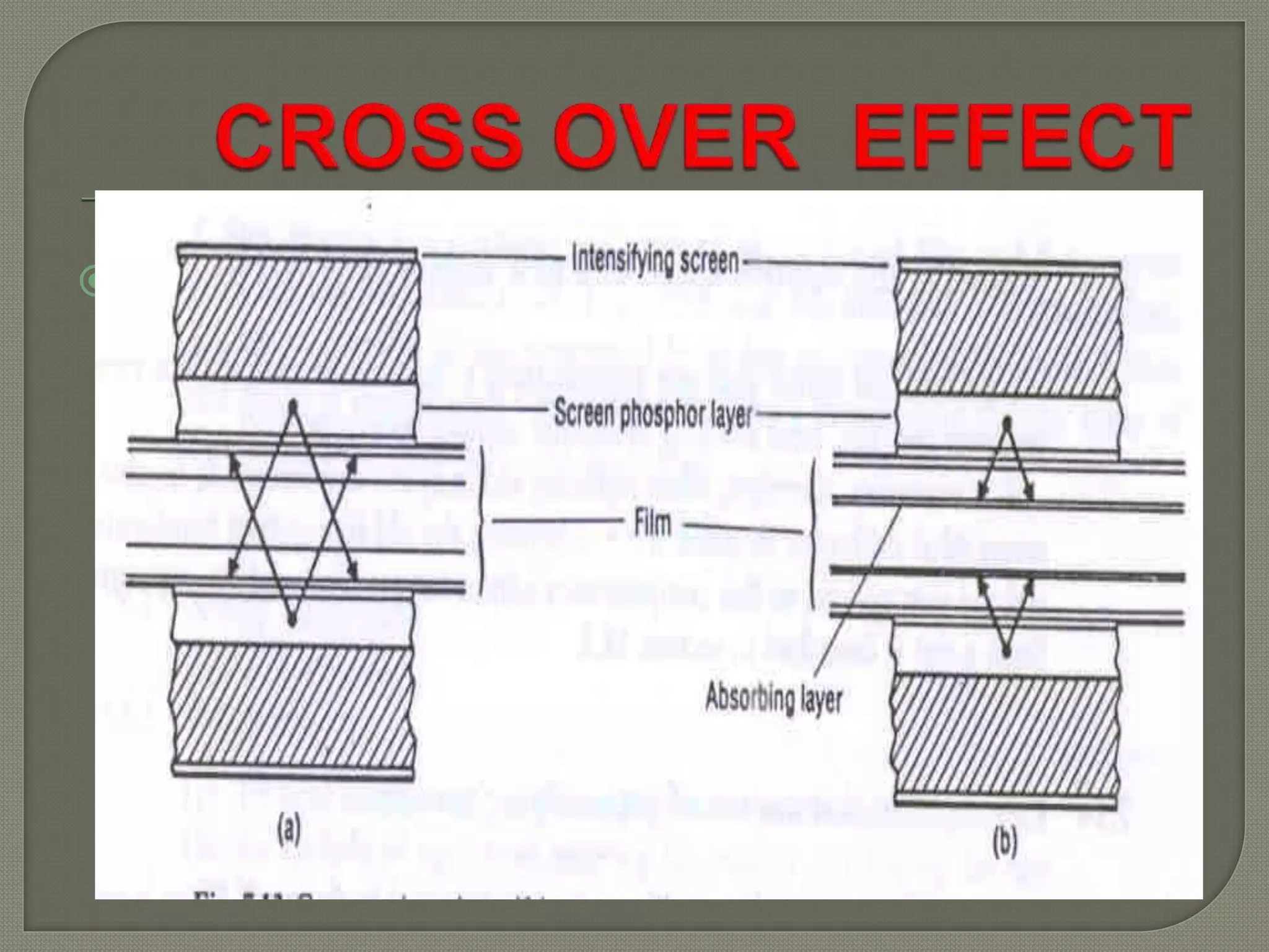
X-Ray
After processing Convert the x-ray
energy into a
visible
light spectrum
Light spectrum
X-Ray Film
X ray film is a photographic film
consisting of photographically
active or radiation-sensitive
DIRECT EXPOSURE FILMS : exposed to x- rays
only e.g.- dental film.
SCREEN FILMS : exposed to x-rays and light.
Film exposed to light only (fluorographic ,
duplicating films)
This is single emulsion film
DUPLITIZED FILM
SINGLE SIDED EMULSION FILM
 Polyester made from polyethylene terephthlate
resin
 THICKNESS OF FILM BASE
 Medical x-ray : 0.18mm
 Fluorographic roll film : 0.08mm
 FUNCTION OF FILM BASE
 Support for emulsion layer.
 To transmit light so that an image can be viewed .
 Transparent and uniform in transmitting light.
 Strong but flexible.
 Uniform in thickness.
 Impermeable to water and processing solution
 Dimensionally Stable throughout processing.
 Inflammable
 Chemically inactive
 Uniform in colour
 Mixture of gelatin solution + solvent of the film base.
 Prevent separation of emulsion layer from the base
 Without subbing layer , the emulsion swells and
contracts during the processing cycle
Consists of silver halide crystals suspended
in gelatin.
Thickness < 0.0005 inch.
Latent image will form in this layer .
Ingredients :
- Gelatin – Suspension medium
- Silver Halide - photosensitive
 Photographic Gelatin - Cattle bone
 Ideal suspension medium – keeps Silver Halide
grains well dispersed & prevents clumping
 Processing solutions penetrate gelatin without
change in strength / performance
 Easy availability
 Uniform quality
Light sensitive material
Contains AgBr : 90 – 99%
AgI : 1 – 10%
Small crystals suspended in Gelatin
Crystal size : 1.0-1.5 microns with 6.3x10⁹
grains per cubic centimeter of emulsion.
NEW FLAT CRYSTALS
OLDER IRREGULAR CRYSTALS
 The grainoffer a larger surface area
to the imaging source , this increase
the filmsensitivityand speed
 Added a lightabsorbingmagenta dye
to the surface of the grains ,known
as optical sensitizing dye
 Advantages:
 Greater sensitivityto light, increased
speed
 Less cross-over effect
Thin layer of clear gelatin which is
applied to the emulsion .
functions
 Protect the emulsion from light pressure
which might occur during use .
 To provide suitable surface characteristics
 Using only in single sided emulsion sheet film
 Because the film emulsion layers swells during
processing
 The film as a whole will have tendency to curl
 To prevent this ,the emulsion layer is balanced by
coating the base on the opposite side to the
emulsion
HALATION : It is a phenomenon
characterised by formation of a diffuse
image or halo around the proper image.
This occurs mainly in the single sided
emulsion film.
Adding a dye to non- curl backing
Adding a dye to base.
• Preferred for single sided emulsion film.
• The dye colour selected is opposite or
complementary to exposing light.
• These dyes are removed during development.
Some times called as anti halo layer or gel
coat
 Using in 35mm still and cine film
 Dyes cannot be removed during development
Crossover is image degradation
caused by light produced in one
intensifying screen passing through
the film base and producing an image
in the opposite emulsion layer
It is a type of halation which occurs
when film is used with intensifying
screen.
Occurs only with double emulsion films
and two screens.
Coloured subbing layer is used.
Addition of magenta dye also reduces
cross over effect.
 It is the sideway scattering of light within the
emulsion
 This contributes to unsharpness (blurring) of image.

based on
- Single emulsion - Screen
type
- Double emulsion - Non
screen
TYPES OF X RAY FILMS
STRUCTURE
PRESENCE OF
SCREEN
DUPLITIZED EMULSION FILM
SINGLE EMULSION FILM
1. Direct- Exposure (non- screen type) film
Intra-oral dental film, kidney surgery film,
radiation monitoring film.
2. Screen-type film
(for use with intensifying screen)
• Screen-type film
• Photofluorographic film
• Cathod-ray-tube (CRT) photography : polaride film
• Duplication film
• Subtraction film
• Laser imaging film
 Also known as non-screen film.
 Some times know as enveloped- wrapped
 exposure to x ray only
 Thicker emulsion is used containing higher
concentration of silver halide.
 Non-screen film is of low speed type.
 A higher radiation dose is used.
PERI-APICAL- 31x41mm film(for single or
groups of teeth)
OCCLUSAL-57X76mm film(mandible or maxillae)
BITE-WING : similar to the periapical ,
positioned vertically behind the upper and lower
teeth .
Patent bite on the flap
Used to demonstrating crown
BITEWING
OCCLUSAL
PERI-APICAL
It enables radiographic exposure of kidney to
be made extra-abdominally during surgery for
stone removal.
Two film feature allows wider range of renal
stones to be demonstrated.
The film are enclosed in a light tight, water
proof polythene packet , the whole packet
is capable of being cold sterilized
The film can be processed in an automatic
processer
 Similar to dental films in appearance.
 On one side of the base is a high speed emulsion, whilst
on the other side is a slower emulsion
 This permits a wide range of exposure level to be
recorded.
 A large exposure to radiation may result in
complete blackening of the fast emulsion
 It is the most widely used image receptor.
 It is used in conjunction with pair of intensifying
screen.
 Good screen film combination provide good quality
radiograph with high speed.
 Screen type
 Cassette fitted with a single intensifying
screen
 Using in mammography
 The films are medium to high contrast with
high definition, capable of demonstrating
micro calcification in soft tissue
Ultrasound
Computerized tomography
Magnetic resonance imaging
Nuclear medicine
Digital subtraction imaging
Single emulsion films used in radiology
 Used in ultrasound imaging.
 It is made up of positive and negative film sheets
with a pod of jellified processing chemistry.
Films should be protected from
 Physical damage
 Light
 High temperature
 High relative humidity
 Harmful gases and fumes
 X-rays and radioactive source
 Fire
CHESNEY S RADIOGRAPHIC IMAGING
RADIOLOGIC SCIENCE FOR
TECHNOLOGIST (BUSHONG)
X  ray film

X ray film

  • 2.
    X-Ray After processing Convertthe x-ray energy into a visible light spectrum Light spectrum X-Ray Film
  • 3.
    X ray filmis a photographic film consisting of photographically active or radiation-sensitive
  • 4.
    DIRECT EXPOSURE FILMS: exposed to x- rays only e.g.- dental film. SCREEN FILMS : exposed to x-rays and light. Film exposed to light only (fluorographic , duplicating films) This is single emulsion film
  • 5.
  • 6.
     Polyester madefrom polyethylene terephthlate resin  THICKNESS OF FILM BASE  Medical x-ray : 0.18mm  Fluorographic roll film : 0.08mm  FUNCTION OF FILM BASE  Support for emulsion layer.  To transmit light so that an image can be viewed .
  • 7.
     Transparent anduniform in transmitting light.  Strong but flexible.  Uniform in thickness.  Impermeable to water and processing solution  Dimensionally Stable throughout processing.  Inflammable  Chemically inactive  Uniform in colour
  • 8.
     Mixture ofgelatin solution + solvent of the film base.  Prevent separation of emulsion layer from the base  Without subbing layer , the emulsion swells and contracts during the processing cycle
  • 9.
    Consists of silverhalide crystals suspended in gelatin. Thickness < 0.0005 inch. Latent image will form in this layer . Ingredients : - Gelatin – Suspension medium - Silver Halide - photosensitive
  • 10.
     Photographic Gelatin- Cattle bone  Ideal suspension medium – keeps Silver Halide grains well dispersed & prevents clumping  Processing solutions penetrate gelatin without change in strength / performance  Easy availability  Uniform quality
  • 11.
    Light sensitive material ContainsAgBr : 90 – 99% AgI : 1 – 10% Small crystals suspended in Gelatin Crystal size : 1.0-1.5 microns with 6.3x10⁹ grains per cubic centimeter of emulsion.
  • 12.
    NEW FLAT CRYSTALS OLDERIRREGULAR CRYSTALS
  • 13.
     The grainoffera larger surface area to the imaging source , this increase the filmsensitivityand speed  Added a lightabsorbingmagenta dye to the surface of the grains ,known as optical sensitizing dye  Advantages:  Greater sensitivityto light, increased speed  Less cross-over effect
  • 14.
    Thin layer ofclear gelatin which is applied to the emulsion . functions  Protect the emulsion from light pressure which might occur during use .  To provide suitable surface characteristics
  • 15.
     Using onlyin single sided emulsion sheet film  Because the film emulsion layers swells during processing  The film as a whole will have tendency to curl  To prevent this ,the emulsion layer is balanced by coating the base on the opposite side to the emulsion
  • 16.
    HALATION : Itis a phenomenon characterised by formation of a diffuse image or halo around the proper image. This occurs mainly in the single sided emulsion film.
  • 17.
    Adding a dyeto non- curl backing Adding a dye to base.
  • 18.
    • Preferred forsingle sided emulsion film. • The dye colour selected is opposite or complementary to exposing light. • These dyes are removed during development. Some times called as anti halo layer or gel coat
  • 19.
     Using in35mm still and cine film  Dyes cannot be removed during development
  • 20.
    Crossover is imagedegradation caused by light produced in one intensifying screen passing through the film base and producing an image in the opposite emulsion layer
  • 21.
    It is atype of halation which occurs when film is used with intensifying screen. Occurs only with double emulsion films and two screens. Coloured subbing layer is used. Addition of magenta dye also reduces cross over effect.
  • 22.
     It isthe sideway scattering of light within the emulsion  This contributes to unsharpness (blurring) of image. 
  • 23.
    based on - Singleemulsion - Screen type - Double emulsion - Non screen TYPES OF X RAY FILMS STRUCTURE PRESENCE OF SCREEN
  • 24.
  • 25.
    1. Direct- Exposure(non- screen type) film Intra-oral dental film, kidney surgery film, radiation monitoring film. 2. Screen-type film (for use with intensifying screen)
  • 26.
    • Screen-type film •Photofluorographic film • Cathod-ray-tube (CRT) photography : polaride film • Duplication film • Subtraction film • Laser imaging film
  • 27.
     Also knownas non-screen film.  Some times know as enveloped- wrapped  exposure to x ray only  Thicker emulsion is used containing higher concentration of silver halide.  Non-screen film is of low speed type.  A higher radiation dose is used.
  • 28.
    PERI-APICAL- 31x41mm film(forsingle or groups of teeth) OCCLUSAL-57X76mm film(mandible or maxillae) BITE-WING : similar to the periapical , positioned vertically behind the upper and lower teeth . Patent bite on the flap Used to demonstrating crown
  • 29.
  • 30.
    It enables radiographicexposure of kidney to be made extra-abdominally during surgery for stone removal. Two film feature allows wider range of renal stones to be demonstrated.
  • 31.
    The film areenclosed in a light tight, water proof polythene packet , the whole packet is capable of being cold sterilized The film can be processed in an automatic processer
  • 32.
     Similar todental films in appearance.  On one side of the base is a high speed emulsion, whilst on the other side is a slower emulsion  This permits a wide range of exposure level to be recorded.  A large exposure to radiation may result in complete blackening of the fast emulsion
  • 33.
     It isthe most widely used image receptor.  It is used in conjunction with pair of intensifying screen.  Good screen film combination provide good quality radiograph with high speed.
  • 34.
     Screen type Cassette fitted with a single intensifying screen  Using in mammography  The films are medium to high contrast with high definition, capable of demonstrating micro calcification in soft tissue
  • 35.
    Ultrasound Computerized tomography Magnetic resonanceimaging Nuclear medicine Digital subtraction imaging Single emulsion films used in radiology
  • 36.
     Used inultrasound imaging.  It is made up of positive and negative film sheets with a pod of jellified processing chemistry.
  • 37.
    Films should beprotected from  Physical damage  Light  High temperature  High relative humidity  Harmful gases and fumes  X-rays and radioactive source  Fire
  • 38.
    CHESNEY S RADIOGRAPHICIMAGING RADIOLOGIC SCIENCE FOR TECHNOLOGIST (BUSHONG)