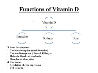
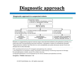
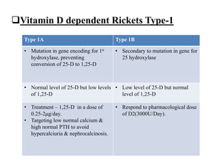
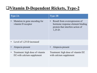
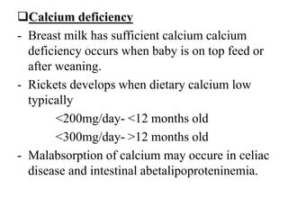
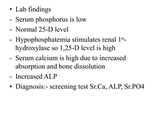
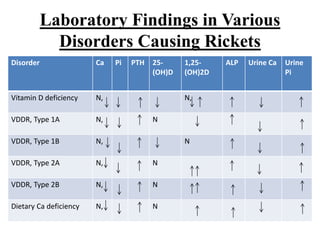
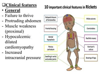
Rickets
Dr. Sachin Subhash Wagh
Dr Avinash Sangle Sir
Vitamin D
• Vitamin D can be synthesized in skin epithelial
cells from cholesterol
• Vitamin D is transported bound to vitamin D-
binding protein to liver where 25-hydroxylase
converts Vitamin D into 25-hydroxyvitamin D,
most abundant circulating form of Vitamin D.
• Final step in activation occurs in kidney, where
enzyme 1α -hydroxylase adds second hydroxyl
group, resulting 1,25-D.
Vitamin D binding protein25-hydroxylase
1α-Hydroxylase
• Most 1,25-D circulates bound to vitamin D-binding
protein.
• 1α-hydroxylase regulation
Up regulation
PTH
Hypophosphatemia
Down regulation
Hyperphosphatemia
1,25-D
• The following standards for defining vitamin
D status in healthy children and adolescents,
based on serum concentrations of 25OHD:
• ●Vitamin D sufficiency: 20 to 100 ng/mL (50
to 250 nmol/L)
• ●Vitamin D insufficiency: 12 to 20 ng/mL (30
to 50 nmol/L)
• ●Vitamin D deficiency:
<12 ng/mL (<30 nmol/L)
Functions of Vitamin D
Vitamin D
Intestine
Kidney Bone
 Bone Development
- Calcium absorption (small Intestine)
- Calcium Resorption ( Bone & Kidneys)
- Maintain blood calcium levels
- Phosphorus absorption
 Hormones
- Regulation of gene expression
- Cell Growth.
• Immunological function of vitamin D
Deficiency
 Rickets – disease of growing bone caused by
unmineralised matrix at growth plates in children only before
fusion of epiphysis.
 Osteomalcia - inadequet mineralization of bone in
children and adults.
 Osteoporosis- Proportionate loss of bone volume and
minerals
Rickets
Introduction
• Rickets is a condition that affects bone development in
children. It causes the bones to become soft and weak,
which can lead to bone deformities.
• Rickets in adults is known as osteomalacia or soft
bones.
• Rickets can cause bone pain, poor growth and
deformities of the skeleton, such as bowed legs,
curvature of the spine, and thickening of the ankles,
wrists and knees.
• Children with rickets are also more likely to fracture
their bones.
Rickets
 Causes
• Vitamin D Disorder
- Nutritional vitamin D deficiency
- Congenital vitamin D deficiency
- Secondary vitamin D deficiency
- Malabsorption
- Increased degradation
- Decreased liver 25-hydroxylase
- Vitamin D-dependent rickets types 1A and 2B
- Vitamin D-dependent rickets type 2A types 2B
- Chronic kidney disease
• Calcium deficiency
- Low intake
- Premature infants
- Malabsorption
- Primary disease
- Dietary inhibitors of calcium absorption
• Phosphorus deficiency
- Inadequate intake
- Premature infants
- Aluminum containing antacid
• Renal losses
- McCune-albright syndrome
- Fanconi syndrome
- X-linked hypophosphatemic rickets
Diagnostic approach
Causes of Rickets
Nutritional vitamin D deficicency
- Symptoms of hypocalcaemia
- Pneumonia, muscle weakness
- Increased PTH & hypophosphatemia
- Bicarbonate wasting
- Treatment:- Stoss therapy, Vitamin D (300000-
600000IU) orally or IM as 2-4 doses over 1 day. OR
- Vitamin D 2000IU/day for 3 months
- Prevention <1 year 400IU/day >1 year 600IU/day.
Vitamin D dependent Rickets Type-1
Type 1A Type 1B
• Mutation in gene encoding for 1α
hydroxylase, preventing
conversion of 25-D to 1,25-D
• Secondary to mutation in gene for
25 hydroxylase
• Normal level of 25-D but low levels
of 1,25-D
• Low level of 25-D but normal
level of 1,25-D
• Treatment – 1,25-D in a dose of
0.25-2μg/day.
• Targeting low normal calcium &
high normal PTH to avoid
hypercalciuria & nephrocalcinosis.
• Respond to pharmacological dose
of D2(3000U/Day).
Vitamin D-Dependent Rickets, Type-2
Type 2A Type 2B
• Mutation in gene encoding for
vitamin D receptor
• Result from overexpression of
hormone response element binding
protein that interfere action of
1,25-D .
• Level of 1,25-D increased
• Alopecia present • Alopecia present
• Treatment: high dose of vitamin
D2 with calcium supplement
Treatment: high dose of vitamin D2
with calcium supplement
Chronic kidney disease
- Decreased activity of 1α-hydroxylase
- Decreased level of 1,25-D associated with
Hyperphosphatemia as result of decreased
renal excretion.
- Treatment:- Vitamin D3 which causes
absorption of calcium, decreases level of PTH.
- Hyperphosphatemia is stimulus for PTH, low
phosphorus in diet and phosphate binder
adviced
Calcium deficiency
- Breast milk has sufficient calcium calcium
deficiency occurs when baby is on top feed or
after weaning.
- Rickets develops when dietary calcium low
typically
<200mg/day- <12 months old
<300mg/day- >12 months old
- Malabsorption of calcium may occure in celiac
disease and intestinal abetalipoproteninemia.
-Diagnosis:- increased ALP, increased PTH,
increased 1,25-D,
Calcium low or normal.
Serum phosphorus is low
Decreased urinary calcium & increased
phosphorus excretion.
-Treatment:- Adequate calcium diet
1-3 years – 700mg/day
4-8years – 1000mg/day
9-18 years – 1300mg/day.
Rickets of Prematurity
- Rickets in VLBW has become significant
problem as survival rate of VLBW increased
- Transfer of calcium & phosphorus from
mother to fetus occurs throughout pregnancy
but 80% occurs in 3rd trimester
- Rickets occurs after 1-4 months after birth can
have non traumatic fracture
- Respiratory distress from atelectasis & poor
ventilation
- Respiratory distress develops after >5 weeks
of birth
• Lab findings
- Serum phosphorus is low
- Normal 25-D level
- Hypophosphatemia stimulates renal 1α-
hydroxylase so 1,25-D level is high
- Serum calcium is high due to increased
absorption and bone dissolution
- Increased ALP
• Diagnosis:- screening test Sr.Ca, ALP, Sr.PO4
• Prevention:- provision of adequate amount of
calcium, phosphorus and vitamin D.
- Avoid Soy formula feed because decreased
bioavailability of calcium and phosphorus.
- 400 IU/day of Vit-D
Laboratory Findings in Various
Disorders Causing Rickets
Disorder Ca Pi PTH 25-
(OH)D
1,25-
(OH)2D
ALP Urine Ca Urine
Pi
Vitamin D deficiency N, N,
VDDR, Type 1A N, N
VDDR, Type 1B N, N
VDDR, Type 2A N, N
VDDR, Type 2B N, N
Dietary Ca deficiency N, N
Clinical features
• General
- Failure to thrive
- Protruding abdomen
- Muscle weakness
(proximal)
- Hypocalcemic
dilated
cardiomyopathy
- Increased
intracranial pressure
• Head
- Craniotabes
- Frontal bossing
- Delayed fontanel
closure
- Delayed dentition
- Caries
- Craniosynostosis
• Chest
- Rachiatic rosary
- Harrison grove
- Respiratory
atelectasis and
infection
• Back
- Scoliosis
- Kyphosis
- Lordosis
• Extremities
- Enlargement of wrist and
ankles
- Valgus or varus deformity
- Windswept deformity
- Anterior bowing of tibia &
femur
- Coxa vara
Radiographic features
Message to parents
THANK YOU

Rickets

  • 1.
    Rickets Dr. Sachin SubhashWagh Dr Avinash Sangle Sir
  • 2.
    Vitamin D • VitaminD can be synthesized in skin epithelial cells from cholesterol • Vitamin D is transported bound to vitamin D- binding protein to liver where 25-hydroxylase converts Vitamin D into 25-hydroxyvitamin D, most abundant circulating form of Vitamin D. • Final step in activation occurs in kidney, where enzyme 1α -hydroxylase adds second hydroxyl group, resulting 1,25-D.
  • 3.
    Vitamin D bindingprotein25-hydroxylase 1α-Hydroxylase
  • 4.
    • Most 1,25-Dcirculates bound to vitamin D-binding protein. • 1α-hydroxylase regulation Up regulation PTH Hypophosphatemia Down regulation Hyperphosphatemia 1,25-D
  • 5.
    • The followingstandards for defining vitamin D status in healthy children and adolescents, based on serum concentrations of 25OHD: • ●Vitamin D sufficiency: 20 to 100 ng/mL (50 to 250 nmol/L) • ●Vitamin D insufficiency: 12 to 20 ng/mL (30 to 50 nmol/L) • ●Vitamin D deficiency: <12 ng/mL (<30 nmol/L)
  • 6.
    Functions of VitaminD Vitamin D Intestine Kidney Bone  Bone Development - Calcium absorption (small Intestine) - Calcium Resorption ( Bone & Kidneys) - Maintain blood calcium levels - Phosphorus absorption  Hormones - Regulation of gene expression - Cell Growth.
  • 7.
  • 8.
    Deficiency  Rickets –disease of growing bone caused by unmineralised matrix at growth plates in children only before fusion of epiphysis.  Osteomalcia - inadequet mineralization of bone in children and adults.  Osteoporosis- Proportionate loss of bone volume and minerals
  • 9.
    Rickets Introduction • Rickets isa condition that affects bone development in children. It causes the bones to become soft and weak, which can lead to bone deformities. • Rickets in adults is known as osteomalacia or soft bones. • Rickets can cause bone pain, poor growth and deformities of the skeleton, such as bowed legs, curvature of the spine, and thickening of the ankles, wrists and knees. • Children with rickets are also more likely to fracture their bones.
  • 10.
    Rickets  Causes • VitaminD Disorder - Nutritional vitamin D deficiency - Congenital vitamin D deficiency - Secondary vitamin D deficiency - Malabsorption - Increased degradation - Decreased liver 25-hydroxylase - Vitamin D-dependent rickets types 1A and 2B - Vitamin D-dependent rickets type 2A types 2B - Chronic kidney disease
  • 11.
    • Calcium deficiency -Low intake - Premature infants - Malabsorption - Primary disease - Dietary inhibitors of calcium absorption • Phosphorus deficiency - Inadequate intake - Premature infants - Aluminum containing antacid
  • 12.
    • Renal losses -McCune-albright syndrome - Fanconi syndrome - X-linked hypophosphatemic rickets
  • 13.
  • 15.
    Causes of Rickets Nutritionalvitamin D deficicency - Symptoms of hypocalcaemia - Pneumonia, muscle weakness - Increased PTH & hypophosphatemia - Bicarbonate wasting - Treatment:- Stoss therapy, Vitamin D (300000- 600000IU) orally or IM as 2-4 doses over 1 day. OR - Vitamin D 2000IU/day for 3 months - Prevention <1 year 400IU/day >1 year 600IU/day.
  • 16.
    Vitamin D dependentRickets Type-1 Type 1A Type 1B • Mutation in gene encoding for 1α hydroxylase, preventing conversion of 25-D to 1,25-D • Secondary to mutation in gene for 25 hydroxylase • Normal level of 25-D but low levels of 1,25-D • Low level of 25-D but normal level of 1,25-D • Treatment – 1,25-D in a dose of 0.25-2μg/day. • Targeting low normal calcium & high normal PTH to avoid hypercalciuria & nephrocalcinosis. • Respond to pharmacological dose of D2(3000U/Day).
  • 17.
    Vitamin D-Dependent Rickets,Type-2 Type 2A Type 2B • Mutation in gene encoding for vitamin D receptor • Result from overexpression of hormone response element binding protein that interfere action of 1,25-D . • Level of 1,25-D increased • Alopecia present • Alopecia present • Treatment: high dose of vitamin D2 with calcium supplement Treatment: high dose of vitamin D2 with calcium supplement
  • 18.
    Chronic kidney disease -Decreased activity of 1α-hydroxylase - Decreased level of 1,25-D associated with Hyperphosphatemia as result of decreased renal excretion. - Treatment:- Vitamin D3 which causes absorption of calcium, decreases level of PTH. - Hyperphosphatemia is stimulus for PTH, low phosphorus in diet and phosphate binder adviced
  • 19.
    Calcium deficiency - Breastmilk has sufficient calcium calcium deficiency occurs when baby is on top feed or after weaning. - Rickets develops when dietary calcium low typically <200mg/day- <12 months old <300mg/day- >12 months old - Malabsorption of calcium may occure in celiac disease and intestinal abetalipoproteninemia.
  • 20.
    -Diagnosis:- increased ALP,increased PTH, increased 1,25-D, Calcium low or normal. Serum phosphorus is low Decreased urinary calcium & increased phosphorus excretion. -Treatment:- Adequate calcium diet 1-3 years – 700mg/day 4-8years – 1000mg/day 9-18 years – 1300mg/day.
  • 21.
    Rickets of Prematurity -Rickets in VLBW has become significant problem as survival rate of VLBW increased - Transfer of calcium & phosphorus from mother to fetus occurs throughout pregnancy but 80% occurs in 3rd trimester - Rickets occurs after 1-4 months after birth can have non traumatic fracture - Respiratory distress from atelectasis & poor ventilation - Respiratory distress develops after >5 weeks of birth
  • 22.
    • Lab findings -Serum phosphorus is low - Normal 25-D level - Hypophosphatemia stimulates renal 1α- hydroxylase so 1,25-D level is high - Serum calcium is high due to increased absorption and bone dissolution - Increased ALP • Diagnosis:- screening test Sr.Ca, ALP, Sr.PO4
  • 23.
    • Prevention:- provisionof adequate amount of calcium, phosphorus and vitamin D. - Avoid Soy formula feed because decreased bioavailability of calcium and phosphorus. - 400 IU/day of Vit-D
  • 24.
    Laboratory Findings inVarious Disorders Causing Rickets Disorder Ca Pi PTH 25- (OH)D 1,25- (OH)2D ALP Urine Ca Urine Pi Vitamin D deficiency N, N, VDDR, Type 1A N, N VDDR, Type 1B N, N VDDR, Type 2A N, N VDDR, Type 2B N, N Dietary Ca deficiency N, N
  • 25.
    Clinical features • General -Failure to thrive - Protruding abdomen - Muscle weakness (proximal) - Hypocalcemic dilated cardiomyopathy - Increased intracranial pressure
  • 26.
    • Head - Craniotabes -Frontal bossing - Delayed fontanel closure - Delayed dentition - Caries - Craniosynostosis • Chest - Rachiatic rosary - Harrison grove - Respiratory atelectasis and infection
  • 27.
    • Back - Scoliosis -Kyphosis - Lordosis • Extremities - Enlargement of wrist and ankles - Valgus or varus deformity - Windswept deformity - Anterior bowing of tibia & femur - Coxa vara
  • 28.
  • 29.
  • 30.