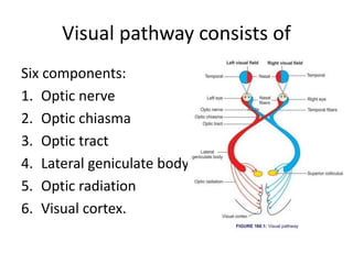
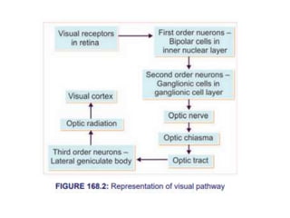
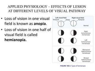
SlideShare a Scribd company logo
1 of 16
VISUAL PATHWAY
SCK
Visual pathway consists of
Six components:
1. Optic nerve
2. Optic chiasma
3. Optic tract
4. Lateral geniculate body
5. Optic radiation
6. Visual cortex.
OPTIC NERVE
• Optic nerve is formed by the axons of ganglionic
cells.
• Optic nerve leaves the eye through optic disk.
• The fibers from temporal part of retina are in
lateral part of the nerve and carry the impulses
from nasal half of visual field of same eye.
• The fibers from nasal part of retina are in medial
part of the nerve and carry the impulses from
temporal half of visual field of same eye.
OPTIC CHIASMA
• Medial fibers of each optic nerve cross the
midline and join the uncrossed lateral fibers of
opposite side, to form the optic tract.
• This area of crossing of the optic nerve fibers
is called optic chiasma.
OPTIC TRACT
• Optic tract is formed by uncrossed fibers of optic nerve on the same side and
crossed fibers of optic nerve from the opposite side.
• All the fibers of optic tract run backward, outward and towards the cerebral
peduncle.
• While reaching the peduncle, the fibers pass between tuber cinereum and
anterior perforated substance.
• Then, the fibers turn around the peduncle to reach the lateral geniculate body in
thalamus. Here, many fibers synapse while few fibers just pass through this and
run towards superior colliculus in midbrain.
• Fibers from fovea do not enter superior colliculus. Some fibers from fovea of each
side pass through the optic tract of same side and others through the optic tract of
opposite side.
• Due to crossing of medial fibers in optic chiasma, the left optic tract carries
impulses from temporal part of left retina and nasal part of right retina, i.e. it is
responsible for vision in nasal half of left visual field and temporal half of right
visual field.
• The right optic tract contains fibers from nasal half of left retina and temporal half
of right retina. It is responsible for vision in temporal half of left visual field and
nasal half of right visual field.
• The right optic tract contains fibers from nasal half of left retina and temporal half
of right retina. It is responsible for vision in temporal half of left visual field and
nasal half of right visual field.
LATERAL GENICULATE BODY
• Majority of the fibers of optic tract terminate in lateral
geniculate body, which forms the subcortical center for
visual sensation. From here, the geniculocalcarine tract or
optic radiation arises. This tract is the last relay of visual
pathway.
• Some of the fibers from optic tract do not synapse in lateral
geniculate body, but pass through it and terminate in one of
the following centers:
I. Superior colliculus: It is concerned with reflex
movements of eyeballs and head, in response to optic
stimulus
II. Pretectal nucleus: It is concerned with light reflexes
III. Supraoptic nucleus of hypothalamus: It is concerned
with the retinal control of pituitary in animals. But in
human, it does not play any important role.
OPTIC RADIATION
• Fibers from lateral geniculate body pass through
internal capsule and form optic radiation.
• The fibers between lateral geniculate body and
visual cortex are also called geniculocalcarine
fibers.
• Optic radiation ends in visual cortex
VISUAL CORTEX
• Primary cortical center for vision is called visual
cortex, which is located on the medial surface of
occipital lobe.
• It forms the walls and lips of calcarine fissure in
medial surface of occipital lobe.
• There is a definite localization of retinal
projections upon visual cortex.
• The peripheral retinal representation occupies the
anterior part of visual cortex. Macular
representation occupies the posterior part of
visual cortex near occipital pole.
Areas of Visual Cortex and their
Function
• Three areas are present in visual cortex:
1. Primary visual area (area 17), which is
concerned with the perception of visual
impulses.
2. Secondary visual area or visual association
area (area 18), which is concerned with the
interpretation of visual impulses.
3. Occipital eye field (area 19), which is
concerned with the movement of eyes
APPLIED PHYSIOLOGY – EFFECTS OF LESION
AT DIFFERENT LEVELS OF VISUAL PATHWAY
• Loss of vision in one visual
field is known as anopia.
• Loss of vision in one half of
visual field is called
hemianopia.
Hemianopia is classified into two types:
1. Homonymous hemianopia
2. Heteronymous hemianopia.
Homonymous hemianopia: Homonymous hemianopia means loss of vision in the same halves
of both the visual fields. Loss of vision in right half of visual field of both eyes is known as right
homonymous hemianopia. Similarly, left homonymous hemianopia means loss of vision in left
half of visual field of both eyes.
Heteronymous hemianopia: Heteronymous hemianopia means loss of vision in opposite halves
of visual field. For example, binasal heteronymous hemianopia means loss of vision in right half
of left visual field and left half of right visual field (nasal half of both visual fields). Bitemporal
heteronymous hemianopia is the loss of sight in left side of left visual field and right side of right
visual field (temporal half of both visual fields).
Effects of Lesion of Optic Nerve
• Lesion in one optic nerve will cause total
blindness or anopia in the corresponding
visual field. Lesion occurs due to increased
intracranial pressure.
Effects of Lesion of Visual Cortex
• Macular sparing In all the conditions mentioned above, total blindness
does not occur because, the macular vision is not lost.
• This phenomenon in which the macular vision is retained (unaffected) in
conditions of hemianopia is called macular sparing.
• Macular sparing occurs because of the following reasons:
1. Fibers from macula project into the visual cortex of both sides.
2. Fibers from macular region are projected into both anterior and posterior
parts of each visual cortex.
Only the bilateral lesion of visual cortex causes total blindness.
VISUAL PATHWAY.pptx
VISUAL PATHWAY.pptx

More Related Content

What's hot

Abnormalities of pupillary reaction
Abnormalities of pupillary reactionAbnormalities of pupillary reaction
Abnormalities of pupillary reaction
Sameeksha Alva
 
Extra ocu.movement by pushkar dhir
Extra ocu.movement by pushkar dhirExtra ocu.movement by pushkar dhir
Extra ocu.movement by pushkar dhir
Pushkar Dhir
 

What's hot (20)

Abnormalities of pupillary reaction
Abnormalities of pupillary reactionAbnormalities of pupillary reaction
Abnormalities of pupillary reaction
 
Anatomy of the orbit (nerves and vessels)
Anatomy of the orbit (nerves and vessels)Anatomy of the orbit (nerves and vessels)
Anatomy of the orbit (nerves and vessels)
 
Visual pathway ppt
Visual pathway pptVisual pathway ppt
Visual pathway ppt
 
Anatomy of eyeball
Anatomy of eyeballAnatomy of eyeball
Anatomy of eyeball
 
Visual pathway and lesion
Visual pathway and lesionVisual pathway and lesion
Visual pathway and lesion
 
Extraocular muscles
Extraocular musclesExtraocular muscles
Extraocular muscles
 
Color vision : introduction, classification, causes
Color vision : introduction, classification, causesColor vision : introduction, classification, causes
Color vision : introduction, classification, causes
 
Visual cortex
Visual cortexVisual cortex
Visual cortex
 
The orbit of eye
The orbit of eyeThe orbit of eye
The orbit of eye
 
Frames and frame measurements__optometry_india_eyenirvaan.com
Frames and frame measurements__optometry_india_eyenirvaan.comFrames and frame measurements__optometry_india_eyenirvaan.com
Frames and frame measurements__optometry_india_eyenirvaan.com
 
Extra ocu.movement by pushkar dhir
Extra ocu.movement by pushkar dhirExtra ocu.movement by pushkar dhir
Extra ocu.movement by pushkar dhir
 
Color vision : Physiology ,Defects, Detection, Diagnosis & Management
Color vision :  Physiology ,Defects, Detection, Diagnosis & ManagementColor vision :  Physiology ,Defects, Detection, Diagnosis & Management
Color vision : Physiology ,Defects, Detection, Diagnosis & Management
 
Orbit and extra ocular muscles
Orbit and extra ocular musclesOrbit and extra ocular muscles
Orbit and extra ocular muscles
 
Orbital blood supply
Orbital blood supplyOrbital blood supply
Orbital blood supply
 
EXTRA OCULAR MUSCLES ANATOMY
EXTRA OCULAR MUSCLES ANATOMYEXTRA OCULAR MUSCLES ANATOMY
EXTRA OCULAR MUSCLES ANATOMY
 
Anatomy of orbital cavity
Anatomy of orbital cavityAnatomy of orbital cavity
Anatomy of orbital cavity
 
Ethics
EthicsEthics
Ethics
 
Extraocular muscles
Extraocular muscles Extraocular muscles
Extraocular muscles
 
Oculomotor nerve
Oculomotor nerveOculomotor nerve
Oculomotor nerve
 
Extraocular muscles dr.gosai
Extraocular muscles dr.gosaiExtraocular muscles dr.gosai
Extraocular muscles dr.gosai
 

Similar to VISUAL PATHWAY.pptx

Visual pathway and defects
Visual pathway and defectsVisual pathway and defects
Visual pathway and defects
Prashanth Reddy
 
Anatomy of visual pathway and its lesions.
Anatomy of visual pathway and its lesions.Anatomy of visual pathway and its lesions.
Anatomy of visual pathway and its lesions.
Ruchi Pherwani
 

Similar to VISUAL PATHWAY.pptx (20)

Presentation (4).pptx
Presentation (4).pptxPresentation (4).pptx
Presentation (4).pptx
 
Central visual pathways
Central visual pathwaysCentral visual pathways
Central visual pathways
 
Generator of vep
Generator of vepGenerator of vep
Generator of vep
 
Visual pathway and defects
Visual pathway and defectsVisual pathway and defects
Visual pathway and defects
 
Retina and visual tract
Retina and visual tractRetina and visual tract
Retina and visual tract
 
NEURO-OPHTHALMOLOGY
NEURO-OPHTHALMOLOGY NEURO-OPHTHALMOLOGY
NEURO-OPHTHALMOLOGY
 
Neuro-Ophthalmology_Dr. Bastola.pptx
Neuro-Ophthalmology_Dr. Bastola.pptxNeuro-Ophthalmology_Dr. Bastola.pptx
Neuro-Ophthalmology_Dr. Bastola.pptx
 
Anatomy of visual pathway and its lesions.
Anatomy of visual pathway and its lesions.Anatomy of visual pathway and its lesions.
Anatomy of visual pathway and its lesions.
 
Pns
PnsPns
Pns
 
visual pathway & its lesions.pptx
visual pathway & its lesions.pptxvisual pathway & its lesions.pptx
visual pathway & its lesions.pptx
 
Optic
OpticOptic
Optic
 
Visual pathway
Visual pathwayVisual pathway
Visual pathway
 
Eye optic nerve and visal pathway.pdf
Eye optic nerve and visal pathway.pdfEye optic nerve and visal pathway.pdf
Eye optic nerve and visal pathway.pdf
 
Special sensory pathways
Special sensory pathwaysSpecial sensory pathways
Special sensory pathways
 
optic pathway and its lesions (2).pptx
optic pathway and its lesions (2).pptxoptic pathway and its lesions (2).pptx
optic pathway and its lesions (2).pptx
 
Cranial Nerve II and Visual Pathways.pptx
Cranial Nerve II and Visual Pathways.pptxCranial Nerve II and Visual Pathways.pptx
Cranial Nerve II and Visual Pathways.pptx
 
THE OPTIC NERVE
THE OPTIC NERVETHE OPTIC NERVE
THE OPTIC NERVE
 
The Eye
The EyeThe Eye
The Eye
 
Visual pathway
Visual pathwayVisual pathway
Visual pathway
 
visualpathway-170222022827 (1).pdf
visualpathway-170222022827 (1).pdfvisualpathway-170222022827 (1).pdf
visualpathway-170222022827 (1).pdf
 

More from Changez1993 (11)

Human Ear.pptx
Human Ear.pptxHuman Ear.pptx
Human Ear.pptx
 
Blood Pathology BS Cardiology.pptx
Blood Pathology BS Cardiology.pptxBlood Pathology BS Cardiology.pptx
Blood Pathology BS Cardiology.pptx
 
THE AXILLA.pptx
THE AXILLA.pptxTHE AXILLA.pptx
THE AXILLA.pptx
 
axillary artery-vein-nerve.ppt
axillary artery-vein-nerve.pptaxillary artery-vein-nerve.ppt
axillary artery-vein-nerve.ppt
 
The Muscles of the Thoracic Cage.pptx
The Muscles of the Thoracic Cage.pptxThe Muscles of the Thoracic Cage.pptx
The Muscles of the Thoracic Cage.pptx
 
Neuromuscular Junction, Skeletal Muscle contraction and Motor Unite.pptx
Neuromuscular Junction, Skeletal Muscle contraction  and Motor Unite.pptxNeuromuscular Junction, Skeletal Muscle contraction  and Motor Unite.pptx
Neuromuscular Junction, Skeletal Muscle contraction and Motor Unite.pptx
 
BIOMECHANICS OF ELBOW COMPLEX 1.pptx
BIOMECHANICS OF ELBOW COMPLEX 1.pptxBIOMECHANICS OF ELBOW COMPLEX 1.pptx
BIOMECHANICS OF ELBOW COMPLEX 1.pptx
 
parkinson.pptx
parkinson.pptxparkinson.pptx
parkinson.pptx
 
facialnerveanatomy-180629040852.pdf
facialnerveanatomy-180629040852.pdffacialnerveanatomy-180629040852.pdf
facialnerveanatomy-180629040852.pdf
 
STROKE.pptx
STROKE.pptxSTROKE.pptx
STROKE.pptx
 
Neuroanatomy Semester 4.docx
Neuroanatomy Semester 4.docxNeuroanatomy Semester 4.docx
Neuroanatomy Semester 4.docx
 

Recently uploaded

Call Girls Aurangabad Just Call 8250077686 Top Class Call Girl Service Available
Call Girls Aurangabad Just Call 8250077686 Top Class Call Girl Service AvailableCall Girls Aurangabad Just Call 8250077686 Top Class Call Girl Service Available
Call Girls Aurangabad Just Call 8250077686 Top Class Call Girl Service Available
Dipal Arora
 
College Call Girls in Haridwar 9667172968 Short 4000 Night 10000 Best call gi...
College Call Girls in Haridwar 9667172968 Short 4000 Night 10000 Best call gi...College Call Girls in Haridwar 9667172968 Short 4000 Night 10000 Best call gi...
College Call Girls in Haridwar 9667172968 Short 4000 Night 10000 Best call gi...
perfect solution
 

Recently uploaded (20)

Night 7k to 12k Navi Mumbai Call Girl Photo 👉 BOOK NOW 9833363713 👈 ♀️ night ...
Night 7k to 12k Navi Mumbai Call Girl Photo 👉 BOOK NOW 9833363713 👈 ♀️ night ...Night 7k to 12k Navi Mumbai Call Girl Photo 👉 BOOK NOW 9833363713 👈 ♀️ night ...
Night 7k to 12k Navi Mumbai Call Girl Photo 👉 BOOK NOW 9833363713 👈 ♀️ night ...
 
Call Girls Guntur Just Call 8250077686 Top Class Call Girl Service Available
Call Girls Guntur  Just Call 8250077686 Top Class Call Girl Service AvailableCall Girls Guntur  Just Call 8250077686 Top Class Call Girl Service Available
Call Girls Guntur Just Call 8250077686 Top Class Call Girl Service Available
 
Premium Call Girls In Jaipur {8445551418} ❤️VVIP SEEMA Call Girl in Jaipur Ra...
Premium Call Girls In Jaipur {8445551418} ❤️VVIP SEEMA Call Girl in Jaipur Ra...Premium Call Girls In Jaipur {8445551418} ❤️VVIP SEEMA Call Girl in Jaipur Ra...
Premium Call Girls In Jaipur {8445551418} ❤️VVIP SEEMA Call Girl in Jaipur Ra...
 
VIP Service Call Girls Sindhi Colony 📳 7877925207 For 18+ VIP Call Girl At Th...
VIP Service Call Girls Sindhi Colony 📳 7877925207 For 18+ VIP Call Girl At Th...VIP Service Call Girls Sindhi Colony 📳 7877925207 For 18+ VIP Call Girl At Th...
VIP Service Call Girls Sindhi Colony 📳 7877925207 For 18+ VIP Call Girl At Th...
 
Top Rated Bangalore Call Girls Mg Road ⟟ 9332606886 ⟟ Call Me For Genuine S...
Top Rated Bangalore Call Girls Mg Road ⟟   9332606886 ⟟ Call Me For Genuine S...Top Rated Bangalore Call Girls Mg Road ⟟   9332606886 ⟟ Call Me For Genuine S...
Top Rated Bangalore Call Girls Mg Road ⟟ 9332606886 ⟟ Call Me For Genuine S...
 
Book Paid Powai Call Girls Mumbai 𖠋 9930245274 𖠋Low Budget Full Independent H...
Book Paid Powai Call Girls Mumbai 𖠋 9930245274 𖠋Low Budget Full Independent H...Book Paid Powai Call Girls Mumbai 𖠋 9930245274 𖠋Low Budget Full Independent H...
Book Paid Powai Call Girls Mumbai 𖠋 9930245274 𖠋Low Budget Full Independent H...
 
Call Girls in Delhi Triveni Complex Escort Service(🔝))/WhatsApp 97111⇛47426
Call Girls in Delhi Triveni Complex Escort Service(🔝))/WhatsApp 97111⇛47426Call Girls in Delhi Triveni Complex Escort Service(🔝))/WhatsApp 97111⇛47426
Call Girls in Delhi Triveni Complex Escort Service(🔝))/WhatsApp 97111⇛47426
 
Call Girls Aurangabad Just Call 8250077686 Top Class Call Girl Service Available
Call Girls Aurangabad Just Call 8250077686 Top Class Call Girl Service AvailableCall Girls Aurangabad Just Call 8250077686 Top Class Call Girl Service Available
Call Girls Aurangabad Just Call 8250077686 Top Class Call Girl Service Available
 
Call Girls Kochi Just Call 8250077686 Top Class Call Girl Service Available
Call Girls Kochi Just Call 8250077686 Top Class Call Girl Service AvailableCall Girls Kochi Just Call 8250077686 Top Class Call Girl Service Available
Call Girls Kochi Just Call 8250077686 Top Class Call Girl Service Available
 
Top Rated Bangalore Call Girls Ramamurthy Nagar ⟟ 9332606886 ⟟ Call Me For G...
Top Rated Bangalore Call Girls Ramamurthy Nagar ⟟  9332606886 ⟟ Call Me For G...Top Rated Bangalore Call Girls Ramamurthy Nagar ⟟  9332606886 ⟟ Call Me For G...
Top Rated Bangalore Call Girls Ramamurthy Nagar ⟟ 9332606886 ⟟ Call Me For G...
 
Call Girls Agra Just Call 8250077686 Top Class Call Girl Service Available
Call Girls Agra Just Call 8250077686 Top Class Call Girl Service AvailableCall Girls Agra Just Call 8250077686 Top Class Call Girl Service Available
Call Girls Agra Just Call 8250077686 Top Class Call Girl Service Available
 
Call Girls Coimbatore Just Call 9907093804 Top Class Call Girl Service Available
Call Girls Coimbatore Just Call 9907093804 Top Class Call Girl Service AvailableCall Girls Coimbatore Just Call 9907093804 Top Class Call Girl Service Available
Call Girls Coimbatore Just Call 9907093804 Top Class Call Girl Service Available
 
Best Rate (Guwahati ) Call Girls Guwahati ⟟ 8617370543 ⟟ High Class Call Girl...
Best Rate (Guwahati ) Call Girls Guwahati ⟟ 8617370543 ⟟ High Class Call Girl...Best Rate (Guwahati ) Call Girls Guwahati ⟟ 8617370543 ⟟ High Class Call Girl...
Best Rate (Guwahati ) Call Girls Guwahati ⟟ 8617370543 ⟟ High Class Call Girl...
 
Russian Call Girls Service Jaipur {8445551418} ❤️PALLAVI VIP Jaipur Call Gir...
Russian Call Girls Service  Jaipur {8445551418} ❤️PALLAVI VIP Jaipur Call Gir...Russian Call Girls Service  Jaipur {8445551418} ❤️PALLAVI VIP Jaipur Call Gir...
Russian Call Girls Service Jaipur {8445551418} ❤️PALLAVI VIP Jaipur Call Gir...
 
Call Girls Gwalior Just Call 8617370543 Top Class Call Girl Service Available
Call Girls Gwalior Just Call 8617370543 Top Class Call Girl Service AvailableCall Girls Gwalior Just Call 8617370543 Top Class Call Girl Service Available
Call Girls Gwalior Just Call 8617370543 Top Class Call Girl Service Available
 
Call Girls Visakhapatnam Just Call 9907093804 Top Class Call Girl Service Ava...
Call Girls Visakhapatnam Just Call 9907093804 Top Class Call Girl Service Ava...Call Girls Visakhapatnam Just Call 9907093804 Top Class Call Girl Service Ava...
Call Girls Visakhapatnam Just Call 9907093804 Top Class Call Girl Service Ava...
 
College Call Girls in Haridwar 9667172968 Short 4000 Night 10000 Best call gi...
College Call Girls in Haridwar 9667172968 Short 4000 Night 10000 Best call gi...College Call Girls in Haridwar 9667172968 Short 4000 Night 10000 Best call gi...
College Call Girls in Haridwar 9667172968 Short 4000 Night 10000 Best call gi...
 
Call Girls Faridabad Just Call 9907093804 Top Class Call Girl Service Available
Call Girls Faridabad Just Call 9907093804 Top Class Call Girl Service AvailableCall Girls Faridabad Just Call 9907093804 Top Class Call Girl Service Available
Call Girls Faridabad Just Call 9907093804 Top Class Call Girl Service Available
 
♛VVIP Hyderabad Call Girls Chintalkunta🖕7001035870🖕Riya Kappor Top Call Girl ...
♛VVIP Hyderabad Call Girls Chintalkunta🖕7001035870🖕Riya Kappor Top Call Girl ...♛VVIP Hyderabad Call Girls Chintalkunta🖕7001035870🖕Riya Kappor Top Call Girl ...
♛VVIP Hyderabad Call Girls Chintalkunta🖕7001035870🖕Riya Kappor Top Call Girl ...
 
Call Girls Siliguri Just Call 8250077686 Top Class Call Girl Service Available
Call Girls Siliguri Just Call 8250077686 Top Class Call Girl Service AvailableCall Girls Siliguri Just Call 8250077686 Top Class Call Girl Service Available
Call Girls Siliguri Just Call 8250077686 Top Class Call Girl Service Available
 

VISUAL PATHWAY.pptx

  • 2. Visual pathway consists of Six components: 1. Optic nerve 2. Optic chiasma 3. Optic tract 4. Lateral geniculate body 5. Optic radiation 6. Visual cortex.
  • 3. OPTIC NERVE • Optic nerve is formed by the axons of ganglionic cells. • Optic nerve leaves the eye through optic disk. • The fibers from temporal part of retina are in lateral part of the nerve and carry the impulses from nasal half of visual field of same eye. • The fibers from nasal part of retina are in medial part of the nerve and carry the impulses from temporal half of visual field of same eye.
  • 4. OPTIC CHIASMA • Medial fibers of each optic nerve cross the midline and join the uncrossed lateral fibers of opposite side, to form the optic tract. • This area of crossing of the optic nerve fibers is called optic chiasma.
  • 5. OPTIC TRACT • Optic tract is formed by uncrossed fibers of optic nerve on the same side and crossed fibers of optic nerve from the opposite side. • All the fibers of optic tract run backward, outward and towards the cerebral peduncle. • While reaching the peduncle, the fibers pass between tuber cinereum and anterior perforated substance. • Then, the fibers turn around the peduncle to reach the lateral geniculate body in thalamus. Here, many fibers synapse while few fibers just pass through this and run towards superior colliculus in midbrain. • Fibers from fovea do not enter superior colliculus. Some fibers from fovea of each side pass through the optic tract of same side and others through the optic tract of opposite side. • Due to crossing of medial fibers in optic chiasma, the left optic tract carries impulses from temporal part of left retina and nasal part of right retina, i.e. it is responsible for vision in nasal half of left visual field and temporal half of right visual field. • The right optic tract contains fibers from nasal half of left retina and temporal half of right retina. It is responsible for vision in temporal half of left visual field and nasal half of right visual field. • The right optic tract contains fibers from nasal half of left retina and temporal half of right retina. It is responsible for vision in temporal half of left visual field and nasal half of right visual field.
  • 6. LATERAL GENICULATE BODY • Majority of the fibers of optic tract terminate in lateral geniculate body, which forms the subcortical center for visual sensation. From here, the geniculocalcarine tract or optic radiation arises. This tract is the last relay of visual pathway. • Some of the fibers from optic tract do not synapse in lateral geniculate body, but pass through it and terminate in one of the following centers: I. Superior colliculus: It is concerned with reflex movements of eyeballs and head, in response to optic stimulus II. Pretectal nucleus: It is concerned with light reflexes III. Supraoptic nucleus of hypothalamus: It is concerned with the retinal control of pituitary in animals. But in human, it does not play any important role.
  • 7. OPTIC RADIATION • Fibers from lateral geniculate body pass through internal capsule and form optic radiation. • The fibers between lateral geniculate body and visual cortex are also called geniculocalcarine fibers. • Optic radiation ends in visual cortex
  • 8. VISUAL CORTEX • Primary cortical center for vision is called visual cortex, which is located on the medial surface of occipital lobe. • It forms the walls and lips of calcarine fissure in medial surface of occipital lobe. • There is a definite localization of retinal projections upon visual cortex. • The peripheral retinal representation occupies the anterior part of visual cortex. Macular representation occupies the posterior part of visual cortex near occipital pole.
  • 9. Areas of Visual Cortex and their Function • Three areas are present in visual cortex: 1. Primary visual area (area 17), which is concerned with the perception of visual impulses. 2. Secondary visual area or visual association area (area 18), which is concerned with the interpretation of visual impulses. 3. Occipital eye field (area 19), which is concerned with the movement of eyes
  • 10.
  • 11. APPLIED PHYSIOLOGY – EFFECTS OF LESION AT DIFFERENT LEVELS OF VISUAL PATHWAY • Loss of vision in one visual field is known as anopia. • Loss of vision in one half of visual field is called hemianopia.
  • 12. Hemianopia is classified into two types: 1. Homonymous hemianopia 2. Heteronymous hemianopia. Homonymous hemianopia: Homonymous hemianopia means loss of vision in the same halves of both the visual fields. Loss of vision in right half of visual field of both eyes is known as right homonymous hemianopia. Similarly, left homonymous hemianopia means loss of vision in left half of visual field of both eyes. Heteronymous hemianopia: Heteronymous hemianopia means loss of vision in opposite halves of visual field. For example, binasal heteronymous hemianopia means loss of vision in right half of left visual field and left half of right visual field (nasal half of both visual fields). Bitemporal heteronymous hemianopia is the loss of sight in left side of left visual field and right side of right visual field (temporal half of both visual fields).
  • 13. Effects of Lesion of Optic Nerve • Lesion in one optic nerve will cause total blindness or anopia in the corresponding visual field. Lesion occurs due to increased intracranial pressure.
  • 14. Effects of Lesion of Visual Cortex • Macular sparing In all the conditions mentioned above, total blindness does not occur because, the macular vision is not lost. • This phenomenon in which the macular vision is retained (unaffected) in conditions of hemianopia is called macular sparing. • Macular sparing occurs because of the following reasons: 1. Fibers from macula project into the visual cortex of both sides. 2. Fibers from macular region are projected into both anterior and posterior parts of each visual cortex. Only the bilateral lesion of visual cortex causes total blindness.

Editor's Notes

  1. the part of the hypothalamus to which the pituitary gland is attached. The anterior perforated substance forms of the roof of the sphenoidal compartment of the Sylvian fissure. It extends from the olfactory striae anteriorly to the optic tract and stem of the temporal lobe posteriorly. The superior colliculus is a paired structure in the rostral midbrain that is involved in incorporating environmental stimuli and coordinating gaze shifts involving both eye and head movements.