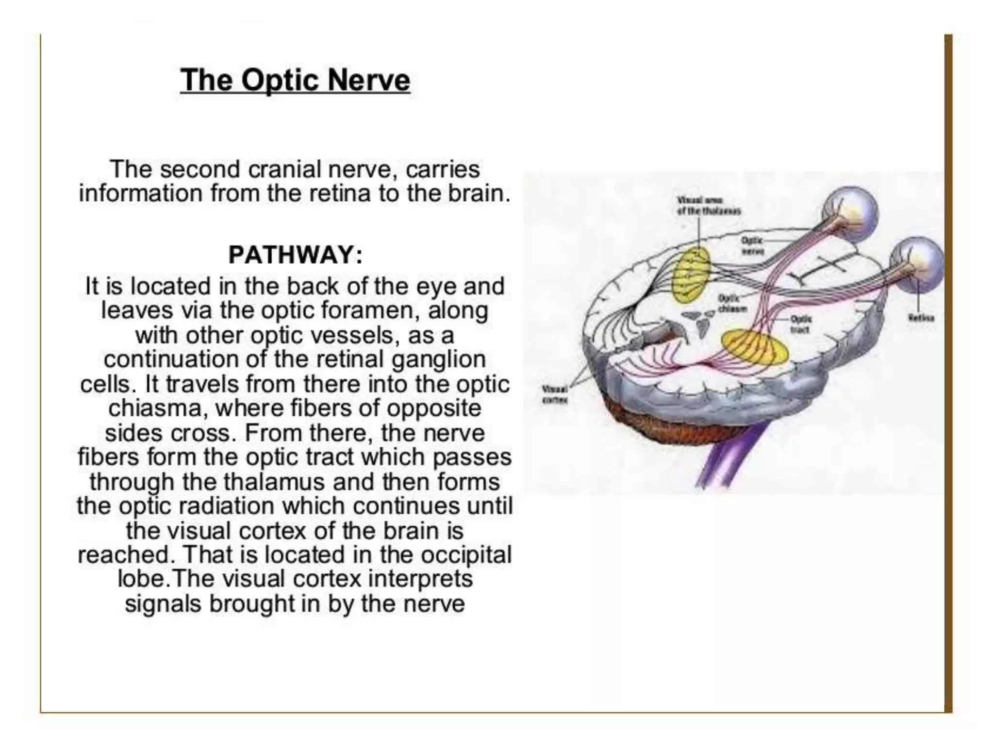
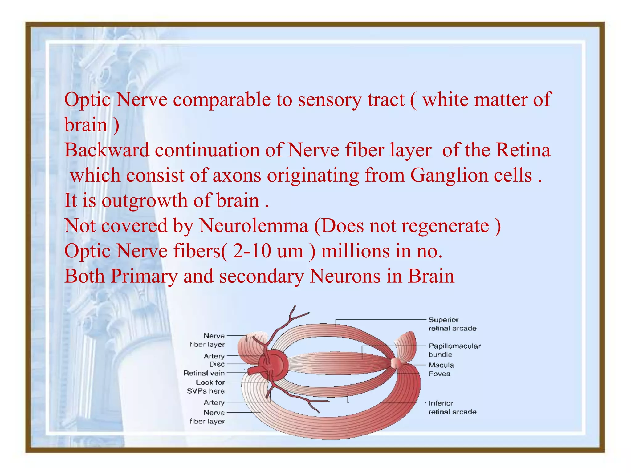
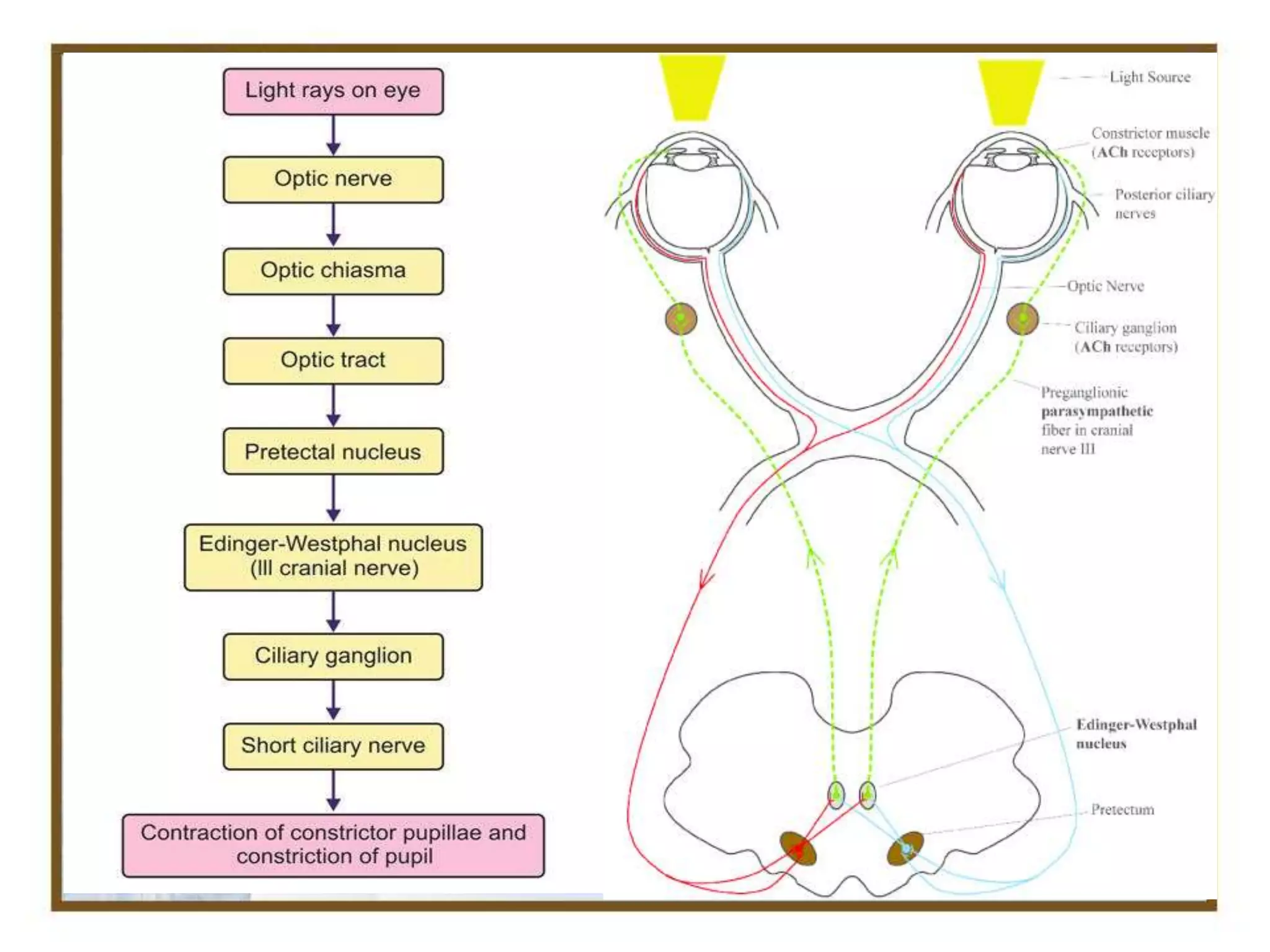
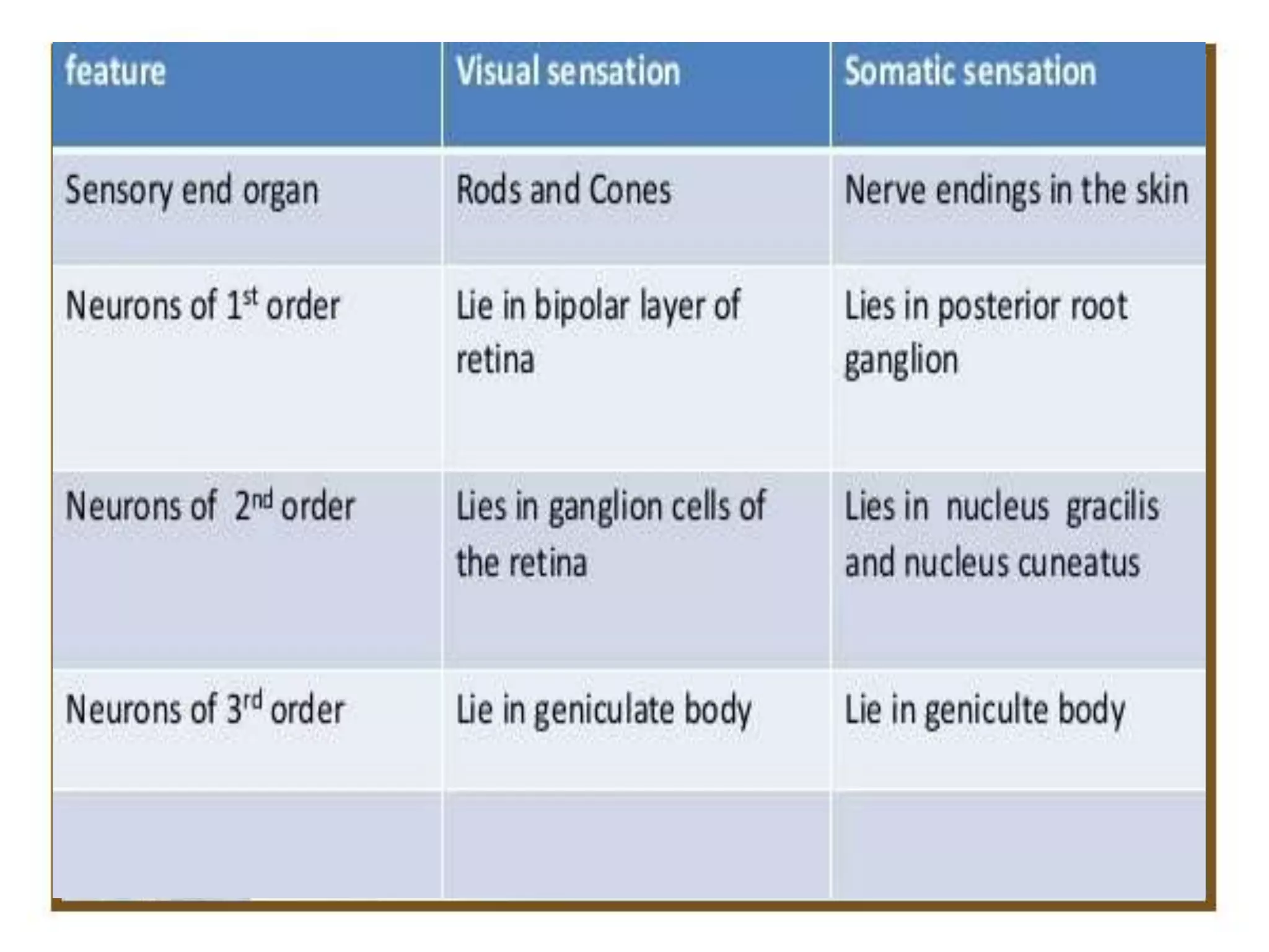
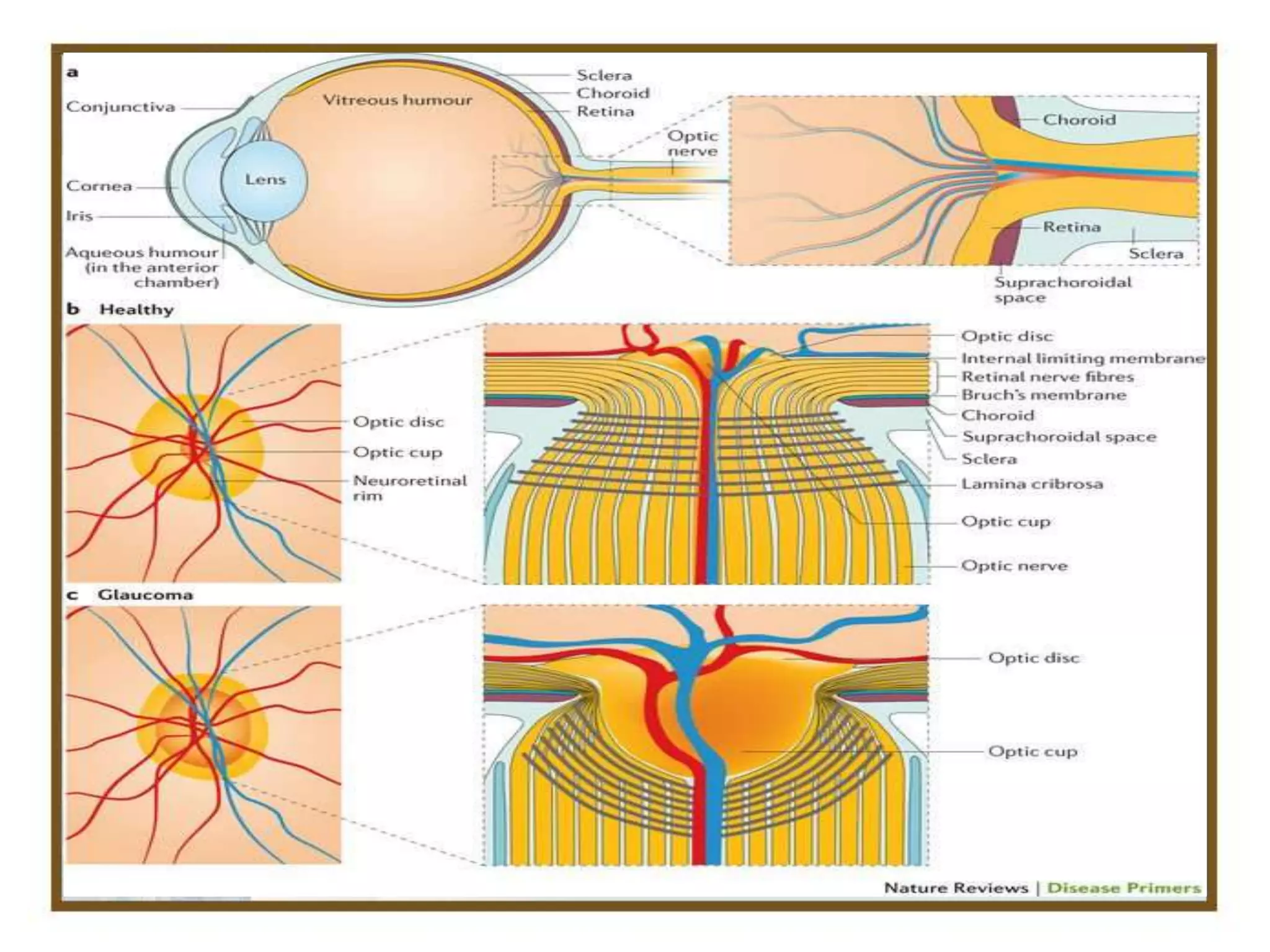
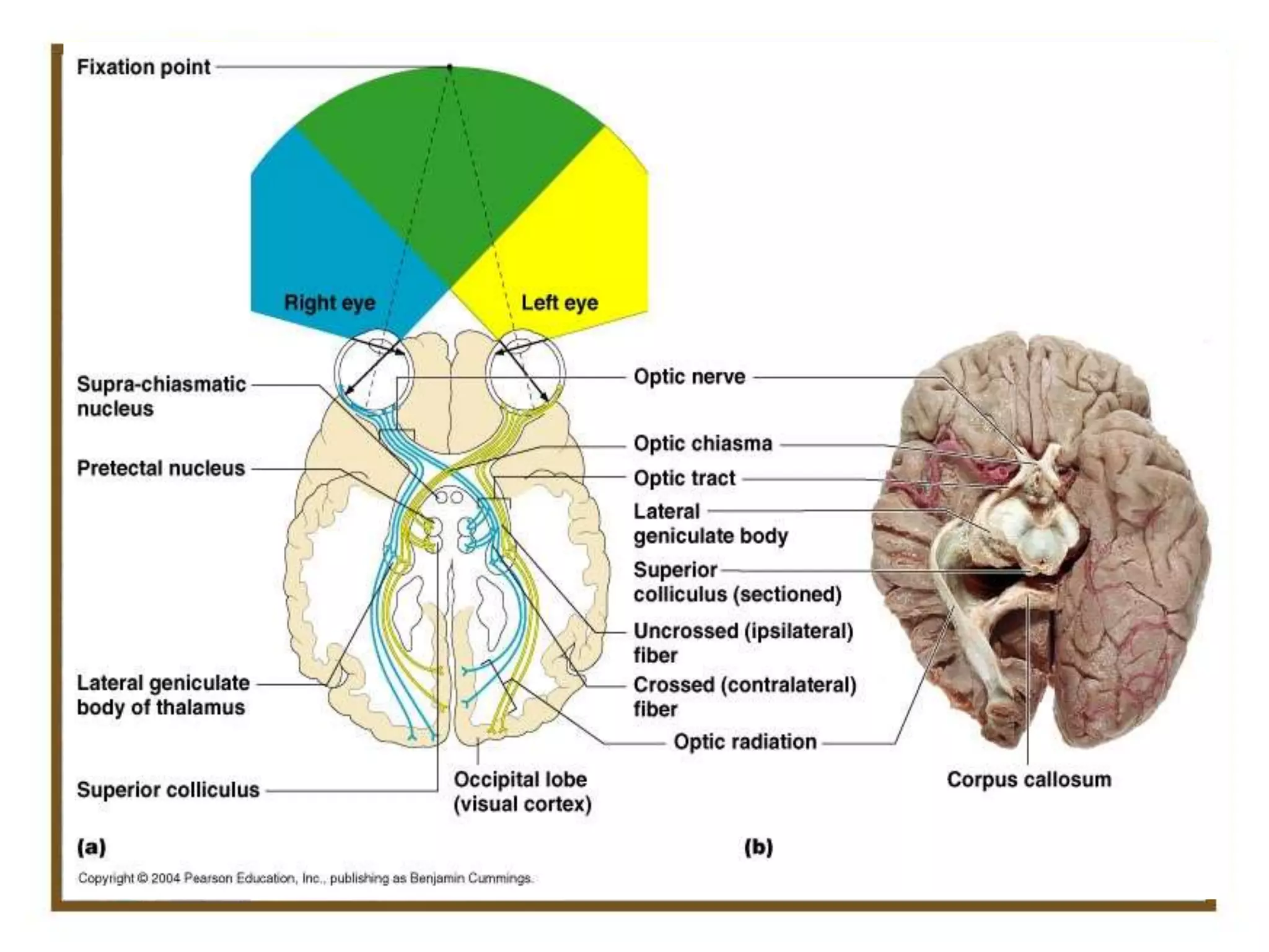
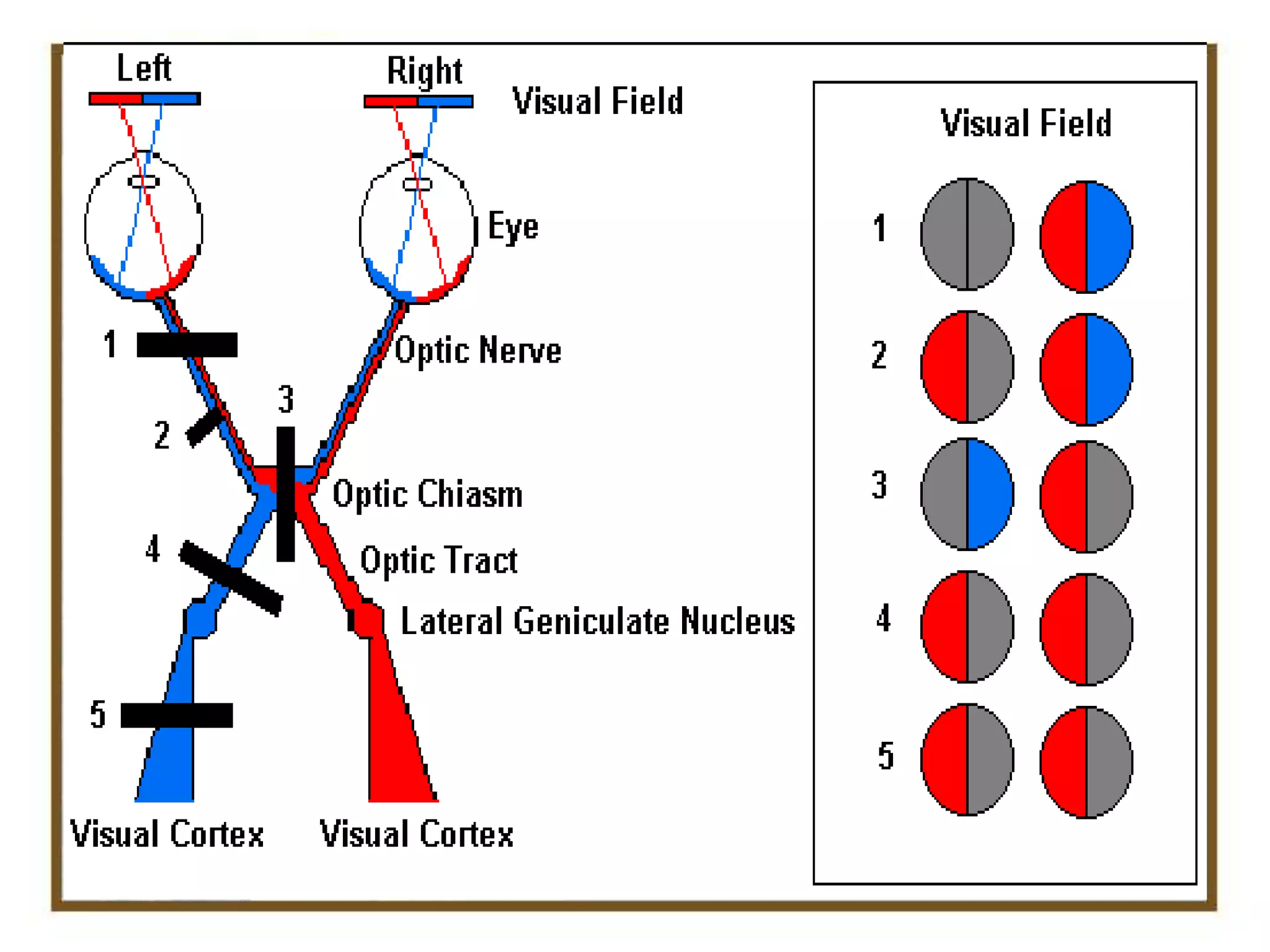
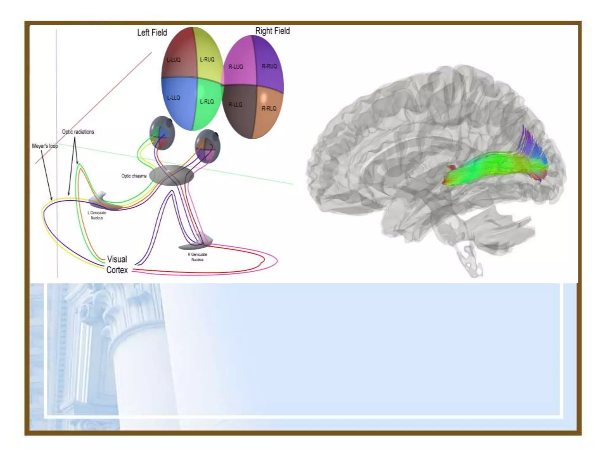
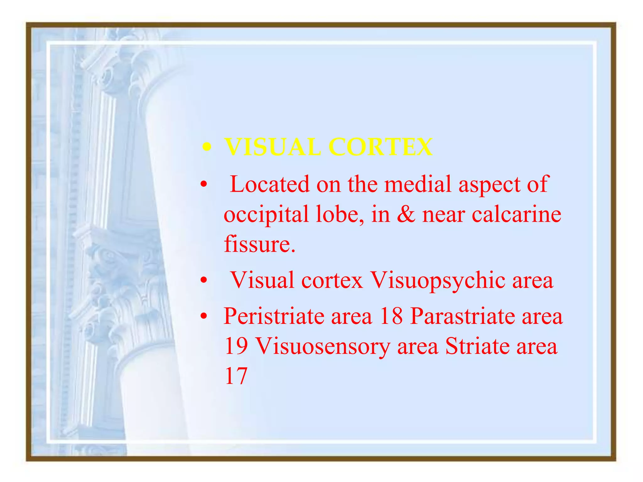
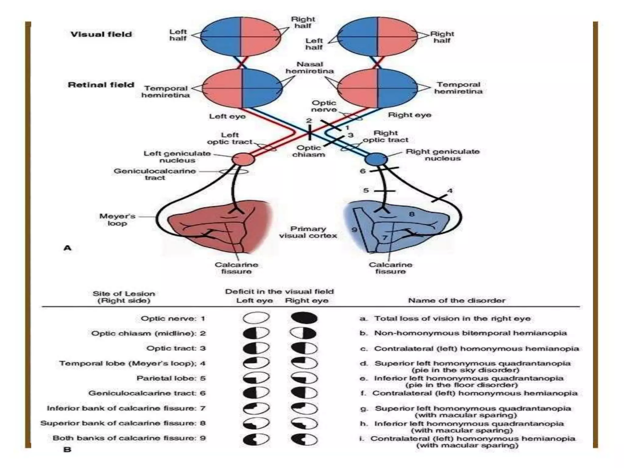
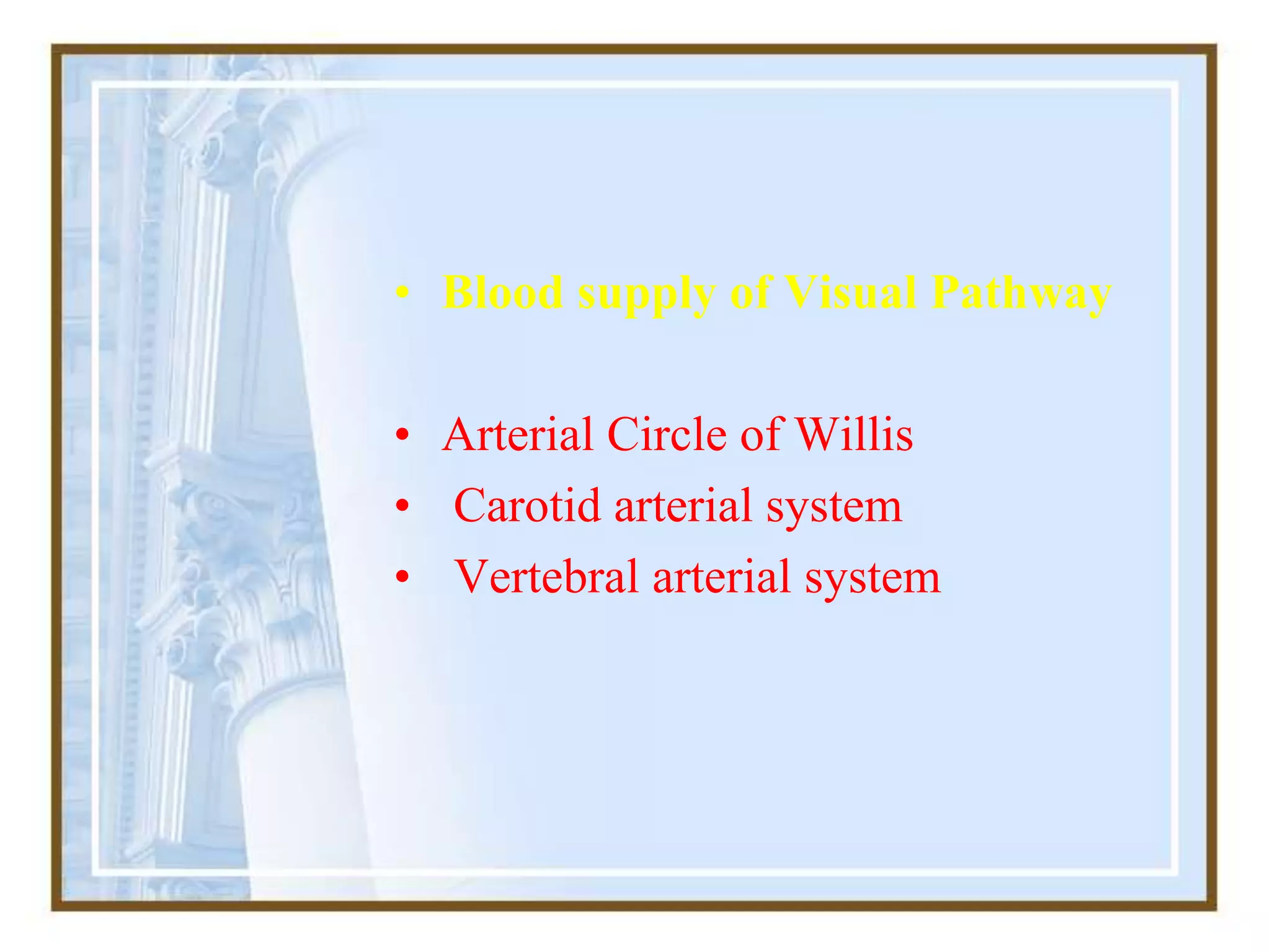
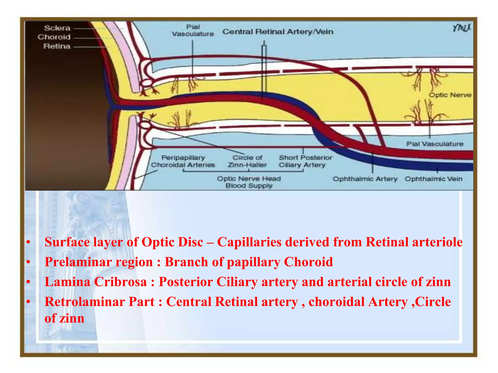
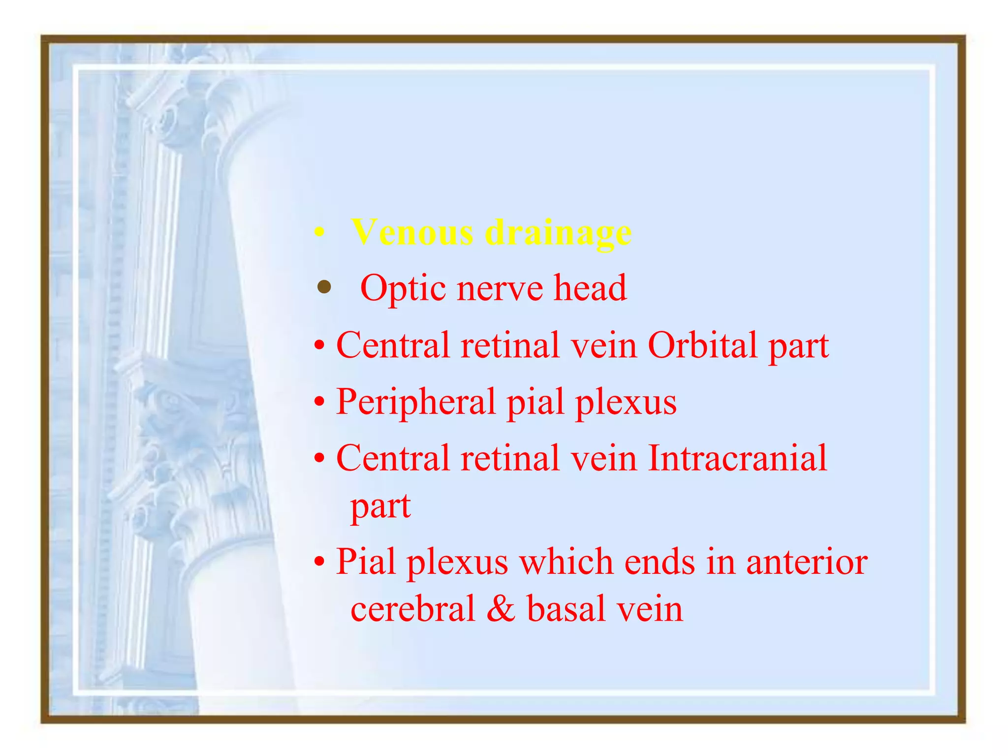
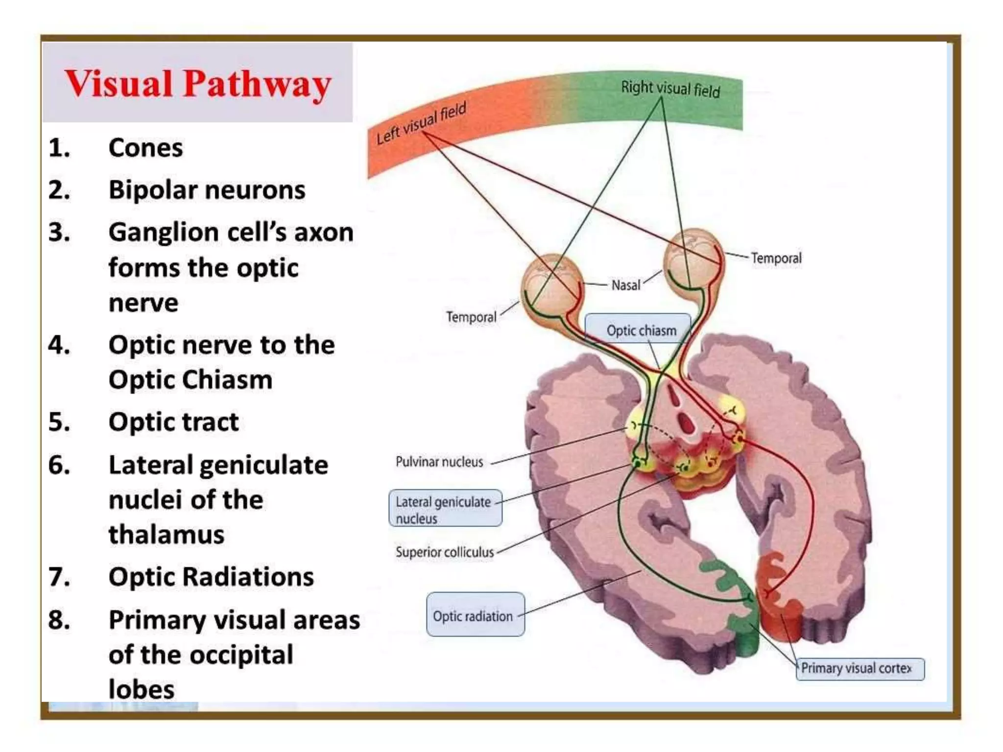
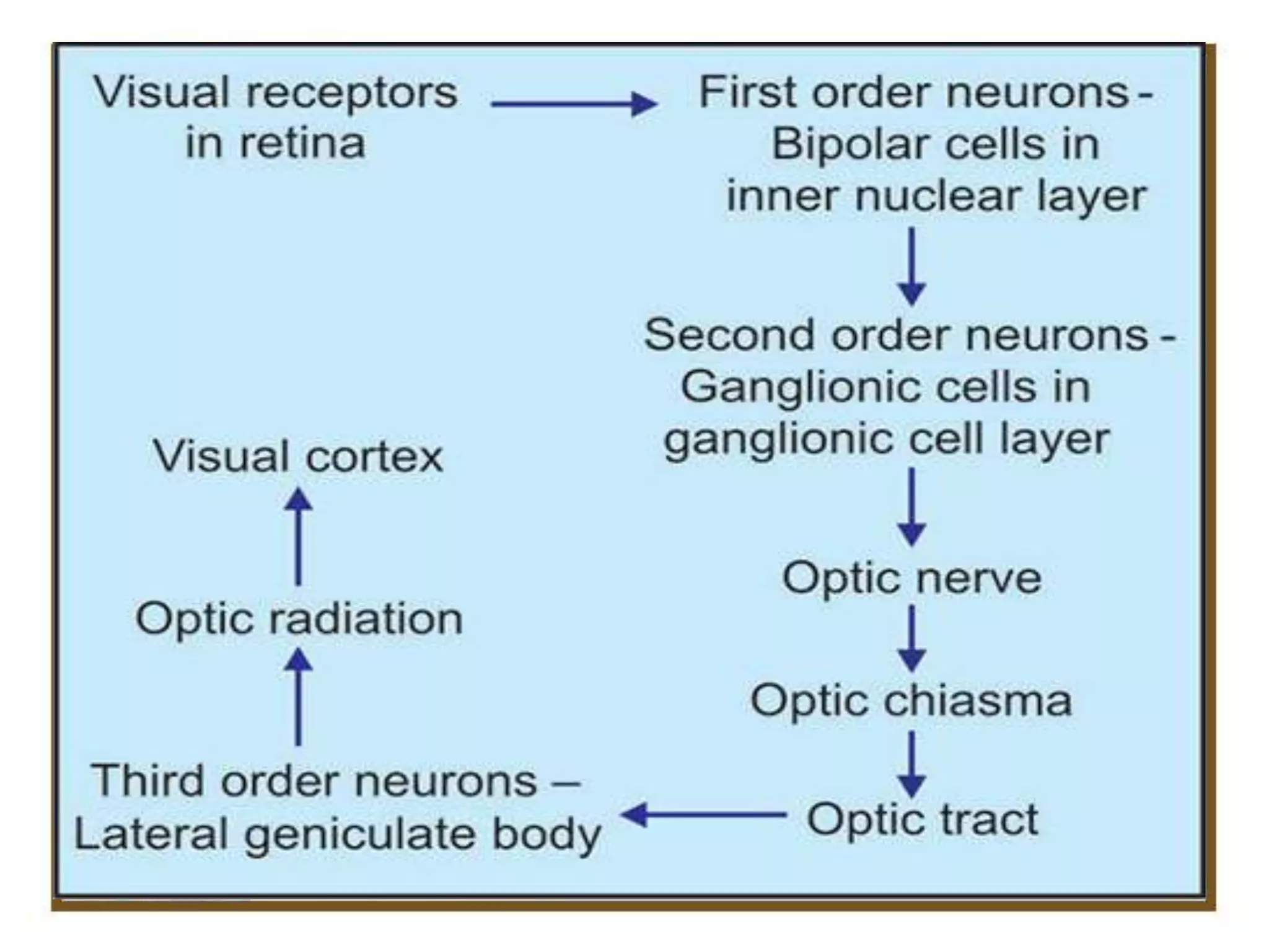
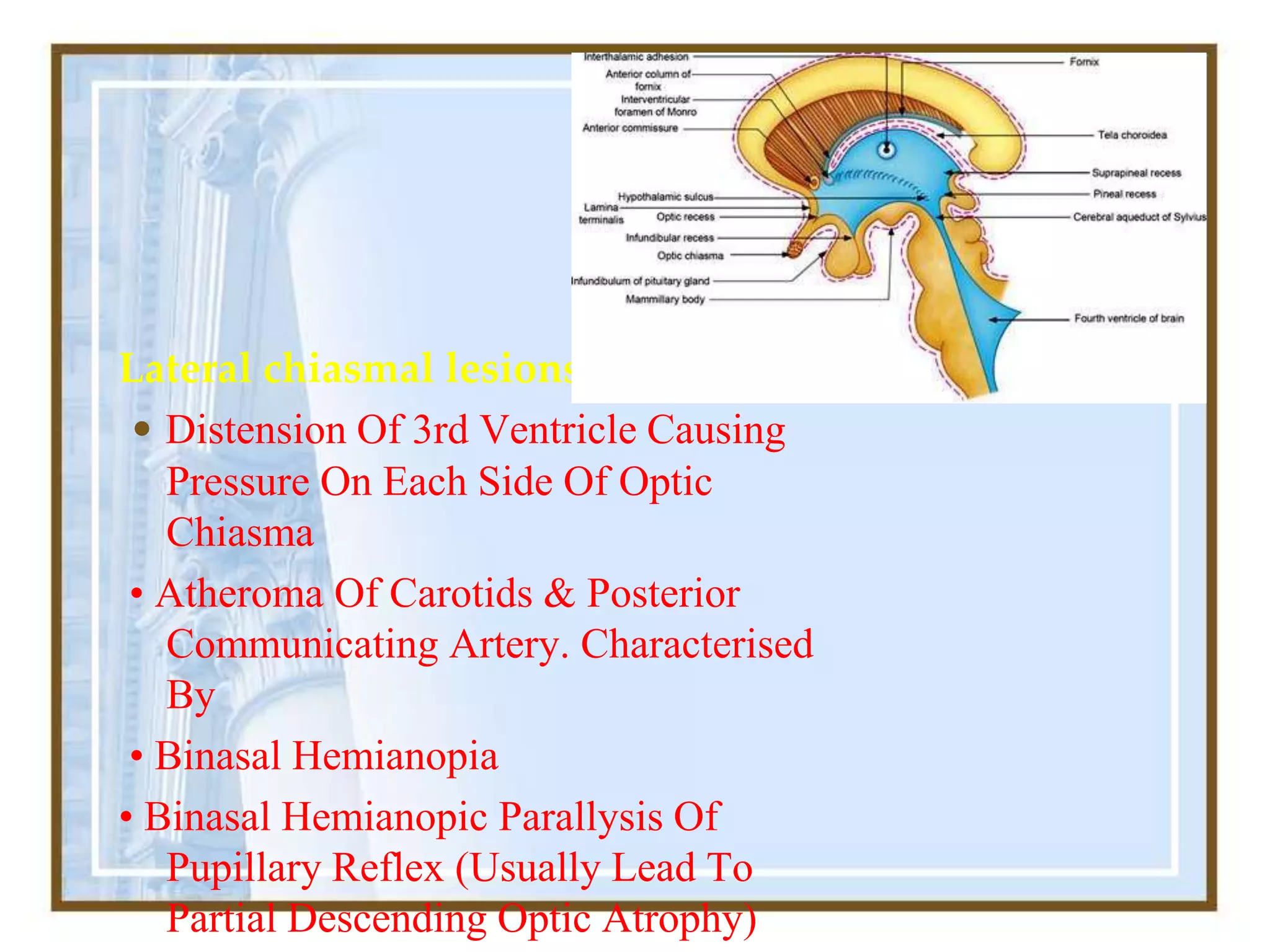
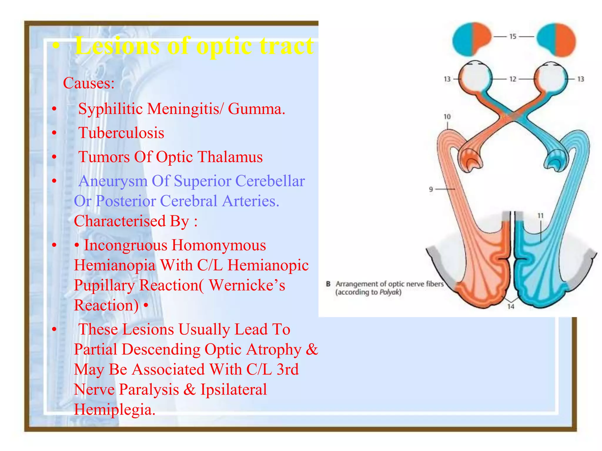
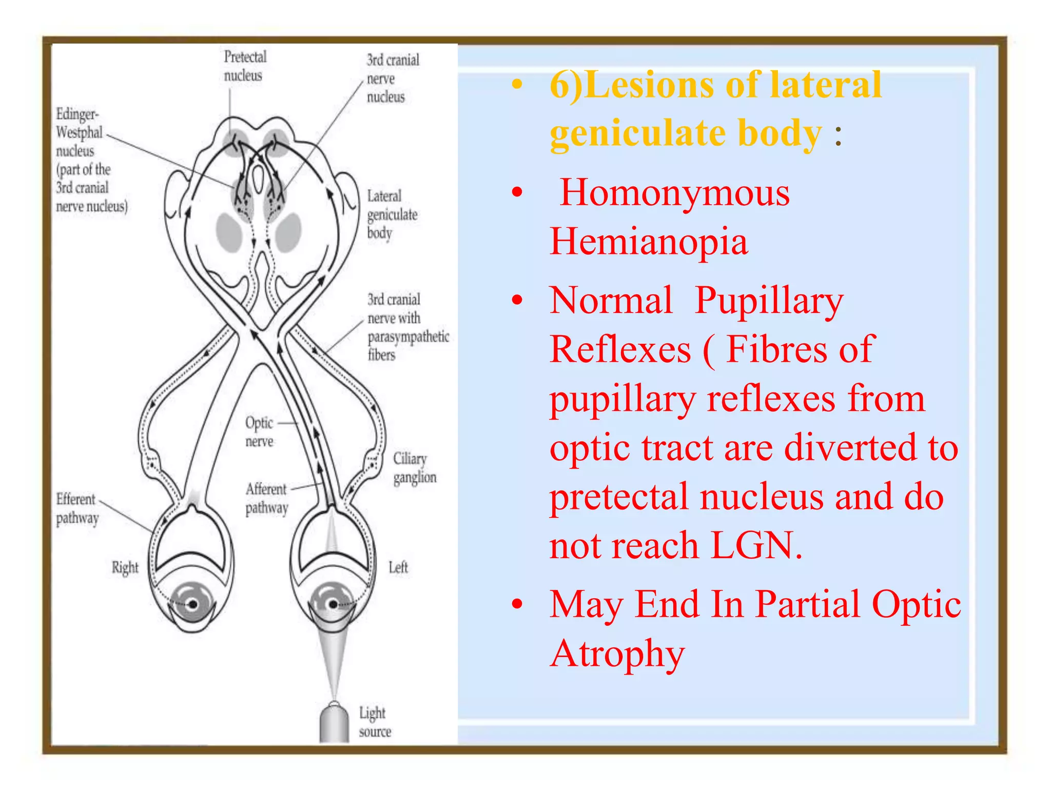
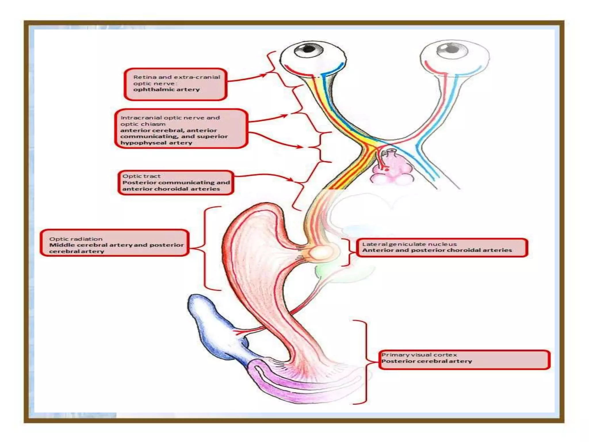
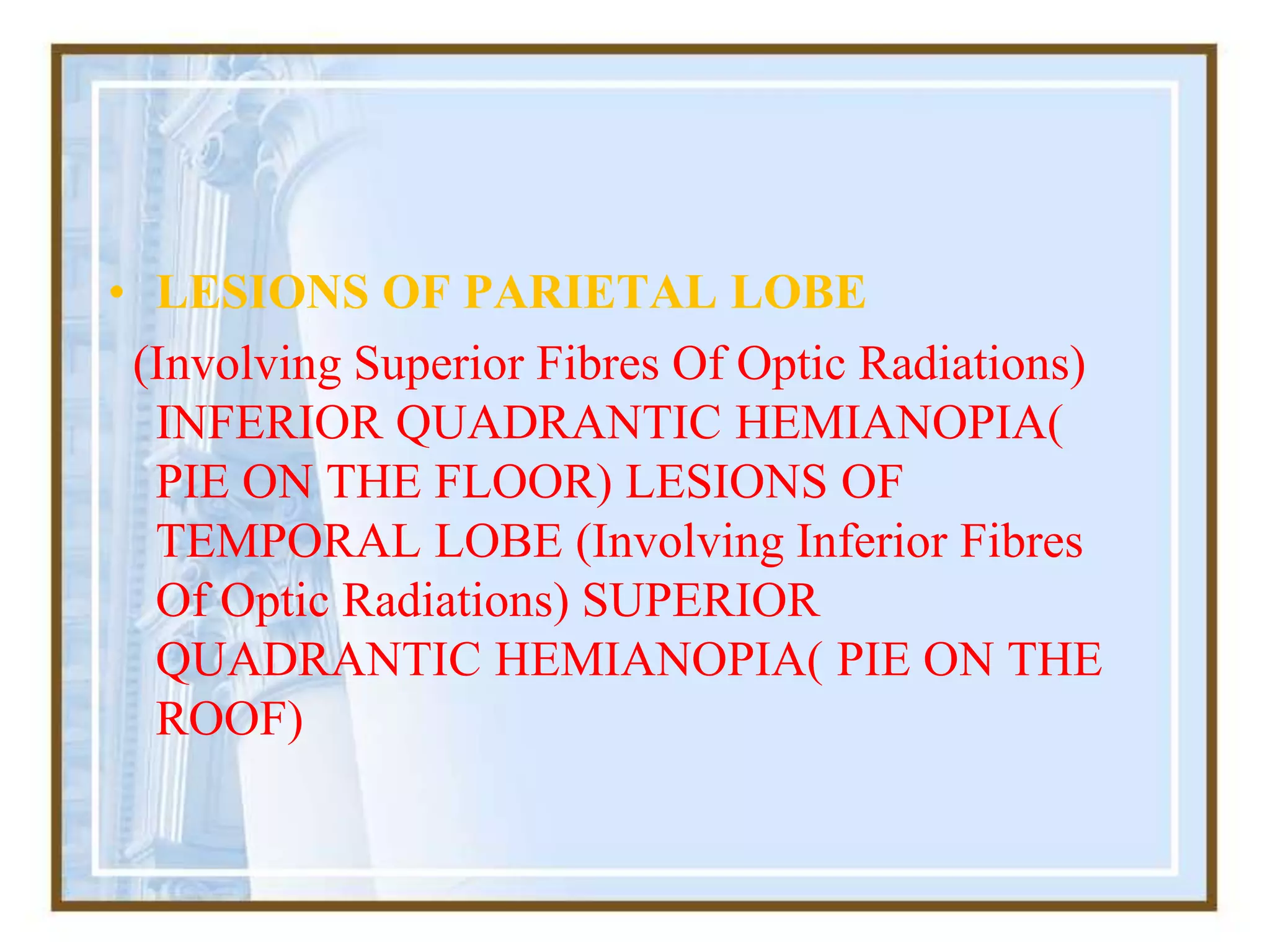
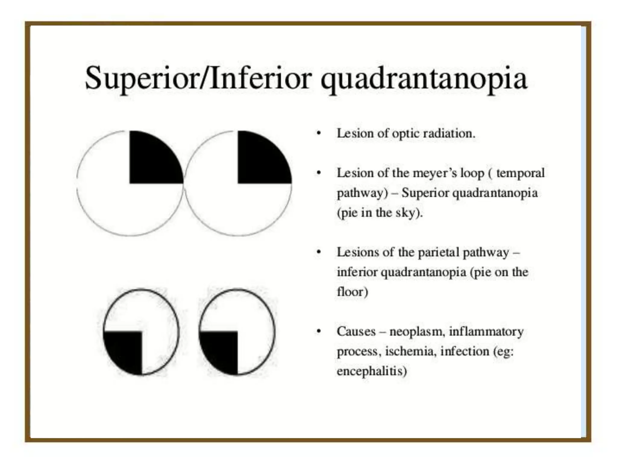
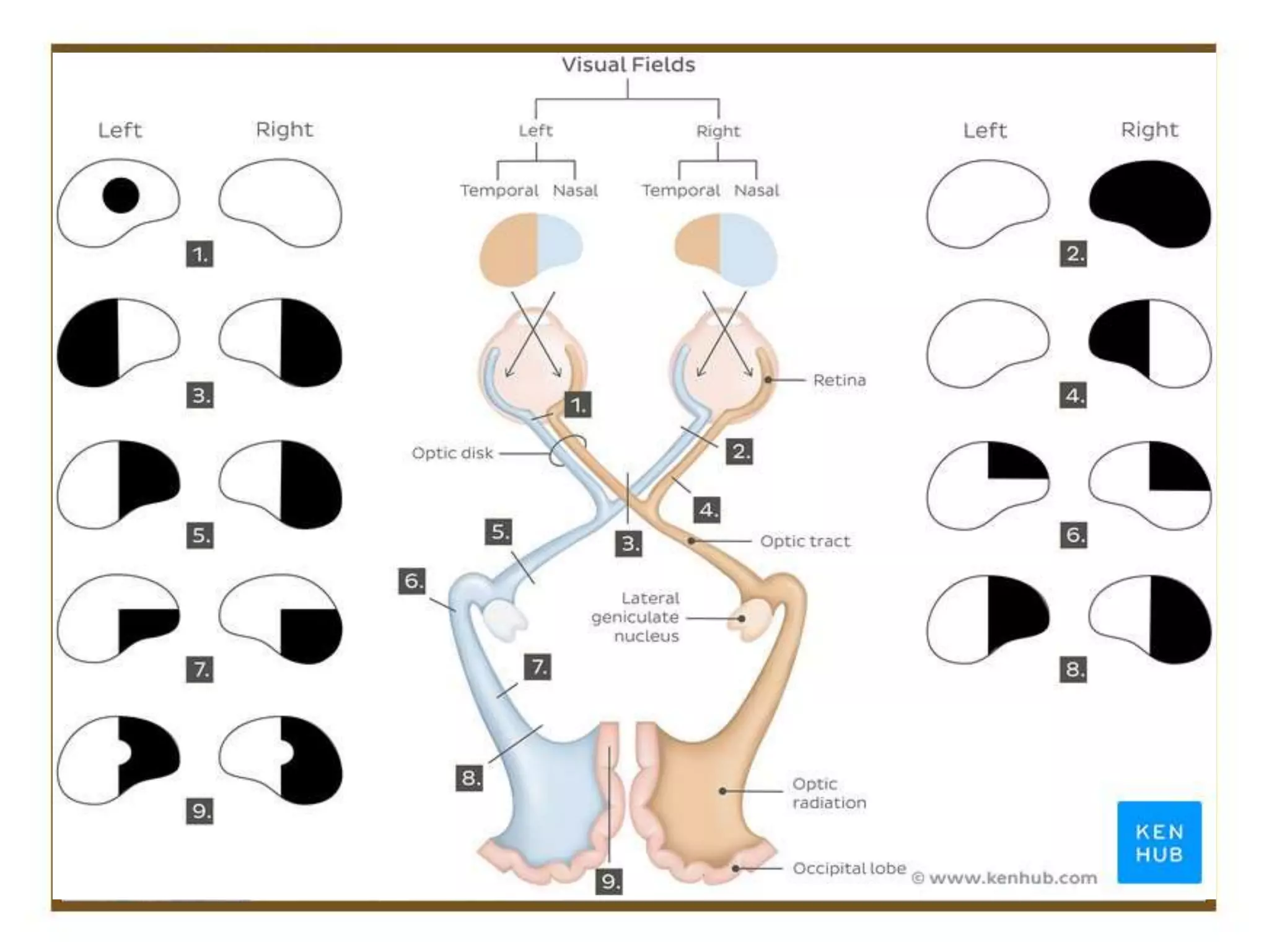
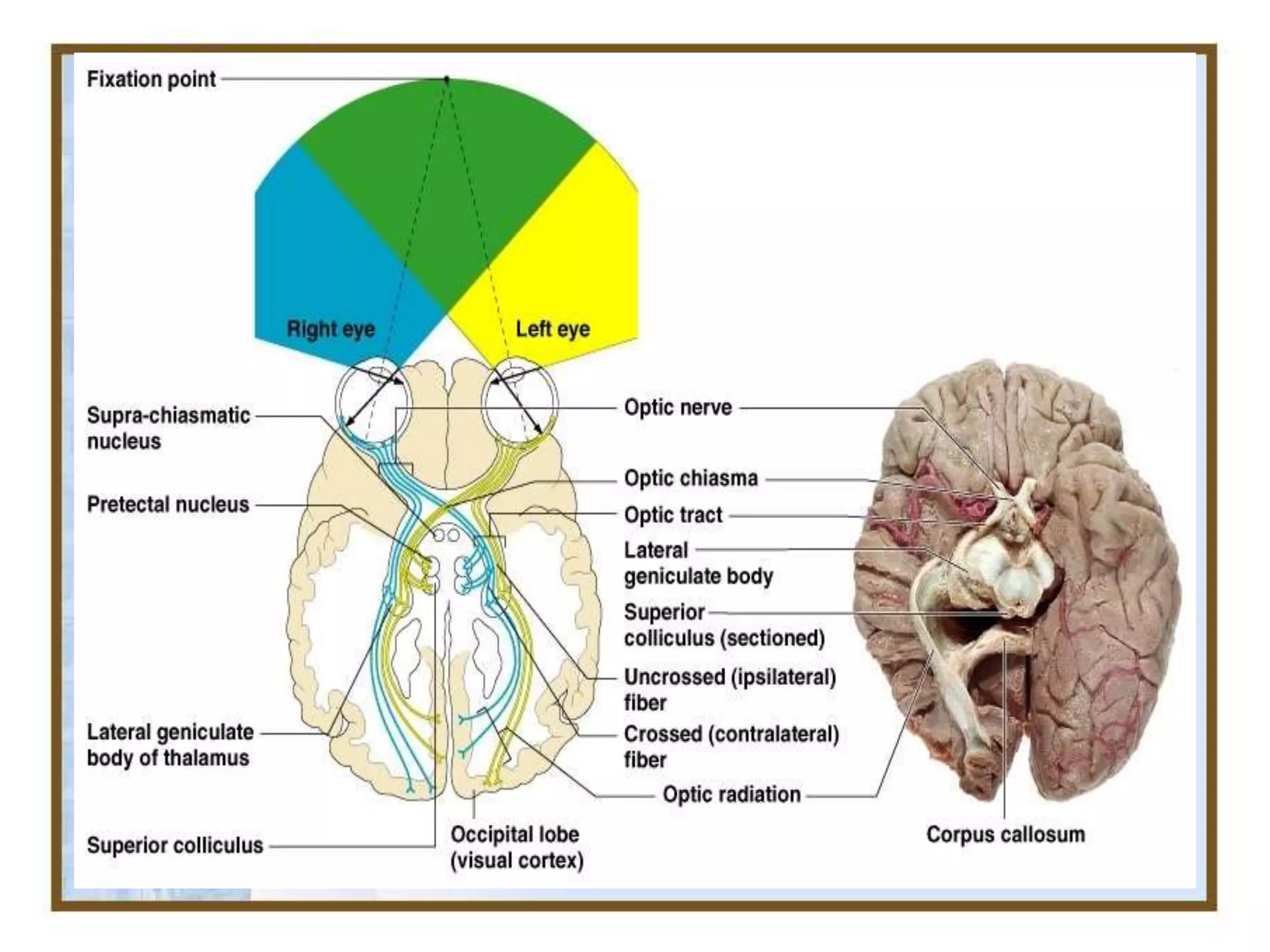
OPTIC NERVE & VISUAL PATHWAY
02 July 2020 ,Thursday 1- 2 Pm
Dr M Saquib
Mbbs,M.S, FSCEH Delhi,
FHVDESAI Pune, Ex Registrar Jnmch, amu
Consultant Ophthalmologist
Hod D/O Ophthalmology
Vice Principal G.S Medical College
G.S .Medical College
Founder Sec: MEDICS India ,
Mail:dms2k5@gmail.Com9634123800
• Visual Acuity – Reduced
• Relative Afferent Pupillary Defect
• Impairment Of Color Vision
• Brightness Sensitivity Decreased
• Contrast Sensitivity Reduced
• Visual Field Defect
Optic Nerve comparable to sensory tract ( white matter of
brain )
Backward continuation of Nerve fiber layer of the Retina
which consist of axons originating from Ganglion cells .
It is outgrowth of brain .
Not covered by Neurolemma (Does not regenerate )
Optic Nerve fibers( 2-10 um ) millions in no.
Both Primary and secondary Neurons in Brain
Optic Nerve – 47-50 mm
Intraocular ( 1 mm)
Intra orbital ( 30 mm)
Intra canalicular ( 6-9 mm)
Intra Cranial ( 10 mm)
Intraorbital part : Extends from back of eyeball to optic
foramina. This part slightly sinuous to give play for the
eye movements. Here optic nerve is surrounded by all 3
layers of meninges & subarachnoid space. The central
retinal artery along with enters the subarachnoid space to
enter the nerve on its inferomedial aspect. Near optic
foramina,optic nerve is closely surronded by annulus of zinn
& the origin of four recti muscles. Some fibres of superior
& medial rectus are adherent to optic nerve
Intracanalicular Part
- This part is closely related to
ophthalmic artery. it crosses the
nerve from medial to lateral side
in dural sheath.
-Sphenoid & posterior
ethmoidal sinuses lie medial to
it & seperated by thin bony
lamina, this relation accounts for
retrobulbar neuritis following
infection of sinuses
Intracranial part
About 10mm
Lies above cavernous sinus &
converges with its fellow to
form chiasma.
Ensheathed in pia mater.
Internal carotid artery runs
below then lateral to it.
OPTIC CHIASMA
• Flattened structure,12 mm horizontally & 8mm
anteroposteriorly.
• Ensheathed by pia & surrounded by CSF.
• Lies over diaphragma sellae so visual field defects
seen in patient with pituitary tumor having
suprasellar extension.
• Posteriorly chiasma continous with the optic
tracts & form the anterior wall of 3rd ventricle.
Nerve fibres arising from nasal half of two retina
decussate at the chiasma.
• Anatomical variation in position
of normal optic chiasma:
• a)central : lies directly over sella,
expanding pituitary tumor
involves chiasma first.
• b)pre-fixed : lies more anteriorly
over tuberculum sellae,pituitary
tumor involves optic tract first.
•
• c) post-fixed : lies more
posterior over dorsum
sellae,pituitary tumor damage
optic nerve
• OPTIC TRACTS
• Cylindrical bundle of nerve
fibres.
• Run outwards & backwards
from posterolateral aspect of
optic chiasma ,between tuber
cinereum & anterior perforated
substance to unite with
cerebral peduncle.
• Fibres from temporal half of
retina of same eye & nasal half
of opposite eye.
• Posteriorly each ends in
Lateral Geniculate Body.
• LATERAL GENICULATE
BODY
• Oval structures situated at
termination of the optic tracts.
• Each consist of 6 layers of
neurons(grey matter) alternating
with white matter (optic fibres)
• Fibres of 2nd order neuron
coming via optic tract relay in
these 3rd Order Neuron .
• OPTIC RADIATIONS (GeniculoCalcarine Pathway)
From LGB to the occipital cortex.
Pass forwards then laterally through the area of wernicke as
optic peduncles.
Anterior to lateral ventricle ,traversing the retrolenticular
part of internal capsule,medial to auditory tract.
Its fibres then spread out fanwise to form medullary optic
lamina.
Inferior fibres subserve upper visual fields & sweep
anteroinferiorly in meyer’s loop & temporal lobe to visual
cortex.
Superior fibres subserve inferior visual field proceed
posteriorly through parietal lobe to visual cortex
• VISUAL CORTEX
• Located on the medial aspect of
occipital lobe, in & near calcarine
fissure.
• Visual cortex Visuopsychic area
• Peristriate area 18 Parastriate area
19 Visuosensory area Striate area
17
• Blood supply of Visual Pathway
• Arterial Circle of Willis
• Carotid arterial system
• Vertebral arterial system
• Surface layer of Optic Disc – Capillaries derived from Retinal arteriole
• Prelaminar region : Branch of papillary Choroid
• Lamina Cribrosa : Posterior Ciliary artery and arterial circle of zinn
• Retrolaminar Part : Central Retinal artery , choroidal Artery ,Circle
of zinn
• Venous drainage
• Optic nerve head
• Central retinal vein Orbital part
• Peripheral pial plexus
• Central retinal vein Intracranial
part
• Pial plexus which ends in anterior
cerebral & basal vein
• 1) LESIONS OF OPTIC
NERVE :
• Causes:
• Optic Atrophy
• Optic Neuropathy
• Acute Optic Neuritis
Traumatic Avulsion Of Optic Nerve.
Characterised By:
Complete Blindness In Affected Eye
With Loss Of Both Direct On Ipsilateral &
Concensual Light Reflex On Contralateral Side.
Near Reflex Is Preserved.
• Lesions through proximal part of optic
nerve :
• Ipsilateral Blindness.
• Contralateral Hemianopia
• Abolition Of Direct Light Reflex On Affected
Side & Concensual Light Reflex On
Contralateral Side.
• Near Reflex Intact.
• Central lesions of chiasma (sagittal) causes:
suprasellar aneurysm
• tumors of pituitary gland
• craniopharyngioma
• suprasellar meningioma & glioma of 3rd ventricle.
• third ventricular dilatation due to obstructive
hydrocephalus.
• chronic chiasmal arachnoiditis.
• Characterised by: Bitemporal hemianopia
• Bitemporal hemianopic paralysis of pupillary reflex.
(usually lead to partial descending optic atrophy)
Lateral chiasmal lesions : causes:
• Distension Of 3rd Ventricle Causing
Pressure On Each Side Of Optic
Chiasma
• Atheroma Of Carotids & Posterior
Communicating Artery. Characterised
By
• Binasal Hemianopia
• Binasal Hemianopic Parallysis Of
Pupillary Reflex (Usually Lead To
Partial Descending Optic Atrophy)
• Lesions of optic tract
Causes:
• Syphilitic Meningitis/ Gumma.
• Tuberculosis
• Tumors Of Optic Thalamus
• Aneurysm Of Superior Cerebellar
Or Posterior Cerebral Arteries.
Characterised By :
• • Incongruous Homonymous
Hemianopia With C/L Hemianopic
Pupillary Reaction( Wernicke’s
Reaction) •
• These Lesions Usually Lead To
Partial Descending Optic Atrophy &
May Be Associated With C/L 3rd
Nerve Paralysis & Ipsilateral
Hemiplegia.
• 6)Lesions of lateral
geniculate body :
• Homonymous
Hemianopia
• Normal Pupillary
Reflexes ( Fibres of
pupillary reflexes from
optic tract are diverted to
pretectal nucleus and do
not reach LGN.
• May End In Partial Optic
Atrophy
• 7)Lesions of optic radiations :
Causes:
• Vascular Occlusion
• Primary & Secondary Tumors
• Trauma
• Characterized By : TOTAL OPTIC
RADIATION INVOLVEMENT
COMPLETE HOMONYMOUS
HEMIANOPIA( Sometimes Sparing
Macula)
• LESIONS OF PARIETAL LOBE
(Involving Superior Fibres Of Optic Radiations)
INFERIOR QUADRANTIC HEMIANOPIA(
PIE ON THE FLOOR) LESIONS OF
TEMPORAL LOBE (Involving Inferior Fibres
Of Optic Radiations) SUPERIOR
QUADRANTIC HEMIANOPIA( PIE ON THE
ROOF)
• Pupillary reactions are normal as
fibres of light reflex leave the
optic tracts to synapse in the
superior colliculi.
Lesions of optic radiations do not
produce optic atrophy as the 1st
order neurons (optic nerve fibres)
synapse in LGB.
• 8)Lesions of visual cortex :
Pupillary Light Reflex Is Normal & Optic
Atrophy Does Not Occur Following Visual
Cortex Lesions.
Congruous Homonymous Hemianopia(sparing
Macula) Occlusion Of Posterior Cerebral Artery
Supplyin Anterior Part Of Occipiatl Cortex
Congruous Homonymous Macular Defect Head
Injury/Gun Shot Injury Leading To Lesions Of
Tip Of Occipital Cortex+
Causes of Hyperaemia of Optic Nerve
• Optic Neuritis
• Papilloedema
• Pseudo –Neuritis
• Neovascularisation of the Optic Disc
a) Diabetic retinopathy
b) CRVO
C) Central Eale’s Disease
d) Neuroretinitis
• Optic Nerve Transmits All Visual Information
• Brightness Perception, Color Perception And Contrast
(Visual Acuity).
• Visual Impulses That Are Responsible For Two Important
Neurological Reflexes: The Light Reflex And
The Accommodation Reflex
• The Light Reflex Refers To The Constriction Of Both
Pupils That Occurs When Light Is Shone Into Either Eye.
• The Accommodation Reflex Change in shape of Lens
That Occurs When One Looks At A Near Object (For
Example, When Reading The Lens Adjusts To Near
Vision.
• The Eye's Blind Spot Is A Result Of The Absence
Of Photoreceptors In The Area Of The Retina Where The
Optic Nerve Leaves The Eye.
Examination of Optic Nerve
• Distance And Near Vision 9 UCVA/BCVA )
• Colour Vision
• Field Of Vision – Central /Peripheral
• Pupillary Reaction
• Intraocular Pressure
• Ophthalmoscopy
• Slit Lamp Biomicroscopy
• Flourescein Angiography
• X Ray Skull /PNS, OPTIC FORAMEN / CANAL AND ORBIT
• ULTRASONOGRAPHY
• CT SCAN
• MRI
• OCT
• VISUAL EVOKED RESPONSE (VER )
THANKS

OPTIC NERVE & VISUAL PATHWAY

  • 1.
    OPTIC NERVE &VISUAL PATHWAY 02 July 2020 ,Thursday 1- 2 Pm Dr M Saquib Mbbs,M.S, FSCEH Delhi, FHVDESAI Pune, Ex Registrar Jnmch, amu Consultant Ophthalmologist Hod D/O Ophthalmology Vice Principal G.S Medical College G.S .Medical College Founder Sec: MEDICS India , Mail:dms2k5@gmail.Com9634123800
  • 2.
    • Visual Acuity– Reduced • Relative Afferent Pupillary Defect • Impairment Of Color Vision • Brightness Sensitivity Decreased • Contrast Sensitivity Reduced • Visual Field Defect
  • 4.
    Optic Nerve comparableto sensory tract ( white matter of brain ) Backward continuation of Nerve fiber layer of the Retina which consist of axons originating from Ganglion cells . It is outgrowth of brain . Not covered by Neurolemma (Does not regenerate ) Optic Nerve fibers( 2-10 um ) millions in no. Both Primary and secondary Neurons in Brain
  • 9.
    Optic Nerve –47-50 mm Intraocular ( 1 mm) Intra orbital ( 30 mm) Intra canalicular ( 6-9 mm) Intra Cranial ( 10 mm)
  • 11.
    Intraorbital part :Extends from back of eyeball to optic foramina. This part slightly sinuous to give play for the eye movements. Here optic nerve is surrounded by all 3 layers of meninges & subarachnoid space. The central retinal artery along with enters the subarachnoid space to enter the nerve on its inferomedial aspect. Near optic foramina,optic nerve is closely surronded by annulus of zinn & the origin of four recti muscles. Some fibres of superior & medial rectus are adherent to optic nerve
  • 12.
    Intracanalicular Part - Thispart is closely related to ophthalmic artery. it crosses the nerve from medial to lateral side in dural sheath. -Sphenoid & posterior ethmoidal sinuses lie medial to it & seperated by thin bony lamina, this relation accounts for retrobulbar neuritis following infection of sinuses
  • 13.
    Intracranial part About 10mm Liesabove cavernous sinus & converges with its fellow to form chiasma. Ensheathed in pia mater. Internal carotid artery runs below then lateral to it.
  • 15.
    OPTIC CHIASMA • Flattenedstructure,12 mm horizontally & 8mm anteroposteriorly. • Ensheathed by pia & surrounded by CSF. • Lies over diaphragma sellae so visual field defects seen in patient with pituitary tumor having suprasellar extension. • Posteriorly chiasma continous with the optic tracts & form the anterior wall of 3rd ventricle. Nerve fibres arising from nasal half of two retina decussate at the chiasma.
  • 16.
    • Anatomical variationin position of normal optic chiasma: • a)central : lies directly over sella, expanding pituitary tumor involves chiasma first. • b)pre-fixed : lies more anteriorly over tuberculum sellae,pituitary tumor involves optic tract first. • • c) post-fixed : lies more posterior over dorsum sellae,pituitary tumor damage optic nerve
  • 17.
    • OPTIC TRACTS •Cylindrical bundle of nerve fibres. • Run outwards & backwards from posterolateral aspect of optic chiasma ,between tuber cinereum & anterior perforated substance to unite with cerebral peduncle. • Fibres from temporal half of retina of same eye & nasal half of opposite eye. • Posteriorly each ends in Lateral Geniculate Body.
  • 19.
    • LATERAL GENICULATE BODY •Oval structures situated at termination of the optic tracts. • Each consist of 6 layers of neurons(grey matter) alternating with white matter (optic fibres) • Fibres of 2nd order neuron coming via optic tract relay in these 3rd Order Neuron .
  • 20.
    • OPTIC RADIATIONS(GeniculoCalcarine Pathway) From LGB to the occipital cortex. Pass forwards then laterally through the area of wernicke as optic peduncles. Anterior to lateral ventricle ,traversing the retrolenticular part of internal capsule,medial to auditory tract. Its fibres then spread out fanwise to form medullary optic lamina. Inferior fibres subserve upper visual fields & sweep anteroinferiorly in meyer’s loop & temporal lobe to visual cortex. Superior fibres subserve inferior visual field proceed posteriorly through parietal lobe to visual cortex
  • 22.
    • VISUAL CORTEX •Located on the medial aspect of occipital lobe, in & near calcarine fissure. • Visual cortex Visuopsychic area • Peristriate area 18 Parastriate area 19 Visuosensory area Striate area 17
  • 24.
    • Blood supplyof Visual Pathway • Arterial Circle of Willis • Carotid arterial system • Vertebral arterial system
  • 25.
    • Surface layerof Optic Disc – Capillaries derived from Retinal arteriole • Prelaminar region : Branch of papillary Choroid • Lamina Cribrosa : Posterior Ciliary artery and arterial circle of zinn • Retrolaminar Part : Central Retinal artery , choroidal Artery ,Circle of zinn
  • 26.
    • Venous drainage •Optic nerve head • Central retinal vein Orbital part • Peripheral pial plexus • Central retinal vein Intracranial part • Pial plexus which ends in anterior cerebral & basal vein
  • 29.
    • 1) LESIONSOF OPTIC NERVE : • Causes: • Optic Atrophy • Optic Neuropathy • Acute Optic Neuritis Traumatic Avulsion Of Optic Nerve. Characterised By: Complete Blindness In Affected Eye With Loss Of Both Direct On Ipsilateral & Concensual Light Reflex On Contralateral Side. Near Reflex Is Preserved.
  • 30.
    • Lesions throughproximal part of optic nerve : • Ipsilateral Blindness. • Contralateral Hemianopia • Abolition Of Direct Light Reflex On Affected Side & Concensual Light Reflex On Contralateral Side. • Near Reflex Intact.
  • 32.
    • Central lesionsof chiasma (sagittal) causes: suprasellar aneurysm • tumors of pituitary gland • craniopharyngioma • suprasellar meningioma & glioma of 3rd ventricle. • third ventricular dilatation due to obstructive hydrocephalus. • chronic chiasmal arachnoiditis. • Characterised by: Bitemporal hemianopia • Bitemporal hemianopic paralysis of pupillary reflex. (usually lead to partial descending optic atrophy)
  • 33.
    Lateral chiasmal lesions: causes: • Distension Of 3rd Ventricle Causing Pressure On Each Side Of Optic Chiasma • Atheroma Of Carotids & Posterior Communicating Artery. Characterised By • Binasal Hemianopia • Binasal Hemianopic Parallysis Of Pupillary Reflex (Usually Lead To Partial Descending Optic Atrophy)
  • 34.
    • Lesions ofoptic tract Causes: • Syphilitic Meningitis/ Gumma. • Tuberculosis • Tumors Of Optic Thalamus • Aneurysm Of Superior Cerebellar Or Posterior Cerebral Arteries. Characterised By : • • Incongruous Homonymous Hemianopia With C/L Hemianopic Pupillary Reaction( Wernicke’s Reaction) • • These Lesions Usually Lead To Partial Descending Optic Atrophy & May Be Associated With C/L 3rd Nerve Paralysis & Ipsilateral Hemiplegia.
  • 35.
    • 6)Lesions oflateral geniculate body : • Homonymous Hemianopia • Normal Pupillary Reflexes ( Fibres of pupillary reflexes from optic tract are diverted to pretectal nucleus and do not reach LGN. • May End In Partial Optic Atrophy
  • 36.
    • 7)Lesions ofoptic radiations : Causes: • Vascular Occlusion • Primary & Secondary Tumors • Trauma • Characterized By : TOTAL OPTIC RADIATION INVOLVEMENT COMPLETE HOMONYMOUS HEMIANOPIA( Sometimes Sparing Macula)
  • 38.
    • LESIONS OFPARIETAL LOBE (Involving Superior Fibres Of Optic Radiations) INFERIOR QUADRANTIC HEMIANOPIA( PIE ON THE FLOOR) LESIONS OF TEMPORAL LOBE (Involving Inferior Fibres Of Optic Radiations) SUPERIOR QUADRANTIC HEMIANOPIA( PIE ON THE ROOF)
  • 40.
    • Pupillary reactionsare normal as fibres of light reflex leave the optic tracts to synapse in the superior colliculi. Lesions of optic radiations do not produce optic atrophy as the 1st order neurons (optic nerve fibres) synapse in LGB.
  • 41.
    • 8)Lesions ofvisual cortex : Pupillary Light Reflex Is Normal & Optic Atrophy Does Not Occur Following Visual Cortex Lesions. Congruous Homonymous Hemianopia(sparing Macula) Occlusion Of Posterior Cerebral Artery Supplyin Anterior Part Of Occipiatl Cortex Congruous Homonymous Macular Defect Head Injury/Gun Shot Injury Leading To Lesions Of Tip Of Occipital Cortex+
  • 44.
    Causes of Hyperaemiaof Optic Nerve • Optic Neuritis • Papilloedema • Pseudo –Neuritis • Neovascularisation of the Optic Disc a) Diabetic retinopathy b) CRVO C) Central Eale’s Disease d) Neuroretinitis
  • 45.
    • Optic NerveTransmits All Visual Information • Brightness Perception, Color Perception And Contrast (Visual Acuity). • Visual Impulses That Are Responsible For Two Important Neurological Reflexes: The Light Reflex And The Accommodation Reflex • The Light Reflex Refers To The Constriction Of Both Pupils That Occurs When Light Is Shone Into Either Eye. • The Accommodation Reflex Change in shape of Lens That Occurs When One Looks At A Near Object (For Example, When Reading The Lens Adjusts To Near Vision. • The Eye's Blind Spot Is A Result Of The Absence Of Photoreceptors In The Area Of The Retina Where The Optic Nerve Leaves The Eye.
  • 46.
    Examination of OpticNerve • Distance And Near Vision 9 UCVA/BCVA ) • Colour Vision • Field Of Vision – Central /Peripheral • Pupillary Reaction • Intraocular Pressure • Ophthalmoscopy • Slit Lamp Biomicroscopy • Flourescein Angiography • X Ray Skull /PNS, OPTIC FORAMEN / CANAL AND ORBIT • ULTRASONOGRAPHY • CT SCAN • MRI • OCT • VISUAL EVOKED RESPONSE (VER )
  • 50.