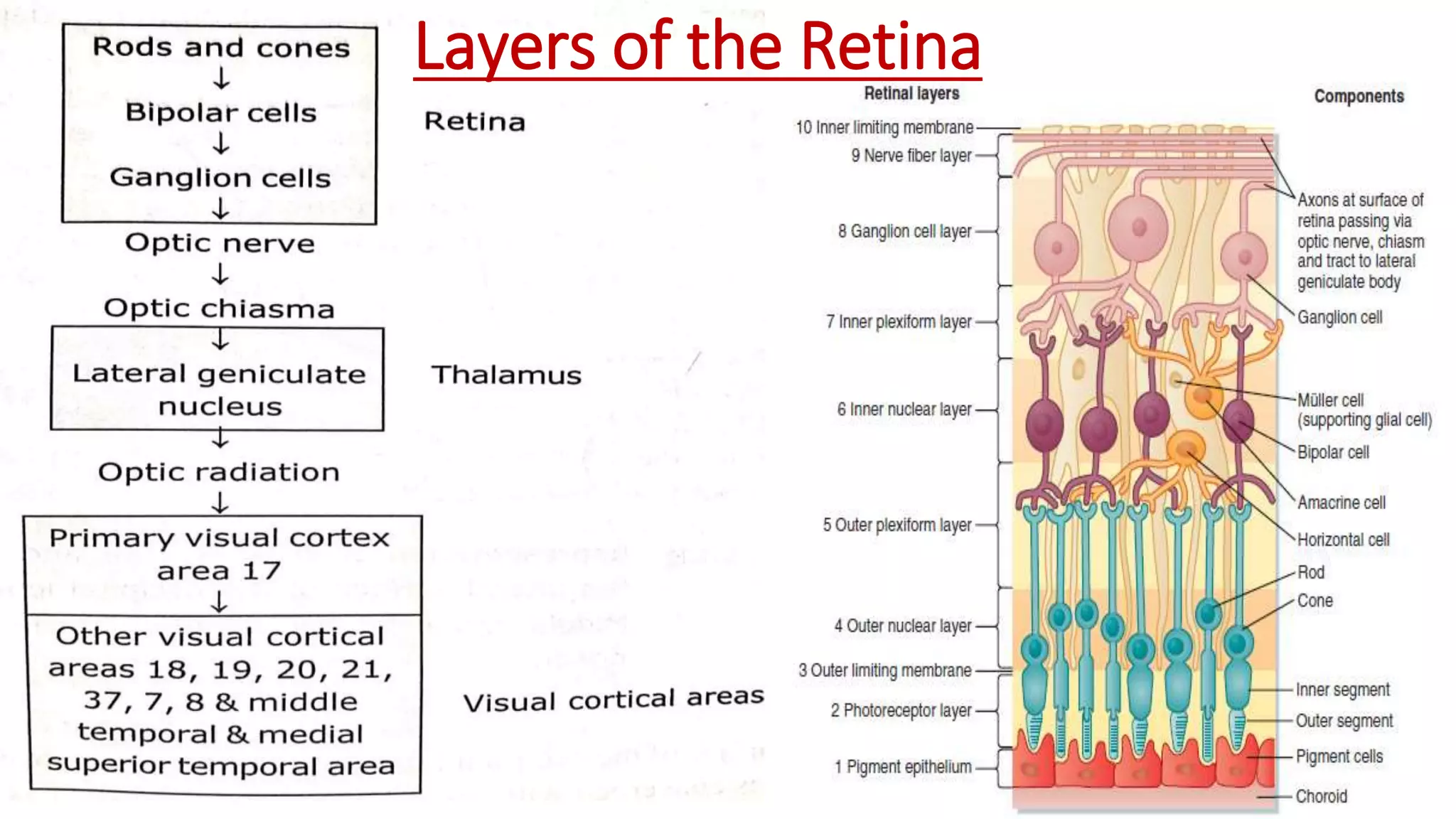
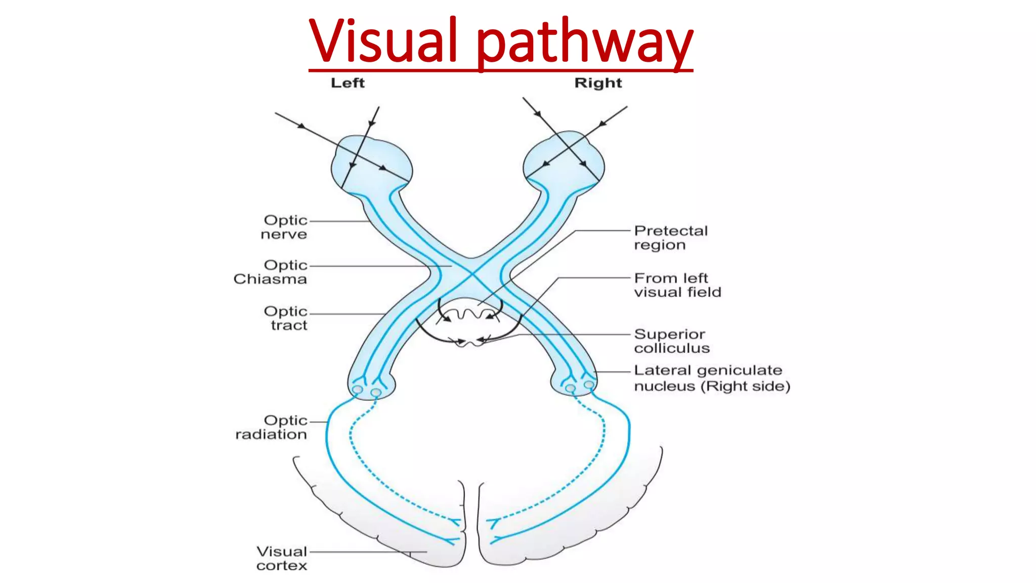
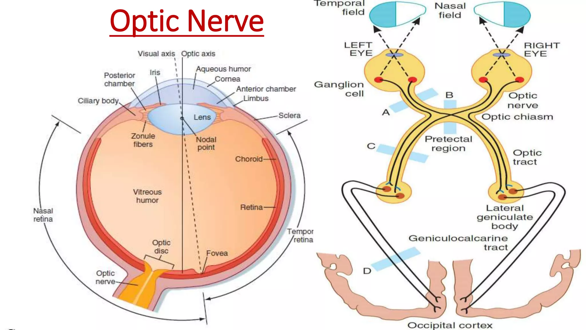
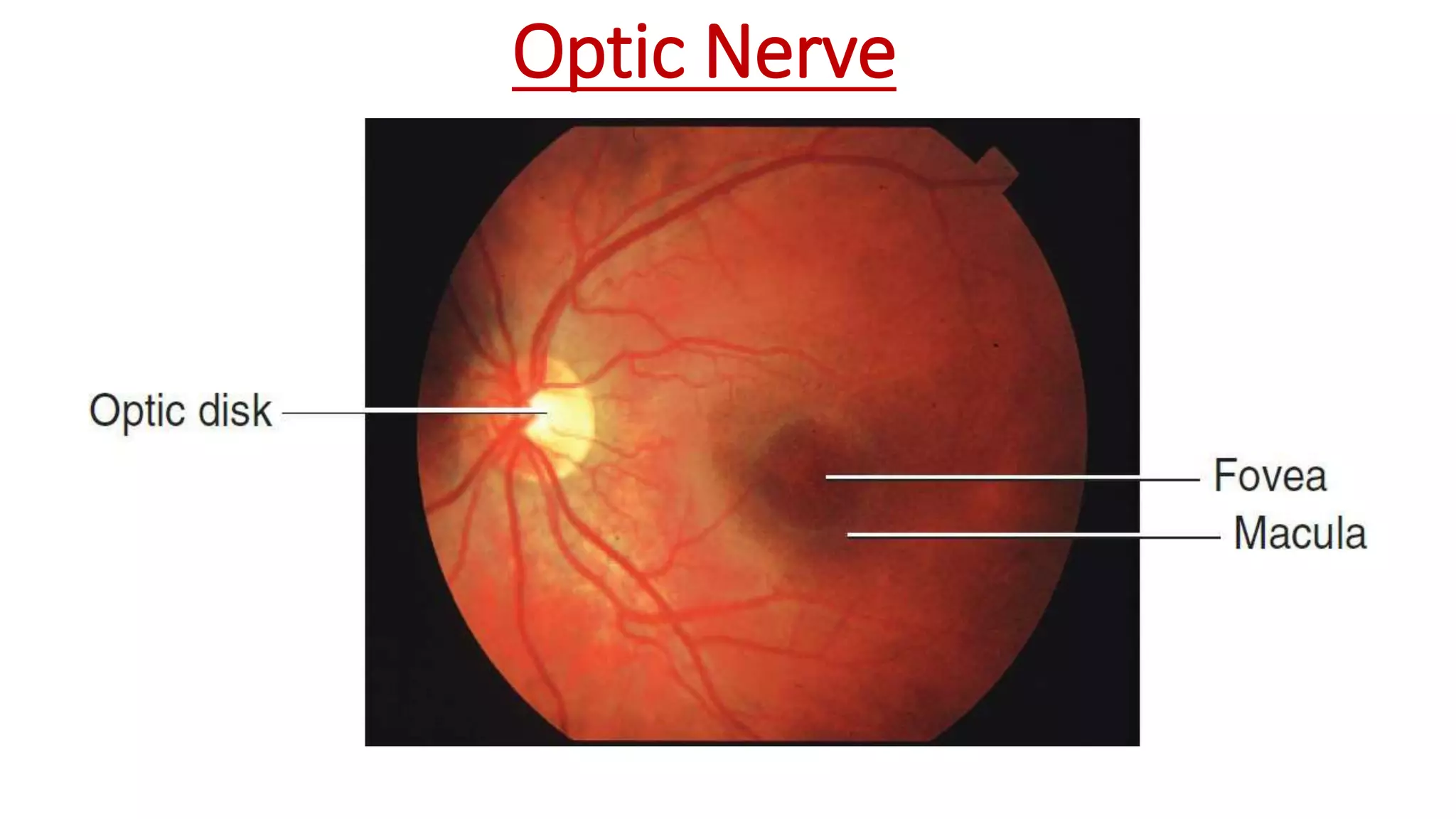
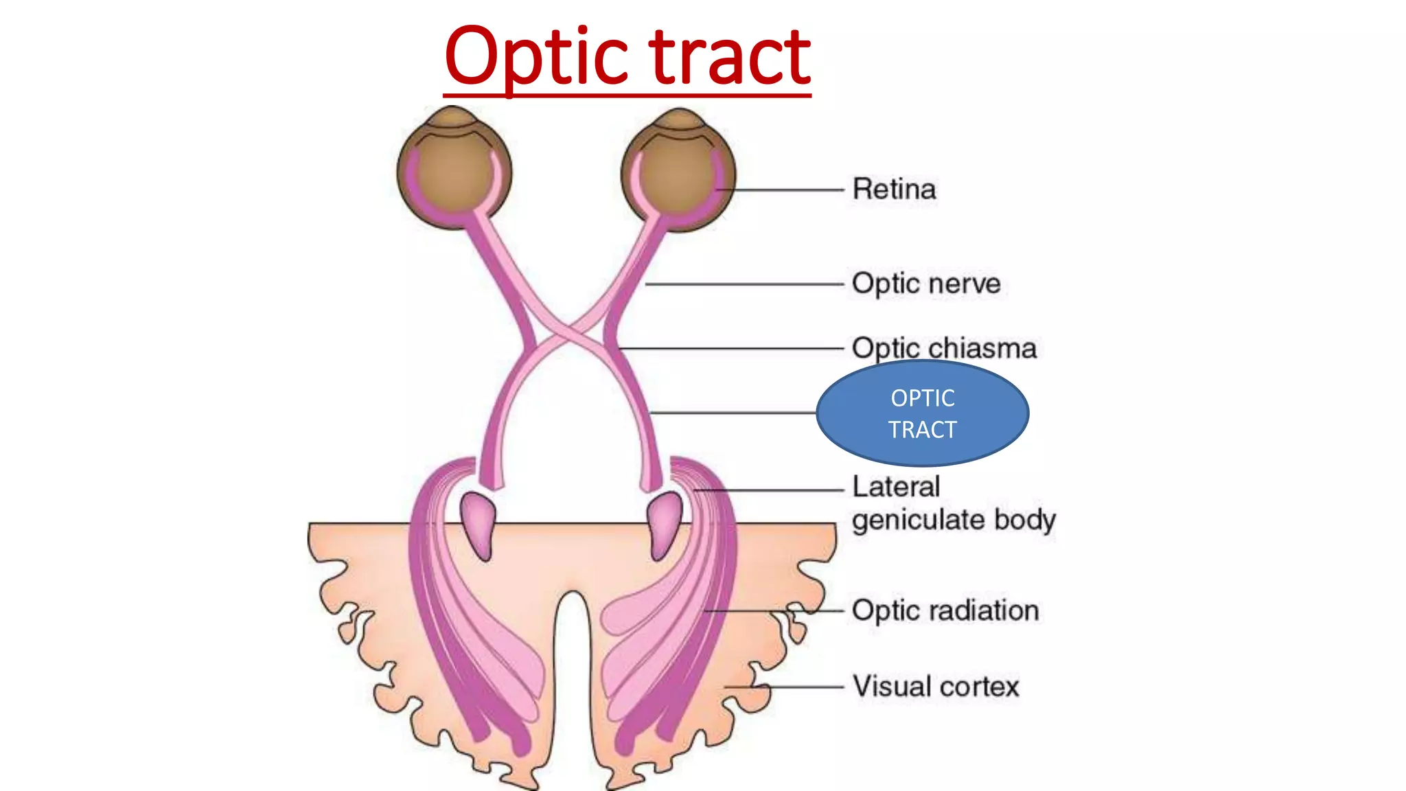
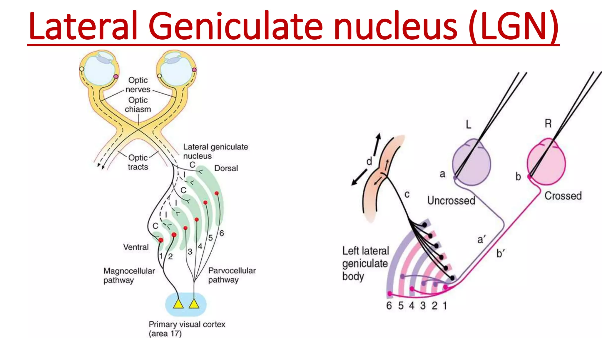
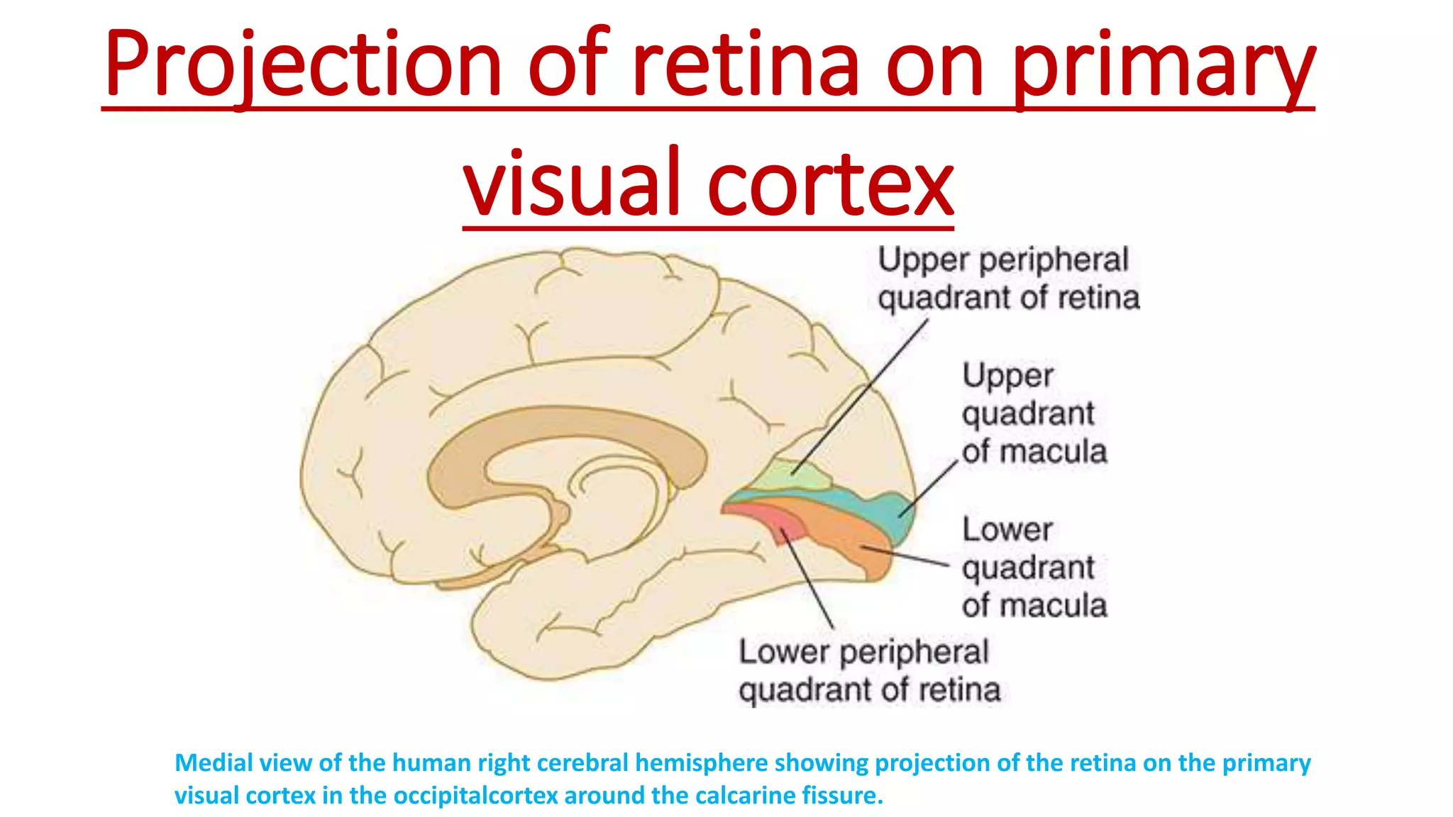
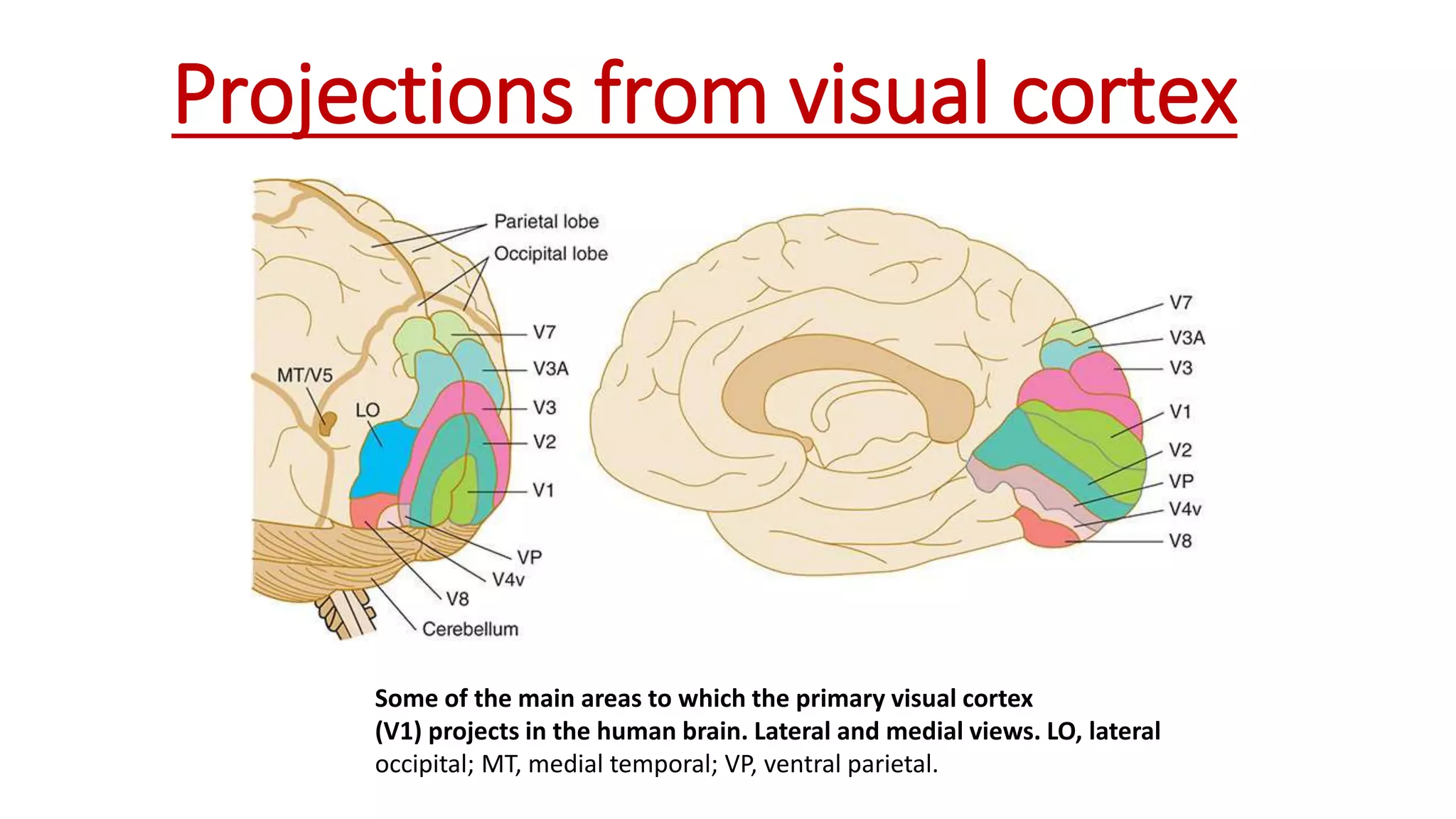
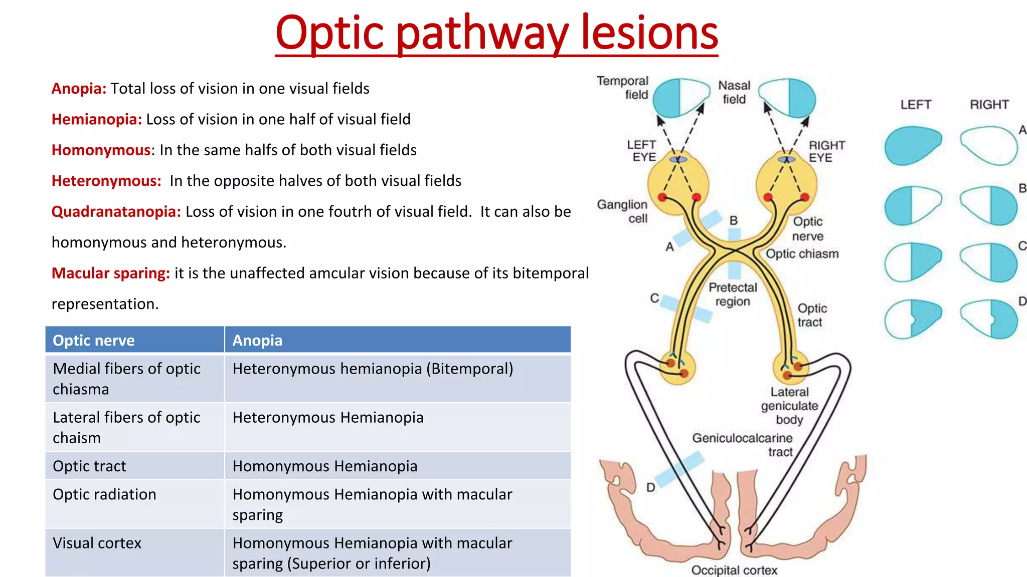
VISUAL
PATHWAYS
Dr. K. Ambareesha, PhD
Assistant Professor, Department of
Physiology, GMC, Secunderabad
SPECIFIC LEARNING OBJECTIVES
Introduction
Layers of Retina
Visual pathway
Order of neurons
Retina
Optic nerve
Optic chaism
Lateral geniculate nucleus
Optic radiation
Visual centres
A schematic of the anatomy of the eye
Layers of the Retina
Three order of neurons
1st order
neurion
•Bipolar cells
2nd order
neuron
•Ganglion cells
3rd order
neuron
•LGN
Visual pathway
Retina
Photosensitive layer that
converts light into electrical
impulse
Optic Nerve
Optic Nerve
Optic Chaisma
Optic tract
OPTIC
TRACT
Lateral Geniculate nucleus (LGN)
Optic Radiation
Optic
Radiation
Projection of retina on primary
visual cortex
Medial view of the human right cerebral hemisphere showing projection of the retina on the primary
visual cortex in the occipitalcortex around the calcarine fissure.
VISUAL CENTRES
Visual Cortex
Representation of retina on the medial side of the occipital cortex (area 17)
Area 18 : visuaopsychic area (V2)
Representation of area 17, 18 and 19 on the lateral surface of the
occiptal lobe . MT, middle temporal, MST, medial superior temporal
Visual orientation,
depth perception
Area 8: Frontal eye field
Located@ Middle
frontal gyrus
It is concerned with
voluntary conjugate
deviation of eye s to
the opposite side
Other areas
Areas 20,21,37,7, Middle temporal and medial superior
temporal areas also participate in visual processing
• Role of Area 20, 21, and 37 involved in analysis of visual details:
1. Recognisation of faces, objects and their texure
2. Recognisation of letters
3. Deatiled colour of objects
4. Understanding the imp of objects and colours.
• Middle temporal and medial superior temporal areas and area 7:
• Analysis of 3dimentional position of objects in the space
• Recognisation of their gross form
• Detection of motion of the objects
Projections from visual cortex
Some of the main areas to which the primary visual cortex
(V1) projects in the human brain. Lateral and medial views. LO, lateral
occipital; MT, medial temporal; VP, ventral parietal.
Functions of visual projection
areas in the human brain.
CONNECTIONS OF VISUAL PATHWAY
Connections
From optic chaisma
to suprachaismtic
nucleus of
hypothalamus and
from there to the
limbic system .
• This may be concerned
with cirtain circadian
rhythms and sexual cycles
in birds and some animals
From the occipital
cortex to the frontal
eye field (area8).
•This is concerned with the
movement of the eye ball
(convergence)
From occipital cortex
to superior collicli and
from there to III, IV, VI
cranial nuclei and to
the spinal cord.
•Fibers also reach the
superior colliculi form the
optic tract.
•These mediate tone,
posture, equlibrium and
visuospinal reflexes.
Connections
Fibers from optic tract before
they end in LGB reach pretectal
nucleus, which inturn send
fibers toIII cranial nerve
nucleus. This mediates the light
reflex
Connections
5
• Some fibers from the optic tract reach vestibulocerebellum
through inferior olivary complex.
• This is responsible for coardination of movements of the eyes
and head
6
• Corticogeniculate fibers from the primary visual cortex and
reticulogeniculate fibers from reticular formation of the midbrain are
inhibitory to the LGN.
• They influence response of LGN cells to the impulses coming from
Ganglion cells
7
• Retinohypothalamic projections may also influence sleep
patters
Optic pathway lesions
A lesion that interrupts one optic nerve
causes blindness in that eye (A).
 A lesion in one optic tract causes blindness in
half of the visual field (C) and is called homonymous (same
side of both visual fields) hemianopia (half-blindness).
Lesions affecting the optic chiasm destroy
fibers from both nasal hemiretinas and produce a
heteronymous (opposite sides of the visual fields) hemianopia (B).
 Occipital lesions may spare the fibers from the macula (as in D)
because of the separation in the brain of these fibers from the
others subserving vision.
Optic pathway lesions
Anopia: Total loss of vision in one visual fields
Hemianopia: Loss of vision in one half of visual field
Homonymous: In the same halfs of both visual fields
Heteronymous: In the opposite halves of both visual fields
Quadranatanopia: Loss of vision in one foutrh of visual field. It can also be
homonymous and heteronymous.
Macular sparing: it is the unaffected amcular vision because of its bitemporal
representation.
Optic nerve Anopia
Medial fibers of optic
chiasma
Heteronymous hemianopia (Bitemporal)
Lateral fibers of optic
chaism
Heteronymous Hemianopia
Optic tract Homonymous Hemianopia
Optic radiation Homonymous Hemianopia with macular
sparing
Visual cortex Homonymous Hemianopia with macular
sparing (Superior or inferior)
Summary
REFERENCES
Berne & Levy
Best and Taylor’s
Boron Boulpaep
G K pal
THANK YOU
“The most pathetic person in the world is some one who
has sight but no vision.”
― Helen Keller

optic pathway and its lesions (2).pptx

  • 1.
    VISUAL PATHWAYS Dr. K. Ambareesha,PhD Assistant Professor, Department of Physiology, GMC, Secunderabad
  • 3.
    SPECIFIC LEARNING OBJECTIVES Introduction Layersof Retina Visual pathway Order of neurons Retina Optic nerve Optic chaism Lateral geniculate nucleus Optic radiation Visual centres
  • 4.
    A schematic ofthe anatomy of the eye
  • 5.
  • 6.
    Three order ofneurons 1st order neurion •Bipolar cells 2nd order neuron •Ganglion cells 3rd order neuron •LGN
  • 7.
  • 8.
    Retina Photosensitive layer that convertslight into electrical impulse
  • 9.
  • 10.
  • 11.
  • 12.
  • 13.
  • 14.
  • 15.
    Projection of retinaon primary visual cortex Medial view of the human right cerebral hemisphere showing projection of the retina on the primary visual cortex in the occipitalcortex around the calcarine fissure.
  • 16.
  • 17.
    Visual Cortex Representation ofretina on the medial side of the occipital cortex (area 17)
  • 18.
    Area 18 :visuaopsychic area (V2) Representation of area 17, 18 and 19 on the lateral surface of the occiptal lobe . MT, middle temporal, MST, medial superior temporal Visual orientation, depth perception
  • 19.
    Area 8: Frontaleye field Located@ Middle frontal gyrus It is concerned with voluntary conjugate deviation of eye s to the opposite side
  • 20.
    Other areas Areas 20,21,37,7,Middle temporal and medial superior temporal areas also participate in visual processing • Role of Area 20, 21, and 37 involved in analysis of visual details: 1. Recognisation of faces, objects and their texure 2. Recognisation of letters 3. Deatiled colour of objects 4. Understanding the imp of objects and colours. • Middle temporal and medial superior temporal areas and area 7: • Analysis of 3dimentional position of objects in the space • Recognisation of their gross form • Detection of motion of the objects
  • 21.
    Projections from visualcortex Some of the main areas to which the primary visual cortex (V1) projects in the human brain. Lateral and medial views. LO, lateral occipital; MT, medial temporal; VP, ventral parietal.
  • 22.
    Functions of visualprojection areas in the human brain.
  • 23.
  • 24.
    Connections From optic chaisma tosuprachaismtic nucleus of hypothalamus and from there to the limbic system . • This may be concerned with cirtain circadian rhythms and sexual cycles in birds and some animals From the occipital cortex to the frontal eye field (area8). •This is concerned with the movement of the eye ball (convergence) From occipital cortex to superior collicli and from there to III, IV, VI cranial nuclei and to the spinal cord. •Fibers also reach the superior colliculi form the optic tract. •These mediate tone, posture, equlibrium and visuospinal reflexes.
  • 25.
    Connections Fibers from optictract before they end in LGB reach pretectal nucleus, which inturn send fibers toIII cranial nerve nucleus. This mediates the light reflex
  • 26.
    Connections 5 • Some fibersfrom the optic tract reach vestibulocerebellum through inferior olivary complex. • This is responsible for coardination of movements of the eyes and head 6 • Corticogeniculate fibers from the primary visual cortex and reticulogeniculate fibers from reticular formation of the midbrain are inhibitory to the LGN. • They influence response of LGN cells to the impulses coming from Ganglion cells 7 • Retinohypothalamic projections may also influence sleep patters
  • 27.
    Optic pathway lesions Alesion that interrupts one optic nerve causes blindness in that eye (A).  A lesion in one optic tract causes blindness in half of the visual field (C) and is called homonymous (same side of both visual fields) hemianopia (half-blindness). Lesions affecting the optic chiasm destroy fibers from both nasal hemiretinas and produce a heteronymous (opposite sides of the visual fields) hemianopia (B).  Occipital lesions may spare the fibers from the macula (as in D) because of the separation in the brain of these fibers from the others subserving vision.
  • 28.
    Optic pathway lesions Anopia:Total loss of vision in one visual fields Hemianopia: Loss of vision in one half of visual field Homonymous: In the same halfs of both visual fields Heteronymous: In the opposite halves of both visual fields Quadranatanopia: Loss of vision in one foutrh of visual field. It can also be homonymous and heteronymous. Macular sparing: it is the unaffected amcular vision because of its bitemporal representation. Optic nerve Anopia Medial fibers of optic chiasma Heteronymous hemianopia (Bitemporal) Lateral fibers of optic chaism Heteronymous Hemianopia Optic tract Homonymous Hemianopia Optic radiation Homonymous Hemianopia with macular sparing Visual cortex Homonymous Hemianopia with macular sparing (Superior or inferior)
  • 29.
  • 30.
    REFERENCES Berne & Levy Bestand Taylor’s Boron Boulpaep G K pal
  • 31.
    THANK YOU “The mostpathetic person in the world is some one who has sight but no vision.” ― Helen Keller