
Recommended
More Related Content
What's hot
What's hot (20)
Viewers also liked
Viewers also liked (20)
Similar to Visual inspection guide for blood compopnents
Similar to Visual inspection guide for blood compopnents (20)
Recently uploaded
Recently uploaded (20)
Visual inspection guide for blood compopnents
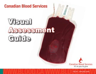
- 1. Visual Assessment Guide Canadian Blood Services T05 021 JANUARY 2009
- 2. CANADIAN BLOOD SERVICES T05 021 JANUARY 2009 i Table of Contents Introduction 1 How to Use 3 Quick Reference Guide Table 1 - Summary of Visual Appearance 4 Table 2 - Summary of Acceptability Criteria 5 Red Cell Components 6 Platelet Components 12 Plasma Components 18 Cryoprecipitate Components 25 References 26 Acknowledgement 27 ISBN 978-1-926581-14-9 © 2009, Canadian Blood Services
- 3. T05 021 JANUARY 2009CANADIAN BLOOD SERVICES 1 INTRODUCTION Typical Platelet (platelet pool from 4 donors using the buffy coat method) Introduction Whole blood donations are collected into a closed bag system that permits aseptic separation into red cells, platelets, plasma and cryoprecipitate. Currently, there are three whole blood processing systems used by the Canadian Blood Services: Buffy Coat (BC) method, Platelet Rich Plasma (PRP) method and Whole Blood (WB) filtration. Platelet and plasma components are also collected by apheresis. Blood components manufactured by Canadian Blood Services are done so under strict manufacturing protocols.
- 4. CANADIAN BLOOD SERVICES INTRODUCTION 2 This guide has been produced for use by both Canadian Blood Services and hospital personnel. The acceptability of a blood component for release from Canadian Blood Services is determined by Canadian Blood Services protocols and procedures. The acceptability of a blood component for transfusion will be determined by local hospital policy and procedures under the direction of the Transfusion Services Medical Director (ref CSA-Z902-04 4.3.6.1). Typical Plasma prepared from whole blood T05 021 JANUARY 2009
- 5. T05 021 JANUARY 2009CANADIAN BLOOD SERVICES HOWTOUSE This guide is divided into four sections covering the four blood components. Each section begins with a brief explanation of the component, a description of the variations in appearance for that component and criteria for acceptability. Inspection of the blood products should take place in a well-lit area. Coloured products have been photographed against a white background, whereas light-coloured (white) products have been photographed against a dark background to obtain a better viewing contrast. Visual Assessments Guides printed by the Canadian Blood Services are laminated to increase their durability; however the quality of the photographs may decrease over time. How to Use This Visual Assessment Guide Prolonged exposure to sunlight or fluorescent lighting may cause fading and to maintain the quality of the photographs, please keep the guide closed when not in use. This guide is available on www.transfusionmedicine.ca and if it is printed locally or viewed on-line the pictures may not be color-true. Table 1 is a quick guide for conditions that affect the visual appearance of components. Not all conditions affect all components. Table 2 is a summary of the acceptability criteria. Note: Storage containers may differ in appearance depending on bag vendor. Labels may or may not appear on the back of products and label formats and locations may be different. 3
- 6. T05 021 JANUARY 2009CANADIAN BLOOD SERVICES QUICKREFERENCEGUIDE Affected Component Condition Red cells Platelets Plasma Cryoprecipitate Hemolysis • Loss of intact red cells results in lower hematocrit and a brighter cherry red color. • Free hemoglobin imparts a light pink tinge to a dark red almost purple color to the supernatant. • Occurs as part of normal aging process n/a • Red cells present in plasma will hemolyze during the freeze/thaw process and will impart a pink to red tinge depending on number of red cells involved • Red cells present in plasma will hemolyze during the freeze/thaw process and will impart a pink to red tinge depending on number of red cells involved Red cell contamination n/a • Varies from a light pink / salmon color tinge to a marked red discoloration • Varies from a light pink / salmon color tinge to a marked red discoloration • Varies from a light pink / salmon color tinge to a marked red discoloration Lipemia • A lighter shade of red and an increased opacity of the unit similar to a ‘strawberry milkshake’ • Increased opacity • ‘Milky’ white appearance • Increased opacity • ‘Milky’ white appearance • Increased opacity • ‘Milky’ white appearance Icterus n/a • Bright yellow to brown • Bright yellow to brown • Bright yellow to brown Bacterial contamination • A dark purple to black discoloration in red cells • Excessive and unusual air bubbles • Clots and fibrin strands • Increased opacity • When assosiated with hemolysis, a pink to red discoloration may be seen in supernatant. • Excessive and unusual air bubbles • Clots and fibrin strands • Increased opacity • Grey discoloration • Excessive and unusual air bubbles • Clots and fibrin strands • Increased opacity • Excessive and unusual air bubbles • Clots and fibrin strands • Increased opacity Particulate matter • Clots appear as small to large dark red or purple masses that do not dissipate with gentle manipulation in red cells. • Cellular aggregates appear as white and opaque masses that do not dissipate with gentle manipulation. • White particulate matter varies from flattened specks to a greasy film and may dissipate with a change in temperature • Cold agglutinins form large red blood cell masses that do not dissipate with gentle manipulation. • Clots and fibrin strands result from the activation of the clotting process and may appear as white/opaque masses or whitish thread like strands that do not dissipate with gentle manipulation • Cellular aggregates may appear as white and opaque masses that do not dissipate with gentle manipulation. • Particulate matter may vary considerably in size. • Clots and fibrin strands result from the activation of the clotting process and may appears as white/opaque masses or whitish thread like strands that do not dissipate with gentle manipulation • Cellular aggregates may appear as white and opaque masses that do not dissipate with gentle manipulation. • Particulate matter may vary considerably in size. • Clots and fibrin strands result from the activation of the clotting process and may appears as white/opaque masses or whitish thread like strands that do not dissipate with gentle manipulation • Cellular aggregates may appear as white and opaque masses that do not dissipate with gentle manipulation. • Particulate matter may vary considerably in size. Discoloration • See hemolysis, lipemia, bacterial contamination • Pink, red, bright orange / yellow, bright green, or brown • Pink, red, bright orange / yellow, bright green, or brown • Pink, red, bright orange / yellow, bright green, or brown Quick Reference Guide Table 1. Visual appearance of blood components n/a=Not applicable 4
- 7. T05 021 JANUARY 2009CANADIAN BLOOD SERVICES 5 QUICKREFERENCEGUIDE Affected Component Condition Red cells Platelets Plasma Cryoprecipitate Hemolysis • Some degree of hemolysis is acceptable and expected. The CSA standard will define acceptable levels of hemolysis as < 0.8% at expiry. n/a • Some degree of hemolysis is possible depending on the number of red cells in the plasma. • Some degree of hemolysis is possible depending on the number of red cells in the plasma. Red cell contamination n/a • Currently there are no standards of acceptability of red cell contamination for platelet units. However, the AABB standards recommend compatibility testing when an apheresis platelet contains more than 2ml of red cells (ref AABB standard 24th ed 5.14.5). • Currently, there are no standards of acceptability for red cell contamination of plasma units. • Currently, there are no standards of acceptability for red cell contamination of plasma units. Lipemia • Blood components with lipemia are acceptable for transfusion. • Blood components with lipemia are acceptable for transfusion. • Blood components with lipemia are acceptable for transfusion. • Blood components with lipemia are acceptable for transfusion. Icterus • Blood components with icterus are acceptable for transfusion. • Blood components with icterus are acceptable for transfusion. • Blood components with icterus are acceptable for transfusion. • Blood components with icterus are acceptable for transfusion. Bacterial contamination • Bacterially contaminated blood components are not acceptable for transfusion • Canadian Blood Services routinely tests platelets derived from whole blood or apheresis for bacterial contamination. Bacterially contaminated blood components are not acceptable for transfusion. • Bacterially contaminated blood components are not acceptable for transfusion • Bacterially contaminated blood components are not acceptable for transfusion Particulate matter • Clots and fibrin - Blood components containing clots and / or fibrin strands should not be transfused. • Cellular aggregates - Blood components containing cellular aggregates should not be transfused. • White Particulate Matter (WPM). Blood components containing WPM are acceptable for transfusion. WPM may dissipate with a change in temperature. • Cold agglutinins - Blood components containing cold agglutinin masses should not be transfused. • Clots and fibrin Strands - Blood components containing clots and/or fibrin strands should not be transfused. • Cellular aggregates- Blood components containing cellular aggregates should not be transfused. • Clots and fibrin - Blood components containing clots and / or fibrin strands should not be transfused. • Cellular Aggregates - Blood components containing cellular aggregates should not be transfused. • White Particulate Matter (WPM) - WPM may be seen in thawed plasma that has been stored in the fridge. Blood components containing WPM are acceptable for transfusion • Clots and fibrin - Blood components containing clots and / or fibrin strands should not be transfused. • Cellular Aggregates - Blood components containing cellular aggregates should not be transfused. • White Particulate Matter (WPM) - WPM may be seen in thawed plasma that has been stored in the fridge. Blood components containing WPM are acceptable for transfusion Discoloration • See hemolysis, lipemia, bacterial contamination • Discoloration due to icterus (yellow), oral contraceptives (green), vitamin A or large quantities of carrots (orange) are all acceptable for transfusion. • Discoloration due to icterus (yellow), oral contraceptives (green), vitamin A or large quantities of carrots (orange) are all acceptable for transfusion. • Discoloration due to icterus (yellow), oral contraceptives (green), vitamin A or large quantities of carrots (orange) are all acceptable for transfusion. Quick Reference Guide Table 2. Acceptability criteria of blood components n/a=Not applicable
- 8. CANADIAN BLOOD SERVICES REDCELLCOMPONENTS Typical Red Blood Cells prepared from whole blood Red Cell Components Red Cell Units Red cell units are an even suspension of red cells in a small amount of residual plasma and additive solution. The visual appearance of segments should not be used to assess a red cell unit as they may not reflect the content of the bag.6 Units should be allowed to settle to permit an assessment of the supernatant. There is a normal variation in the shade of red seen in red cell units that may reflect a difference in the hematocrit of the donor (i.e. a higher hematocrit will result in a darker red). 6T05 021 JANUARY 2009
- 9. CANADIAN BLOOD SERVICES REDCELLCOMPONENTS Appearance of Red Cell Units There are several conditions that may change the appearance of the red cell unit: Hemolysis • Visual appearance – Of particular note, hemolysis caused by bacterial contamination may result in a dark purple to black discoloration (see ‘Bacterial Contamination’). – Free hemoglobin within the supernatant of red cells will cause a discoloration that will vary from a light pink tinge to a dark red almost purple color, depending on the extent of hemolysis (i.e. a higher degree of hemolysis will result in a darker discoloration). • Hemolysis is the break down of red cells and the subsequent release of hemoglobin, the pigmented protein in red cells. Hemolysis of stored red cells is a normal process and increases with storage times. Some degree of hemolysis is acceptable and expected. The CSA standard will define acceptable levels of hemolysis as < 0.8% at expiry. 7 • Visual assessment of supernatant color is the only non-destructive means to estimate hemolysis in a red cell unit. It is very subjective and it is difficult to define a “cut-off” color for determining a unit’s acceptability. The color of the supernatant varies according to the volume of the supernatant and the hematocrit and hemoglobin levels in the red cell unit. The presence of red cells in the supernatant may be mistaken for hemolysis. If hemolysis is suspected, it may be worthwhile to allow the unit to settle for 24 hours or more to minimize red cell contamination of the supernatant prior to inspection for hemolysis. • There are numerous causes of hemolysis. A few examples related to the manufacturing process and subsequent handling of red cell units are: – Temperature extremes – Excessive centrifugation – Tubing stripping – Tubing heat sealers – Sheer force during manufacturing eg. kinked tubing, improperly broken cannula – Bacterial contamination – Mishandling – Incompatible solutions T05 021 JANUARY 2009
- 10. CANADIAN BLOOD SERVICES REDCELLCOMPONENTS 8 Supernatant of RBC Percent Hemolysis = 0.11 % Photographs were taken after allowing the RBC to settle for 4 days to permit an evident visualization of the supernatant. This assessment may be performed earlier or later. Supernatant of RBC Percent Hemolysis = 0.36 % Supernatant of RBC showing higher levels of hemolysis Percent Hemolysis = 1.14 % T05 021 JANUARY 2009 The CSA standard will define acceptable levels of hemolysis as < 0.8% at expiry.
- 11. CANADIAN BLOOD SERVICES REDCELLCOMPONENTS Lipemia • Visual appearance – The appearance of a lipemic red cell unit has been compared to a ‘strawberry milkshake’. • Lipemia is the presence of lipid particles in the blood. The presence of lipemia is acceptable for transfusion. When excessive lipemia interferes with testing all components are discarded at Canadian Blood Services. • Causes of lipemia in a red cell unit are donor related and may be related to: – Fatty meals before blood donation – Chronic conditions such as hypercholesterolemia Bacterial contamination • Visual appearance – There are usually no visible changes to the component, but occasionally it may be possible to detect the following: – Growing bacteria within a red cell unit may consume the oxygen and cause a dark purple to black discoloration in red cells. – The presence of bacteria may produce gas resulting in excessive and unusual air bubbles. Lipemic RBC Unit No picture available at this time 9 – The presence of bacteria may activate clotting resulting in clots and fibrin strands. – Increased opacity – Because bacterial contamination can cause hemolysis, a pink to red discoloration may be seen in the supernatant. • Bacterial contamination is the presence of bacteria in the blood. Blood should be free of bacteria. Blood donations are collected and then processed in a closed system to maintain aseptic conditions. Bacterially contaminated blood components are not acceptable for transfusion. • Bacterial contamination may be related to the donor or subsequent manufacturing processes. – Donor with asymptomatic bacteremia (i.e. bacteria in blood) – Inadequate cleansing of skin prior to venipuncture. To minimize bacterial contamination, CBS uses a diversion pouch for the first portion of the blood donation that should capture the skin plug. – Loss of sterility during component manufacturing or handling. Bacterial Contamination in RBC Unit No picture available at this time T05 021 JANUARY 2009
- 12. CANADIAN BLOOD SERVICES REDCELLCOMPONENTS Particulate matter • Particulate matter, as opposed to foreign objects, can be the result of blood collection and / or component manufacturing processes. Composed of platelets, white and red blood cells, and / or in some instances fibrin, particulate matter may increase during storage of a red cell unit. There are several categories of particulate matter that may be found within in a red cell unit: 1. Clots and fibrin strands – Clots and fibrin strands result from the activation of the clotting processes and can be a mixture of clotting proteins (including fibrin) and platelets. Blood donations are collected and processed in bags that contain anticoagulants to inhibit the clotting processes. Blood components containing clots and / or fibrin strands should not be transfused. – Clots may appear as small to large dark red or purple masses that do not dissipate with gentle manipulation in red cells. 10T05 021 JANUARY 2009 Clot in RBC unit
- 13. CANADIAN BLOOD SERVICES REDCELLCOMPONENTS 11 2. Cellular aggregates – Cellular aggregates may appear as white and opaque masses that do not dissipate with gentle manipulation. Blood components containing cellular aggregates should not be transfused. 3. White particulate matter (WPM) – WPM is a non-cellular material and is a normal constituent of blood. The WPM may dissipate with a change in temperature. – WPM is a lipid-rich material that appears white in color and variable in shape, from flattened specks to a greasy film. Generally there are low numbers of WPM per red cell unit. – The presence of WPM is acceptable for transfusion. 4. Cold agglutinins form large red blood cell masses that do not dissipate with gentle manipulation. Blood components containing cold agglutinin masses should not be transfused. T05 021 JANUARY 2009 White Particulate Matter in RBC unit
- 14. T05 021 JANUARY 2009CANADIAN BLOOD SERVICES 12 REDCELLCOMPONENTS White Particulate Matter in RBC unit
- 15. CANADIAN BLOOD SERVICES PLATELETCOMPONENTS Platelet Units There are three current methods of platelet production: Pooled Platelets from BC production, Random Donor Platelet units from PRP production methods, and Apheresis Platelets (AP). Because platelet units contain cells and plasma, they are opaque and vary in color from beige to yellow. Residual red cells in platelet units may confer a salmon / pink or marked red colouring. 13 Platelet Components Typical Platelet (platelet pool from 4 donors using the buffy coat method) T05 021 JANUARY 2009
- 16. CANADIAN BLOOD SERVICES PLATELETCOMPONENTS 14 Typical Platelet (prepared by Apheresis) Typical Platelet (prepared using the PRP method) T05 021 JANUARY 2009
- 17. CANADIAN BLOOD SERVICES PLATELETCOMPONENTS 15 Appearance of Platelets Units There are several conditions that may change the appearance of platelet units: Red cell contamination Red Cell Contamination in Pooled Platelet Unit (platelets prepared using the Buffy Coat method) RBC contamination = 2.15 mL RBC. • Visual appearance – The degree of red discoloration reflects the amount of red cell contamination and this varies from a light pink / salmon color tinge to a marked red discoloration. • Red cell contamination occurs during production and results from ineffective separation of red cells and platelets. Currently there are no standards of acceptability of red cell contamination for platelet units. However, the AABB standards recommend compatability testing when an apheresis platelet contains more than 2 ml of red cells. (ref AABB standard 24th ed 5.14.5) T05 021 JANUARY 2009
- 18. CANADIAN BLOOD SERVICES PLATELETCOMPONENTS 16 Red Cell Contamination in Platelets (platelets prepared using the PRP method) RBC contamination = 0.50 mL RBC Lipemia • Visual appearance – Lipemia may result in a ‘milky’ white appearance. • Lipemia is the presence of lipid particles in the blood. The presence of lipemia is acceptable for transfusion. When excessive lipemia interferes with testing all components are discarded at Canadian Blood Services. • Causes of lipemia in a platelet unit are donor related and may be related to: – Fatty meals before blood donation – Chronic conditions such as hypercholesterolemia Icterus • Visual appearance – Bright yellow to brown. • Icterus, describes the yellow discoloration due to high bilirubin content in blood. The presence of icterus is acceptable for transfusion. Refer to plasma components for photographs T05 021 JANUARY 2009
- 19. CANADIAN BLOOD SERVICES PLATELETCOMPONENTS 17 When excessive bilirubin interferes with testing all components are discarded at Canadian Blood Services. • Causes of icterus in a platelet unit from eligible donors may be related to: – Inherited liver disorders (e.g. Gilbert’s Syndrome) – Gallstones Bacterial contamination • Visual appearance - There are usually no visible changes to the component, but occasionally it may be possible to detect the following: – The presence of bacteria may produce gas resulting in excessive and unusual air bubbles. – The presence of bacteria may activate clotting resulting in clots and fibrin strands. – Increased opacity – Grey discoloration • Bacterial contamination is the presence of bacteria in the blood. Blood should be free of bacteria. Blood donations are collected and then processed in a closed system to maintain aseptic conditions. Canadian Blood Services routinely tests platelets derived from whole blood or aphereses for bacterial contamination. Bacterial contamination in Platelet unit No picture available at this time Bacterially contaminated blood components are not acceptable for transfusion. • Bacterial contamination may be related to the donor or subsequent manufacturing processes. – Donor with asymptomatic bacteremia (i.e. bacteria in blood) – Inadequate cleansing of skin prior to venipuncture. To minimize bacterial contamination, CBS uses a diversion pouch for the first portion of the blood donation that should capture the skin plug. – Loss of sterility during component manufacturing or handling. Particulate matter • Visual appearance – Clots may appear as white and opaque masses that do not dissipate with gentle manipulation. • Particulate matter, as opposed to foreign objects, can be the result of blood collection and / or component manufacturing processes. There are several categories of particulate matter that may be found within in a platelet unit: 1. Clots and fibrin strands – Clots and fibrin strands result from the activation of the clotting processes and can be a mixture T05 021 JANUARY 2009
- 20. CANADIAN BLOOD SERVICES PLATELETCOMPONENTS 18 of clotting proteins, platelets and fibrin. Blood donations are collected and processed in bags that contain anticoagulants to inhibit the clotting processes. Blood components containing clots and fibrin strands should not be transfused. 2. Cellular aggregates – Cellular aggregates may appear as white and opaque masses that do not dissipate with gentle manipulation. Blood components containing cellular aggregates should not be transfused. Particulate Matter in Platelet Unit (platelets prepared using the PRP method Discoloration • The term ‘discoloration’ refers to a variety of donor factors that alter the plasma portion of platelet units and these can range from metabolic conditions to medications or large doses of vitamins taken by donors. Discoloration from contamination with red cells or bacteria, can also be causes. • Causes of discoloration, and their resulting alterations of plasma color, include: – Contamination 1. Red cell – see section 1, Platelet Components. 2. Bacterial – see section 4, Platelet Components. Bacterially contaminated blood components are not acceptable for transfusion. – bright yellow to greenish brown – Icterus. Acceptable for transfusion. – light green – Oral contraceptive pill. Acceptable for transfusion. – bright orange – Vitamin A and large quantities of carrots. Acceptable for transfusion. T05 021 JANUARY 2009
- 21. CANADIAN BLOOD SERVICES PLASMACOMPONENTS Plasma Components Plasma Units Currently there are four plasma components produced: Plasma, Frozen Plasma, Fresh Frozen Plasma, and Cryosupernatant Plasma. 19 Typical Apheresis Fresh Frozen Plasma T05 021 JANUARY 2009
- 22. PLASMACOMPONENTS CANADIAN BLOOD SERVICES 20 Examples of a Typical Plasma prepared from whole blood T05 021 JANUARY 2009
- 23. CANADIAN BLOOD SERVICES PLASMACOMPONENTS Appearance of Plasma Units There are several conditions that may change the appearance of plasma units: Red cell contamination • Red cell contamination occurs during production and results from ineffective separation of red cells and plasma. • Plasma unit – The degree of red discoloration reflects the amount of red cell contamination and this varies from a light pink / salmon color tinge to a marked red discoloration. – Currently, there are no standards of acceptability for red cell contamination of plasma units. Lipemia • Visual appearance – Lipemia may result in a ‘milky’ white appearance. • Lipemia is the presence of lipid particles in the blood. The presence of lipemia is acceptable for transfusion. Lipemic Plasma 21T05 021 JANUARY 2009
- 24. CANADIAN BLOOD SERVICES PLASMACOMPONENTS When excessive lipemia interferes with testing all components are discarded at Canadian Blood Services. • Causes of lipemia in a plasma unit are donor related and may be related to: – Fatty meals before blood donation – Chronic conditions such as hypercholesterolemia Icterus • Visual appearance – Bright yellow to brown. • Icterus, describes the yellow discoloration due to high bilirubin content in blood. The presence of icterus in a blood component is acceptable for transfusion. When excessive bilirubin interferes with testing all components are discarded at Canadian Blood Services. • Causes of icterus in a plasma from eligible donors may be related to: – Inherited liver disorders (e.g. Gilbert’s Syndrome) – Gallstones Icteric Plasma 22T05 021 JANUARY 2009
- 25. CANADIAN BLOOD SERVICES PLASMACOMPONENTS Bacterial contamination • Visual appearance - There are usually no visible changes to the component, but occasionally it may be possible to detect the following: – The presence of bacteria may produce gas resulting in unusual air bubbles. – The presence of bacteria may activate clotting resulting in clots and fibrin strands. – Increased opacity. • Bacterial contamination is the presence of bacteria in the blood. Blood should be free of bacteria. Blood donations are collected and then processed in a closed system to maintain aseptic conditions. Bacterially contaminated blood components are not acceptable for transfusion. • Bacterial contamination may be related to the donor or subsequent manufacturing processes. Bacterial contamination in Plasma unit No picture available at this time – Donor with asymptomatic bacteremia (i.e. bacteria in blood) – Inadequate cleansing of skin prior to venipuncture. To minimize bacterial contamination, CBS uses a diversion pouch for the first portion of the blood donation that should capture the skin plug. – Loss of sterility during component manufacturing or handling. Particulate matter • Visual appearance – Particulate matter may appear as white and opaque masses that do not dissipate with gentle manipulation. • Particulate matter, as opposed to foreign objects, can be the result of blood collection and / or component manufacturing processes. Composed of platelets, white and red blood cells, and / or in some instances fibrin, particulate matter may increase during storage of a plasma unit. There are several categories of particulate matter that may be found within in a plasma unit: 23T05 021 JANUARY 2009
- 26. CANADIAN BLOOD SERVICES PLASMACOMPONENTS 1. Clots and fibrin strands – Clots and fibrin strands result from the activation of the clotting processes and can be a mixture of clotting proteins (including fibrin) and platelets. Blood donations are collected and processed in bags that contain anticoagulants to inhibit the clotting processes. Blood components containing clots and fibrin strands should not be transfused. 2. Cellular aggregates – Cellular aggregates may appear as white and opaque masses that do not dissipate with gentle manipulation. Blood components containing cellular aggregates should not be transfused. 3. White Particulate Matter (WPM) – WPM may be seen in thawed plasma that has been stored in the fridge. Components with WPM are acceptable for transfusion. Discoloration • The term ‘discoloration’ refers to a variety of donor factors that alter the plasma units and these can range from metabolic conditions to medications or large doses of vitamins taken by donors. Discoloration from contamination with red cells and bacteria (see Appearance of Red Cell Units), can also be causes. • Causes of discoloration, and their resulting alterations of plasma color, include: – Contamination 1. Red cell – see section 1, Plasma Components. 2. Bacterial – see section 4, Plasma Components. Bacterially contaminated blood components are not acceptable for transfusion. – bright yellow to greenish brown – Icterus. Acceptable for transfusion. – light green – Oral contraceptive pill. Acceptable for transfusion. – bright orange – Vitamin A and large quantities of carrots. Acceptable for transfusion. 24T05 021 JANUARY 2009
- 27. CANADIAN BLOOD SERVICES PLASMACOMPONENTS Plasma with Green Colour (green colour is commonly a result of donor taking oral contraceptive pill) 25T05 021 JANUARY 2009
- 28. CANADIAN BLOOD SERVICES CRYOPRECIPITATECOMPONENTS Cryoprecipitate Components Cryoprecipitate Units Cryoprecipitate appears as a concentrated precipitate at the bottom of a bag. It will appear thick, opaque, whitish and paste- like. Upon thawing, the cryoprecipitate mass will dissolve and re-suspend in the small amount of residual plasma and appear as an even, thick, whitish liquid. Appearance of Cryoprecipitate Units There are several conditions that may change the appearance of cryoprecipitate units: The unusual conditions described in the plasma components section may also apply to cryoprecipitate components. 26 Examples of a Typical Cryoprecipitate T05 021 JANUARY 2009
- 29. CANADIAN BLOOD SERVICES REFERENCES 27 References 1. Visual Inspection Reference Guide, American National Red Cross, American Red Cross Biomedical Services, 2006. 2. Standards for Blood Banks and Transfusion Services, 23rd Edition. AABB, 2005. 3. FDA summary basis of approval for red blood cells frozen and red blood cells deglycerolized (Reference number 86-0335). Applicant-Department of the Navy, Navy Hospital, Bethesda, MD, US License Number 635-10 1986. 4. Council of Europe. Guide to the preparation, use and quality assurance of blood components, 9th ed. Strasbourg, France: Council of Europe Publishing, 2003. 5. Investigation of White Particulate Matter Found in Red Cell Concentrates, Devine D, Canadian Blood Services, 2003. 6. Janatpour KA, Paglieroni TG, Crocker VL, et al. Visual assessment of hemolysis in red blood cell units and segments can be deceptive. Transfusion 2004:44:984-989. 7. Houseworth JL, Parks JA, Miguel EA, et al. Iatrogenic green plasma. Transfusion 2005;45:1047. 8. CSA standard, Z 902-04 Blood and Blood components. T05 021 JANUARY 2009
- 30. CANADIAN BLOOD SERVICES T05 021 JANUARY 2009 28 Acknowledgement Canadian Blood Services extends thanks to those who contributed to the preparation of this guide and to those who provided a peer review. Dr. Jason Acker Ryan Andress Dr. Maggie Constantine Shelley Doyle Myriam Hurabielle Craig Jenkins Wanda Lefresne Dr. Katherine Serrano Susan Shimla Dr. Tanya Petraszko Janet Unrau Fraser Health Authority, British Columbia Vancouver Island Health Authority, British Columbia Red Deer Regional Hospital, Alberta Canadian Blood Services Staff, BC & Yukon Canadian Blood Services Staff, Edmonton