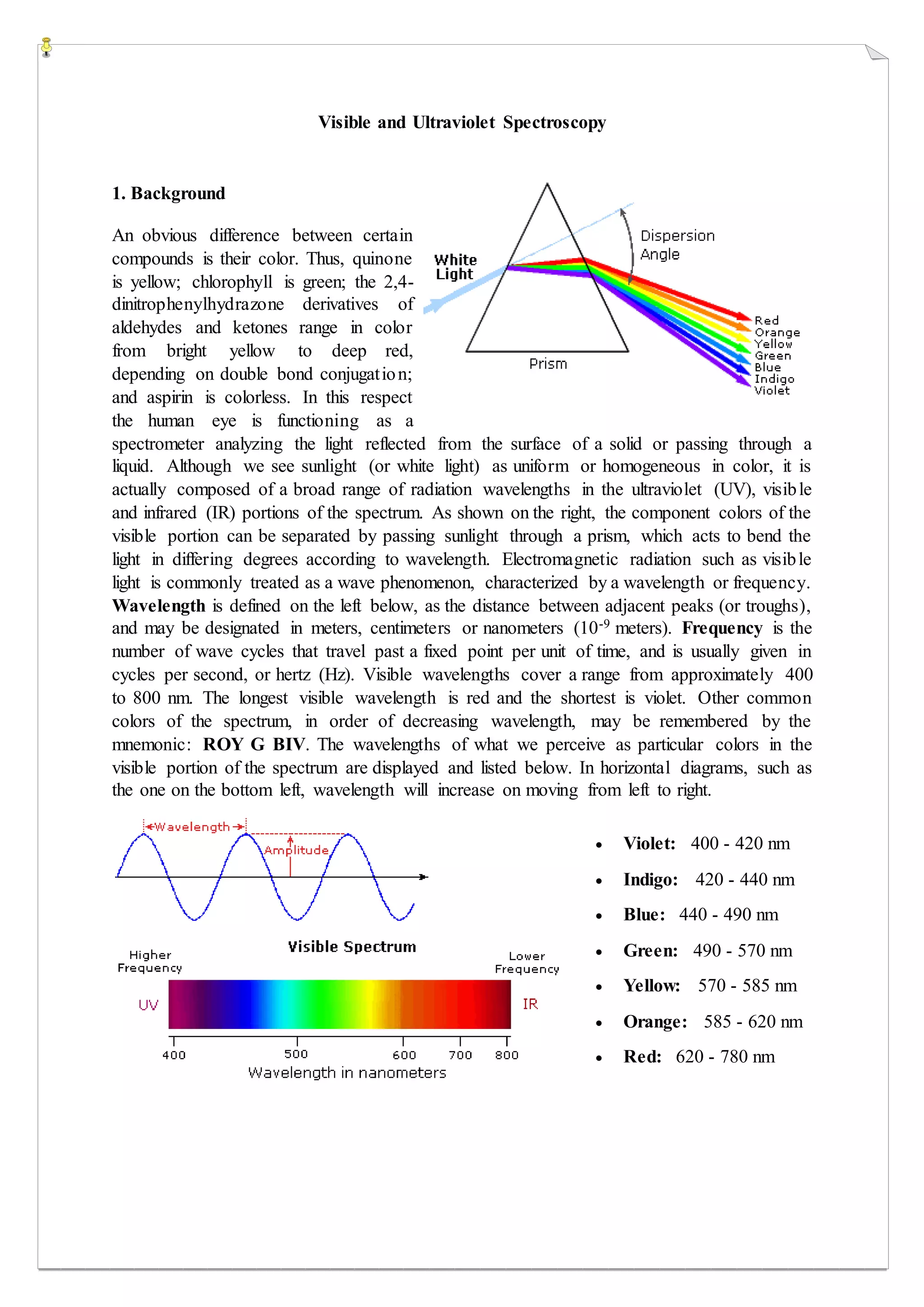
Boost PC performance: How more available memory can improve productivity
Visible and ultraviolet spectroscopy
1. Visible and Ultraviolet Spectroscopy
1. Background
An obvious difference between certain
compounds is their color. Thus, quinone
is yellow; chlorophyll is green; the 2,4-
dinitrophenylhydrazone derivatives of
aldehydes and ketones range in color
from bright yellow to deep red,
depending on double bond conjugation;
and aspirin is colorless. In this respect
the human eye is functioning as a
spectrometer analyzing the light reflected from the surface of a solid or passing through a
liquid. Although we see sunlight (or white light) as uniform or homogeneous in color, it is
actually composed of a broad range of radiation wavelengths in the ultraviolet (UV), visible
and infrared (IR) portions of the spectrum. As shown on the right, the component colors of the
visible portion can be separated by passing sunlight through a prism, which acts to bend the
light in differing degrees according to wavelength. Electromagnetic radiation such as visible
light is commonly treated as a wave phenomenon, characterized by a wavelength or frequency.
Wavelength is defined on the left below, as the distance between adjacent peaks (or troughs),
and may be designated in meters, centimeters or nanometers (10-9 meters). Frequency is the
number of wave cycles that travel past a fixed point per unit of time, and is usually given in
cycles per second, or hertz (Hz). Visible wavelengths cover a range from approximately 400
to 800 nm. The longest visible wavelength is red and the shortest is violet. Other common
colors of the spectrum, in order of decreasing wavelength, may be remembered by the
mnemonic: ROY G BIV. The wavelengths of what we perceive as particular colors in the
visible portion of the spectrum are displayed and listed below. In horizontal diagrams, such as
the one on the bottom left, wavelength will increase on moving from left to right.
Violet: 400 - 420 nm
Indigo: 420 - 440 nm
Blue: 440 - 490 nm
Green: 490 - 570 nm
Yellow: 570 - 585 nm
Orange: 585 - 620 nm
Red: 620 - 780 nm
2. When white light passes through or is reflected by a
colored substance, a characteristic portion of the mixed
wavelengths is absorbed. The remaining light will then
assume the complementary color to the wavelength(s)
absorbed. This relationship is demonstrated by the
color wheel shown on the right. Here, complementary
colors are diametrically opposite each other. Thus,
absorption of 420-430 nm light renders a substance
yellow, and absorption of 500-520 nm light makes it
red. Green is unique in that it can be created by
absoption close to 400 nm as well as absorption near 800 nm.
Early humans valued colored pigments, and used them for decorative purposes. Many of these
were inorganic minerals, but several important organic dyes were also known. These included
the crimson pigment, kermesic acid, the blue dye, indigo, and the yellow saffron pigment,
crocetin. A rare dibromo-indigo derivative, punicin, was used to color the robes of the royal
and wealthy. The deep orange hydrocarbon carotene is widely distributed in plants, but is not
sufficiently stable to be used as permanent pigment, other than for food coloring. A common
feature of all these colored compounds, displayed below, is a system of extensivelyconjugated
pi-electrons.
3. 2. The Electromagnetic Spectrum
The visible spectrum constitutes but a small part of the total radiation spectrum. Most of the
radiation that surrounds us cannot be seen, but can be detected by dedicated sensing
instruments. This electromagnetic spectrum ranges from very short wavelengths (including
gamma and x-rays) to very long wavelengths (including microwaves and broadcast radio
waves).
The energy associated with a given segment of the spectrum is proportional to its frequency.
The bottom equation describes this relationship, which provides the energy carried by a photon
of a given wavelength of radiation.
4. 3. UV-Visible Absorption Spectra
To understand why some compounds are colored and others are not, and to determine the
relationship of conjugation to color, we must make accurate measurements of light absorption
at different wavelengths in and near the visible part of the spectrum. Commercial optical
spectrometers enable such experiments to be conducted with ease, and usually survey both the
near ultraviolet and visible portions of the spectrum.
The visible region of the spectrum comprises photon energies of 36 to 72 kcal/mole, and the
near ultraviolet region, out to 200 nm, extends this energy range to 143 kcal/mole. Ultraviolet
radiation having wavelengths less than 200 nm is difficult to handle, and is seldom used as a
Routine tool for structural analysis.
The energies noted above are sufficient to promote or excite a molecular electron to a higher
energy orbital. Consequently, absorption spectroscopy carried out in this region is sometimes
called "electronic spectroscopy". As a rule, energetically favored electron promotion will be
from the highest occupied molecular orbital (HOMO) to the lowest unoccupied molecular
orbital (LUMO), and the resulting species is called an excited state.
When sample molecules are exposed to light having an energy that matches a possible
electronic transition within the molecule, some of the light energy will be absorbed as the
electron is promoted to a higher energy orbital. An optical spectrometer records the
wavelengths at which absorption occurs, together with the degree of absorption at each
wavelength. The resulting spectrum is presented as a graph of absorbance (A) versus
wavelength, as in the isoprene spectrum shown below. Since isoprene is colorless, it does not
absorb in the visible part of the spectrum and this region is not displayed on the graph.
Absorbance usually ranges from 0 (no absorption) to 2 (99% absorption), and is precisely
defined in context with spectrometer operation.
Because the absorbance of a sample will be proportional to the number of absorbing molecules
in the spectrometer light beam (e.g. their molar concentration in the sample tube), it is necessary
to correct the absorbance value for this and other operational factors if the spectra of different
compounds are to be compared in a meaningful way. The corrected absorption value is called
"molar absorptivity", and is particularly useful when comparing the spectra of different
5. compounds and determining the relative strength of light absorbing functions (chromophores).
Molar absorptivity (ε) is defined as:
Molar Absorptivity, ε =
𝐴
𝑐𝐿
(where A= absorbance, c = sample concentration in moles/liter & l = len
If the isoprene spectrum on the right was obtained from a dilute hexane solution (𝑐 = 4 × 10
may be changed to a molar absorptivity scale once this information about the sample is in hand. Clicking on
Chromophore Example
C=C Ethene
C≡C 1-Hexyne
C=O Ethanal
N=O Nitromethane
C-X X=Br
X=I
Methyl bromide
Methyl Iodide
6. From the chart above it should be clear that the only molecular moieties likely to absorb light
in the 200 to 800 nm region are pi-electron functions and hetero atoms having non-bonding
valence-shell electron pairs. Such light absorbing groups are referred to as chromophores..
The oxygen non-bonding electrons in alcohols and ethers do not give rise to absorption above
160 nm. Consequently, pure alcohol and ether solvents may be used for spectroscopic studies.
The presence of chromophores in a molecule is best documented by UV-Visible spectroscopy,
but the failure of most instruments to provide absorption data for wavelengths below 200 nm
makes the detection of isolated chromophores problematic. Fortunately, conjugation generally
moves the absorption maxima to longer wavelengths, as in the case of isoprene, so conjugation
becomes the major structural feature identified by this technique.
Molar absorptivities may be very large for strongly absorbing chromophores (>10,000) and
very small if absorption is weak (10 to 100). The magnitude of ε reflects both the size of the
chromophore and the probability that light of a given wavelength will be absorbed when it
strikes the chromophore.
7. 4. The Importance of Conjugation
A comparison of the absorption spectrum of
1-pentene, λmax = 178 nm, with that of
isoprene clearly demonstrates the importance
of chromophore conjugation. Further
evidence ofthiseffect is shown below. The
spectrum on the left illustrates that
conjugation of double and triple bonds also
shifts the absorption maximum to longer
wavelengths. From the polyene spectra
displayed in the center diagram, it is clear that
each additional double bond in the conjugated
pi-electron system shifts the absorption
maximum about 30 nm in the same direction.
Also, the molar absorptivity (ε) roughly
doubles with each new conjugated double
bond. Spectroscopists use the terms defined
in the table on the right when describing shifts
in absorption. Thus, extending conjugation
generally results in bathochromic and hyperchromic shifts in absorption.
The appearance of several absorption peaks or shoulders for a given chromophore is common
for highly conjugated systems, and is often solvent dependent. This fine structure reflects not
only the different conformations such systems may assume, but also electronic transitions
between the different vibrational energy levels possible for each electronic state.
Vibrational fine structure of this kind is most pronounced in vapor phase spectra, and is
increasingly broadened and obscured in solution as the solvent is changed from hexane to
methanol.
8. Terminology for Absorption Shifts
Nature of Shift
Descriptive
Term
To Longer
Wavelength
Bathochromic
To Shorter
Wavelength
Hypsochromic
To Greater
Absorbance
Hyperchromic
To Lower
Absorbance
Hypochromic
9. To understand why conjugation should cause bathochromic shifts in the absorption maxima
of chromophores, we need to look at the relative energy levels of the pi-orbitals. When two
double bonds are conjugated, the four p-atomic orbitals combine to generate four pi-
molecular orbitals (two are bonding and two are antibonding). In a similar manner, the three
double bonds of a conjugated triene create six pi-molecular orbitals, half bonding and half
antibonding. The energetically most favorable π π* excitation occurs from the highest
energy bonding pi-orbital (HOMO) to the lowest energy antibonding pi-orbital (LUMO).
Examples of π π* Excitation
10. Many other kinds of conjugated pi-electron systems act as chromophores and absorb light in
the 200 to 800 nm region. These include unsaturated aldehydes and ketones and aromatic ring
compounds. A few examples are displayed below. The spectrum of the unsaturated ketone (on
the left) illustrates the advantage of a logarithmic display of molar absorptivity. The π __> π*
absorption located at 242 nm is very strong, with an ε = 18,000. The weak n __> π* absorption
near 300 nm has an ε = 100.
Benzene exhibits very strong light absorption near 180 nm (ε > 65,000) , weaker absorption at
200 nm (ε = 8,000) and a group of much weaker bands at 254 nm (ε = 240). Only the last group
of absorptions are completely displayed because of the 200 nm cut-off characteristic of most
spectrophotometers. The added conjugation in naphthalene, anthracene and tetracene causes
bathochromic shifts of these absorption bands, as displayed in the chart on the left below. All
the absorptions do not shift by the same amount, so for anthracene (green shaded box) and
tetracene (blue shaded box) the weak absorption is obscured by stronger bands that have
experienced a greater red shift. As might be expected from their spectra, naphthalene and
anthracene are colorless, but tetracene is orange.
11. The spectrum of the bicyclic diene (above right) shows some vibrational fine structure, but in
general is similar in appearance to that of isoprene, shown above. Closer inspection discloses
that the absorption maximum of the more highly substituted diene has moved to a longer
wavelength by about 15 nm. This "substituent effect" is general for dienes and trienes, and is
even more pronounced for enone chromophores.