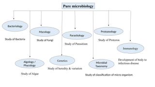
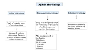
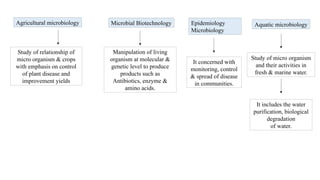
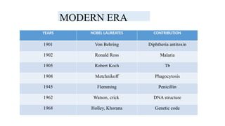
Unit I 10 Hours
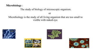
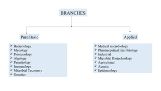
Introduction, history of microbiology, its branches, scope and its
importance.
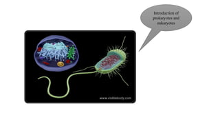
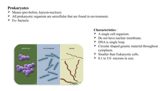
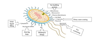
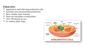
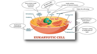
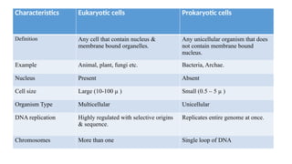
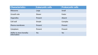
Introduction to Prokaryotes and Eukaryotes
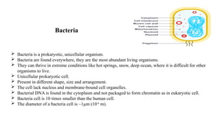
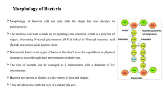
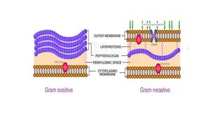
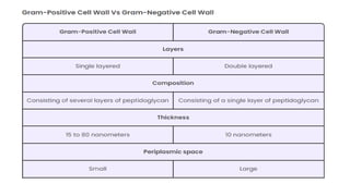
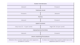
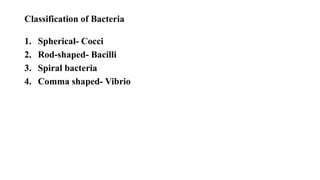
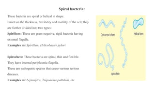
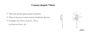
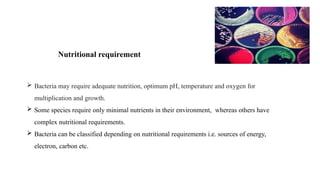
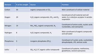
Study of ultra-structure and morphological classification of bacteria,
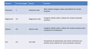
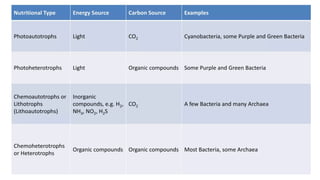
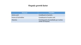
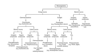
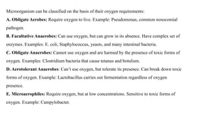
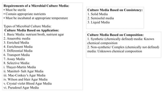
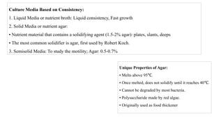
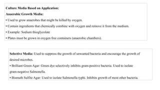
nutritional requirements, raw materials used for culture media and physical
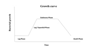
parameters for growth, growth curve, isolation and preservation methods
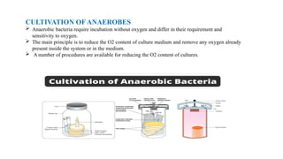
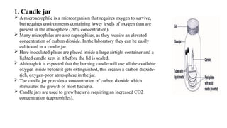
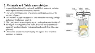
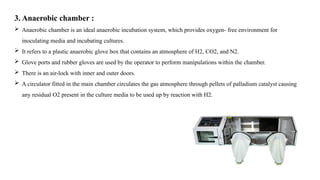
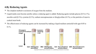
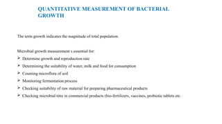
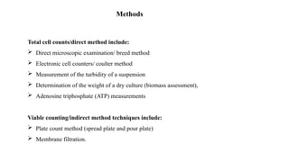
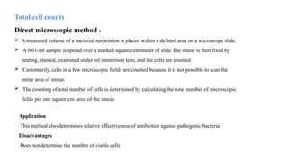
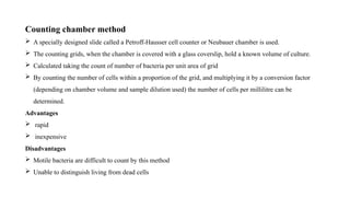
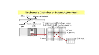
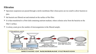
for pure cultures, cultivation of anaerobes, quantitative measurement of
bacterial growth (total & viable count).
Study of different types of phase constrast microscopy, dark field
microscopy and electron microscopy.