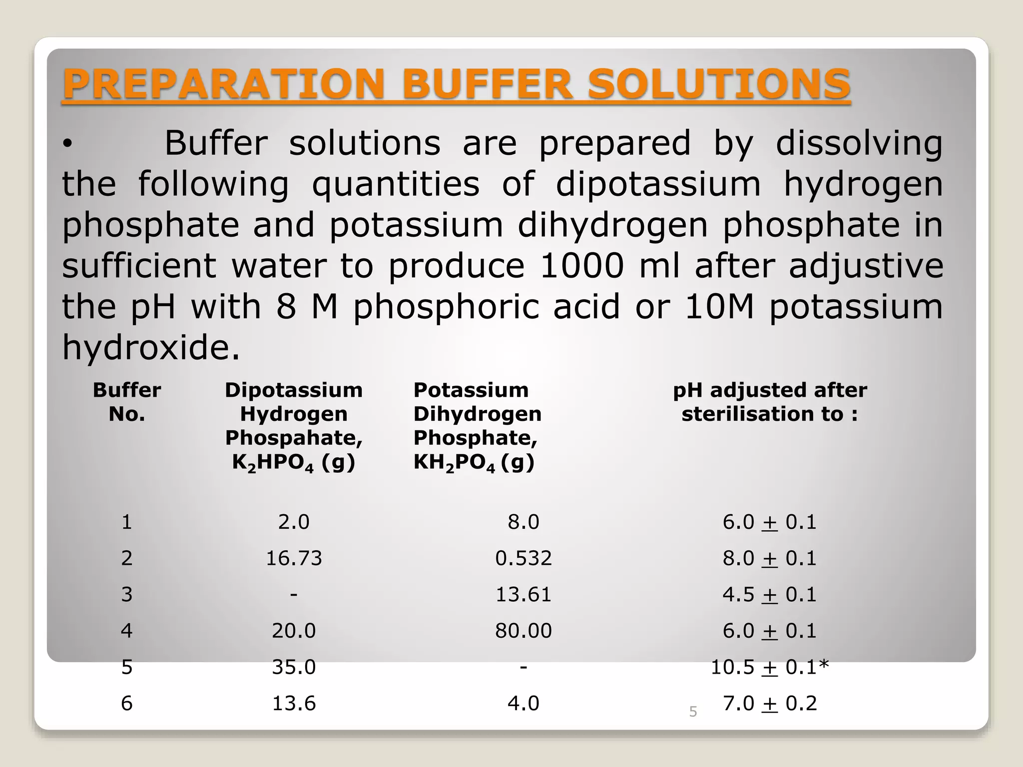
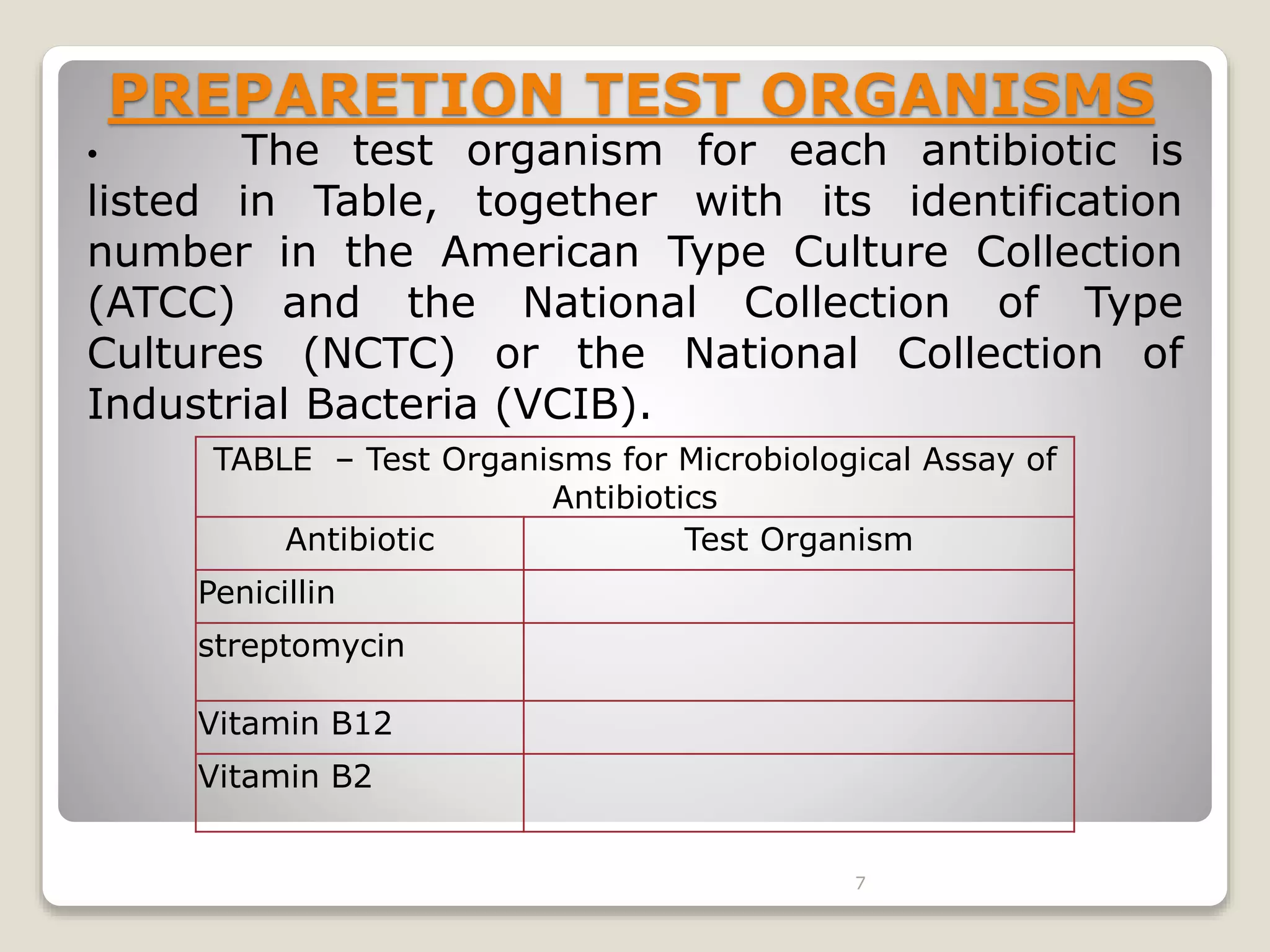
MICROBIOLOGICAL
ASSAY OF ANTIBIOTICS
& VITAMINS.
BY- SANCHIT DHANKHAR
MICROBIOLOGICAL ASSAY OF
ANIBIOTICS
The inhibition of growth under standardized
conditions may be utilized for demonstrating the
therapeutic efficacy of antibiotics.
Any subtle change in the antibiotic molecule which
may not be detected by chemical methods will be
revealed by a change in the antimicrobial activity
and hence microbiological assays are very useful
for resolving doubts regarding possible change in
potency of antibiotics and their preparations.
2
PRINCIPLE
The microbiological assay is based upon a
comparison of the inhibition of growth of micro-
organisms by measured concentration of the
antibiotics to be examined with that produced by
known concentrations of a standard preparation of
the antibiotic having a known activity.
Two general method are usually employed:-
1. The cylinder-plate (or cup-plate) method.
2. The turbidimetric (or tube assay) method.
3
PREPARATION OF MEDIA:
The Media required for the preparation of test
organism are made from the ingredients.
Minor modifications of the individual ingredients
may be made, or reconstituted dehydrated media
may be used provided the resulting media have
equal or better growth-promoting properties and
give a similar standard curve response.
Dissolve the ingredients in sufficient water to
produce 1000 ml and add sufficient 1M Sodium
hydroxide or 1M Hydrochloride acid, as required so
that after sterilization the PH is bw 6.5 to 7.5.
4
PREPARATION BUFFER SOLUTIONS
• Buffer solutions are prepared by dissolving
the following quantities of dipotassium hydrogen
phosphate and potassium dihydrogen phosphate in
sufficient water to produce 1000 ml after adjustive
the pH with 8 M phosphoric acid or 10M potassium
hydroxide.
Buffer
No.
Dipotassium
Hydrogen
Phospahate,
K2HPO4 (g)
Potassium
Dihydrogen
Phosphate,
KH2PO4 (g)
pH adjusted after
sterilisation to :
1 2.0 8.0 6.0 + 0.1
2 16.73 0.532 8.0 + 0.1
3 - 13.61 4.5 + 0.1
4 20.0 80.00 6.0 + 0.1
5 35.0 - 10.5 + 0.1*
6 13.6 4.0 7.0 + 0.2
5
PREPARATION OF THE STANDARD
SOLUTION
• To prepare a stock solution, dissolve a
quantity of the Standard Preparation of a
given antibiotic, accurately weighed and
previously dried where so indicated in
Table, in the solvent specified, and then
dilute to the required concentration as
indicated. Store in a refrigerator and use
within the period indicated.
6
PREPARETION TEST ORGANISMS
• The test organism for each antibiotic is
listed in Table, together with its identification
number in the American Type Culture Collection
(ATCC) and the National Collection of Type
Cultures (NCTC) or the National Collection of
Industrial Bacteria (VCIB).
TABLE – Test Organisms for Microbiological Assay of
Antibiotics
Antibiotic Test Organism
Penicillin
streptomycin
Vitamin B12
Vitamin B2
7
CYLINDER-PLATE OR CUP-PLATE
METHOD
• Inoculate a previously liquified medium appropriate to
the assay, with the requsite quantity of suspension of the micro
organism, add the suspension to the medium at a temperature
between 40° and 50° and immediately pour the inoculated
medium into the petri dishes or large rectangular plates to give
a depth of 3 to 4 mm.
• Ensure that the layers of medium are uniform in
thickness, by placing the dishes or plates on a level surface.
• Using the appropriate buffer solutions, prepare solutions
of known concentrations of the antibiotic to be examined
8
• Apply the solutions to the surface of the solid medium in
sterile cylinders or in cavities prepared in the agar. The volume of
solution added to each cylinder or cavity must be uniform and
sufficient almost to fill the holes when these are used.
• Leave the dishes or plates standing for 1 to 4 hours at
room temperature or at 4°, as appropriate, as a period of pre-
incubation diffusion to minimise the effects of variation in time
between the application of the different solutions. Incubate them
for about 18 hours at the particular temperature.
• Accurately measure the diameters or areas of the circular
inhibition zones and calculate the results.
9
L = 3a + 2b + c – e : H = 3e + 2d + c- a
5 5
Where
L = the calculated zone diameter for the lowest
concentration of the standard curve response line.
H = the calculated zone diameter for the highest
concentration of the standard curve response line.
c = average zone diameter of 36 readings of the reference
point standard solution.
a, b, d, e = corrected average values for the other standard
solutions, lowest to highest concentrations, respectively.
10
TURBIDIMETRIC OR TUBE ASSAY
METHOD
• The method has the advantage of a shorter incubation period for
the growth of the test organism (usually 3 to 4 hours) but the
presence of solvent residues or other inhibitory substances affects
this assay more than the cylinder plates assay.
• Prepare five different concentrations of the standard solution for
preparing the standard curve by diluting the stock solution of the
Standard Preparation of the antibiotic & increasing stepwise in the
ration 4:5.
• Select the median concentration & dilute the solution of the
substance being examined (unknown) to obtain approximately this
concentration.
• Place 1 ml of each concentration of the standard solution and of
the sample solution in each of the tubes in duplicate.
11
• To each tube add 9 ml. of nutrient medium, previously seeded
with the appropriate test organism.
• At the same time prepare three control tubes, one containing
the inoculated culture medium (culture control), another
identical with it but treated immediately with 0.5 ml of dilute
formaldehyde solution (blank) and a third containing
uninoculated culture medium.
•Place all the tubes, randomly distributed or in a randomized
block arrangement, in an incubator or water-bath and maintain
them at the specified temperature, for 3 to 4 hours. After
incubation add 0.5 ml of dilute formaldehyde solution to each
tube. Measure the growth of the test organism by determining
the absorbance at about 530nm of each of the solutions in the
tubes against the blank.
12
L = 3a + 2b + c – e : H = 3e + 2d + c- a
5 5
Where
L = the calculated absorbance for the lowest
concentration of the standard response line.
H = the calculated absorbance for the highest
concentration of the standard response line.
a, c, b, d, e = average absorbance values for
each concentration of the standard response line
lowest to highest respectively.
13
DEFINITION:
 Microbiological assay of vitamins is a type of
biological assay performed with the aid of
microorganisms.
 Many therapeutic agents, which either inhibit the
growth of microorganisms or are essential for the
growth of them are standardized by microbial assay.
 Principles of microbial assay were developed in
1920s.
MICROBIOLOGICAL ASSAY OF
VITAMINS
14
MICROBIOLOGICAL ASSAY OF VITAMINS:
PRINCIPLE:
 Vitamins and amino acids are essential for the
growth of microorganisms.
 The basis of this assay is to measure the
ability of test organism to utilize the substance
being assayed under a proper nutritional
condition.
 The organisms require these growth factors
(vitamins & amino acids) in micro or nano
grams.
15
The response (growth of test organism) is
proportional to the dose (amount of factor)
added to medium.
Materials required for Microbial assay of
vitamins & amino acids:
 A stock solution.
 A inoculum media.
 Assay medium.
 A standard curve.
16
MICROBIAL ASSAY OF VITAMIN B12:
About VitB12:
 Also known as cyanocobalamin. Its a water soluble
vitamin.
 Structure is similar to that of heme where the iron is
replaced with cobalt as a centre of molecule.
 Its main sources are liver, eggs , milk, meat & fish.
 VitB12 deficiency causes Macrolytic anemia,
pernicious anemia.
 National Research Council, USA recommends a
daily intake of about 5mg of vitB12
17
PRINCIPLE OF ASSAY:
 The test organism selected must be capable of
utilizing
free cyanocobalamin.
 Lactobacillus Liechmannii is found to satisfy the
requirements.
 Gram negative bacilli, non-pathogenic, easy to
culture & easily available.
 Isolated from milk, cheese, & other dairy
products.
 Assay is performed by using either titrimetric or
turbidimetric method. 18
PRECAUTIONS:
 Great care must be taken to avoid contamination
 All the glass wares must be free from detergents and
other chemicals.
Glass wares must be heated to 2500C for atleast 1hr
before use.
The whole experiment must be carried out under
proper aseptic condition.
19
Reagents Required :
1) Standard stock solution:
 An accurately weighed amount of
Cyanocobalamin reference Standard is added to
sufficient 25%ethanol (resulting in a solution
containing 1.0 μg of cyanocobalamin per ml).
 Stored in refrigerator. It should be used within
2 months.
 Further dilutions of this stock solution (1 μg/ ml)
are made as follows:
 Add 1 ml stock solution to 99 ml purified water
(1 ml =10 ng).
 Add 1 mL of the above solution to 199 ml
purified water (1 ml = 0.05 ng). 20
2) Test solution to be assayed:-
:
 Accurate amount of Vitamin to be assayed is
taken & dissolved in water, Dil HCl or NaOH is
added to adjust pH at 6.0.
 Make up to volume with water.
21
3) Preparation of Inoculum:-
:
 Transfer a loop full of Lactobacillus
Liechmannii from a recent sub-culture into two
tubes each containing 10ml of sterile culture
medium.
Composition of culture medium :( pH 6.8)
Yeast extract - 0.75gm
Peptone - 0.75gm
Dextrose - 1gm
Pot dihydrogen phosphate - 0.2gm
Tomato juice filtrate - 10ml
Sorbitan mono oleate solution - 1ml
Water up to - 100ml
22
 Incubate for 18 to 24hrs at 37oC.
 Centrifuge the culture .
 Decant the supernatent fluid, under aseptic condition.
 Suspend these cultured cells into 10ml of sterile
suspension of Basal medium stock solution.
 Again centrifuge & decant off supernatent fluid.
 Repeat this for atleast 3times to avoid contamination.
 Finally suspend the cells uniformly in 10ml of sterile
medium.
 Aseptically transfer 1ml of the suspension of cells to
10ml of sterile medium & mix.
This resulting cell suspension is taken as inoculum.
23
Procedure for assay :-
 Clean ten test tubes & add to it 0, 0.5, 1.0, 2.0,
2.5, 2.5, 3.0, 3.5, 4.0, 4.5, & 5ml of standard
cyanocobalamin solution.
 To each tube add 5ml of Basal medium solution
 Volume of each is adjusted to 10ml by water
 In another 4 test tubes add 1, 2, 3, 4ml of test
solution which is to be assayed .
 To each of this also add 5ml of Basal medium
stock solution & adjust volume to 10ml with water.
24
Sterilize all test tubes in autoclave at 121oC for
15mins.
Cool the test tubes at room temperature.
Inoculate a drop of inoculum prepared of lactobacillus
liechmannii.
Incubate the test tubes for 64 to 72hrs at temperature
range of 30 to 37oC.
 After incubation period titrate contents of each test
tube with 0.05N NaOH using bromothymol blue as
indicator until green colour.
Record all the titre readings clearly.
25
Interpretation of results:-
 Determine the average of titration values of each level of
both standard & test solutions.
 Plot a graph considering average titration values(in ml) of
0.05N NaOH against concentration of standard
cyanocobalamin solution.
 A linear graph is obtained.
 By interpolating the standard curve determine the
concentration as activity per ml of vit B12.
 From the graph the concentration of test solution of
cyanocobalamin is found & reported.
26
Theory: The microbiological assay of an antibiotic is based upon a comparison of the inhibition of
growth of micro-organisms by measured concentrations of the antibiotics under examination with
that produced by known concentrations of a standard preparation of the antibiotic having a known
activity.
Two general methods are usually employed, the cylinder-plate (or cup-plate) method and the
turbidimetric (or tube assay) method. The cylinder-plate method (Method A) depends upon
diffusion of the antibiotic from a vertical cylinder through a solidified agar layer in a Petri dish or
plate to an extent such that growth of the added micro-organism is prevented entirely in a zone
around the cylinder containing a solution of the antibiotic. The turbidimetric method (Method B)
depends upon the inhibition of growth of a microbial culture in a uniform solution of the antibiotic
in a fluid medium that is favorable to its rapid growth in the absence of the antibiotic. The assay is
designed in such a way that the mathematical model on which the potency equation is based can be
proved to be valid. If a parallel-line model is chosen, the two log dose response lines of the
preparation under examination and the standard preparation should be parallel; they should be
rectilinear over the range of doses used in the calculation. These conditions should be verified by
validity tests for a given probability. Other mathematical models, such as the slope ratio method,
may be used provided that proof of validity is demonstrated.
Microbiological assay of penicillin
27
Procedure: 1. Dissolve 35mg of benzyl penicillin in 100ml of water. From this, take 1 ml
and dilute to 100ml of sterile water.
2. By using this stock solution, prepare 1, 2, 3, 4 and 5 units/ ml of standard stock
solution.
3. Prepare a solution of unknown or a given sample of antibiotic in the same solvent as
that of standard solution and dilute it till the level of standard preparation of the
antibiotics.
4. Sterile the antibiotic assay medium by autoclave and prepare Petri plates in laminar air
flow.
5. Spread the test microorganism (Bacillus subtilis) on the surface of Petri plates by
spread plate technique.
6. By using flame sterilized cork borer, prepare 5 to 6 cups in each plate keeping adequate
distance from each other.
7. Standard and test diluted antibiotic solutions are added in each labeled cavity of plates.
8. Transfer all the plates in refrigerator for proper diffusion of antibiotics at 4˚ C for 1 to 2
hours. Incubate all the plates in incubator at 32 -35˚ C for 24 to 48 hours.
Observations and Results: Observe the zone of inhibition around the cavity. Measure the
diameter by antibiotics zone reader and record in the observation tables. Plot the graph of
the diameter of the zone of inhibition against log concentration of antibiotic and
determine the concentrations of unknown antibiotic sample from the graph.
28
Microbiological assay of streptomycin
Procedure: 1. Dissolve 256 mg of streptomycin sulphate in 100 ml of sterile water.
From this, take 1 ml and dilute with 100 ml of sterile water. By using this stock
solution, prepare 10,12,14,16 and 18 units/ ml of standard stock solution.
2. Prepare a solution of unknown or given sample of antibiotics in the same solvent
as that of standard solution and dilute it up to the level of standard preparation of
the antibiotics.
3. Sterile the antibiotic assay medium by autoclave and prepare Petri plates in
laminar air flow.
4. Spread the test micro-organisms on the surface of Petri plates by spread plate
technique.
5. Prepare cup or cavities on plate with sterile cork borer keeping adequate distance
from each other.
6. Standard and test dilute antibiotic solutions (0.1 ml to 1 ml depending on size of
cavity or cylinder) are added in each labeled cavity of plates.
7. Transfer all the plates in refrigerator for proper diffusion of antibiotics at 4˚ C for 1
to 2 hours. Incubate all the plates in incubator at 32 -35˚ C for 24 to 48 hours.
Observations and Results:Observe the zone of inhibition around the cavity.
Measure the diameter by antibiotics zone reader and record in the observation
tables. Plot the graph of the diameter of the zone of inhibition against log
concentration of antibiotic and determine the concentrations of unknown antibiotic
sample from the graph.
29
30

Microbiological assays- Pharmacuetical Microbiology

  • 1.
    MICROBIOLOGICAL ASSAY OF ANTIBIOTICS &VITAMINS. BY- SANCHIT DHANKHAR
  • 2.
    MICROBIOLOGICAL ASSAY OF ANIBIOTICS Theinhibition of growth under standardized conditions may be utilized for demonstrating the therapeutic efficacy of antibiotics. Any subtle change in the antibiotic molecule which may not be detected by chemical methods will be revealed by a change in the antimicrobial activity and hence microbiological assays are very useful for resolving doubts regarding possible change in potency of antibiotics and their preparations. 2
  • 3.
    PRINCIPLE The microbiological assayis based upon a comparison of the inhibition of growth of micro- organisms by measured concentration of the antibiotics to be examined with that produced by known concentrations of a standard preparation of the antibiotic having a known activity. Two general method are usually employed:- 1. The cylinder-plate (or cup-plate) method. 2. The turbidimetric (or tube assay) method. 3
  • 4.
    PREPARATION OF MEDIA: TheMedia required for the preparation of test organism are made from the ingredients. Minor modifications of the individual ingredients may be made, or reconstituted dehydrated media may be used provided the resulting media have equal or better growth-promoting properties and give a similar standard curve response. Dissolve the ingredients in sufficient water to produce 1000 ml and add sufficient 1M Sodium hydroxide or 1M Hydrochloride acid, as required so that after sterilization the PH is bw 6.5 to 7.5. 4
  • 5.
    PREPARATION BUFFER SOLUTIONS •Buffer solutions are prepared by dissolving the following quantities of dipotassium hydrogen phosphate and potassium dihydrogen phosphate in sufficient water to produce 1000 ml after adjustive the pH with 8 M phosphoric acid or 10M potassium hydroxide. Buffer No. Dipotassium Hydrogen Phospahate, K2HPO4 (g) Potassium Dihydrogen Phosphate, KH2PO4 (g) pH adjusted after sterilisation to : 1 2.0 8.0 6.0 + 0.1 2 16.73 0.532 8.0 + 0.1 3 - 13.61 4.5 + 0.1 4 20.0 80.00 6.0 + 0.1 5 35.0 - 10.5 + 0.1* 6 13.6 4.0 7.0 + 0.2 5
  • 6.
    PREPARATION OF THESTANDARD SOLUTION • To prepare a stock solution, dissolve a quantity of the Standard Preparation of a given antibiotic, accurately weighed and previously dried where so indicated in Table, in the solvent specified, and then dilute to the required concentration as indicated. Store in a refrigerator and use within the period indicated. 6
  • 7.
    PREPARETION TEST ORGANISMS •The test organism for each antibiotic is listed in Table, together with its identification number in the American Type Culture Collection (ATCC) and the National Collection of Type Cultures (NCTC) or the National Collection of Industrial Bacteria (VCIB). TABLE – Test Organisms for Microbiological Assay of Antibiotics Antibiotic Test Organism Penicillin streptomycin Vitamin B12 Vitamin B2 7
  • 8.
    CYLINDER-PLATE OR CUP-PLATE METHOD •Inoculate a previously liquified medium appropriate to the assay, with the requsite quantity of suspension of the micro organism, add the suspension to the medium at a temperature between 40° and 50° and immediately pour the inoculated medium into the petri dishes or large rectangular plates to give a depth of 3 to 4 mm. • Ensure that the layers of medium are uniform in thickness, by placing the dishes or plates on a level surface. • Using the appropriate buffer solutions, prepare solutions of known concentrations of the antibiotic to be examined 8
  • 9.
    • Apply thesolutions to the surface of the solid medium in sterile cylinders or in cavities prepared in the agar. The volume of solution added to each cylinder or cavity must be uniform and sufficient almost to fill the holes when these are used. • Leave the dishes or plates standing for 1 to 4 hours at room temperature or at 4°, as appropriate, as a period of pre- incubation diffusion to minimise the effects of variation in time between the application of the different solutions. Incubate them for about 18 hours at the particular temperature. • Accurately measure the diameters or areas of the circular inhibition zones and calculate the results. 9
  • 10.
    L = 3a+ 2b + c – e : H = 3e + 2d + c- a 5 5 Where L = the calculated zone diameter for the lowest concentration of the standard curve response line. H = the calculated zone diameter for the highest concentration of the standard curve response line. c = average zone diameter of 36 readings of the reference point standard solution. a, b, d, e = corrected average values for the other standard solutions, lowest to highest concentrations, respectively. 10
  • 11.
    TURBIDIMETRIC OR TUBEASSAY METHOD • The method has the advantage of a shorter incubation period for the growth of the test organism (usually 3 to 4 hours) but the presence of solvent residues or other inhibitory substances affects this assay more than the cylinder plates assay. • Prepare five different concentrations of the standard solution for preparing the standard curve by diluting the stock solution of the Standard Preparation of the antibiotic & increasing stepwise in the ration 4:5. • Select the median concentration & dilute the solution of the substance being examined (unknown) to obtain approximately this concentration. • Place 1 ml of each concentration of the standard solution and of the sample solution in each of the tubes in duplicate. 11
  • 12.
    • To eachtube add 9 ml. of nutrient medium, previously seeded with the appropriate test organism. • At the same time prepare three control tubes, one containing the inoculated culture medium (culture control), another identical with it but treated immediately with 0.5 ml of dilute formaldehyde solution (blank) and a third containing uninoculated culture medium. •Place all the tubes, randomly distributed or in a randomized block arrangement, in an incubator or water-bath and maintain them at the specified temperature, for 3 to 4 hours. After incubation add 0.5 ml of dilute formaldehyde solution to each tube. Measure the growth of the test organism by determining the absorbance at about 530nm of each of the solutions in the tubes against the blank. 12
  • 13.
    L = 3a+ 2b + c – e : H = 3e + 2d + c- a 5 5 Where L = the calculated absorbance for the lowest concentration of the standard response line. H = the calculated absorbance for the highest concentration of the standard response line. a, c, b, d, e = average absorbance values for each concentration of the standard response line lowest to highest respectively. 13
  • 14.
    DEFINITION:  Microbiological assayof vitamins is a type of biological assay performed with the aid of microorganisms.  Many therapeutic agents, which either inhibit the growth of microorganisms or are essential for the growth of them are standardized by microbial assay.  Principles of microbial assay were developed in 1920s. MICROBIOLOGICAL ASSAY OF VITAMINS 14
  • 15.
    MICROBIOLOGICAL ASSAY OFVITAMINS: PRINCIPLE:  Vitamins and amino acids are essential for the growth of microorganisms.  The basis of this assay is to measure the ability of test organism to utilize the substance being assayed under a proper nutritional condition.  The organisms require these growth factors (vitamins & amino acids) in micro or nano grams. 15
  • 16.
    The response (growthof test organism) is proportional to the dose (amount of factor) added to medium. Materials required for Microbial assay of vitamins & amino acids:  A stock solution.  A inoculum media.  Assay medium.  A standard curve. 16
  • 17.
    MICROBIAL ASSAY OFVITAMIN B12: About VitB12:  Also known as cyanocobalamin. Its a water soluble vitamin.  Structure is similar to that of heme where the iron is replaced with cobalt as a centre of molecule.  Its main sources are liver, eggs , milk, meat & fish.  VitB12 deficiency causes Macrolytic anemia, pernicious anemia.  National Research Council, USA recommends a daily intake of about 5mg of vitB12 17
  • 18.
    PRINCIPLE OF ASSAY: The test organism selected must be capable of utilizing free cyanocobalamin.  Lactobacillus Liechmannii is found to satisfy the requirements.  Gram negative bacilli, non-pathogenic, easy to culture & easily available.  Isolated from milk, cheese, & other dairy products.  Assay is performed by using either titrimetric or turbidimetric method. 18
  • 19.
    PRECAUTIONS:  Great caremust be taken to avoid contamination  All the glass wares must be free from detergents and other chemicals. Glass wares must be heated to 2500C for atleast 1hr before use. The whole experiment must be carried out under proper aseptic condition. 19
  • 20.
    Reagents Required : 1)Standard stock solution:  An accurately weighed amount of Cyanocobalamin reference Standard is added to sufficient 25%ethanol (resulting in a solution containing 1.0 μg of cyanocobalamin per ml).  Stored in refrigerator. It should be used within 2 months.  Further dilutions of this stock solution (1 μg/ ml) are made as follows:  Add 1 ml stock solution to 99 ml purified water (1 ml =10 ng).  Add 1 mL of the above solution to 199 ml purified water (1 ml = 0.05 ng). 20
  • 21.
    2) Test solutionto be assayed:- :  Accurate amount of Vitamin to be assayed is taken & dissolved in water, Dil HCl or NaOH is added to adjust pH at 6.0.  Make up to volume with water. 21 3) Preparation of Inoculum:- :  Transfer a loop full of Lactobacillus Liechmannii from a recent sub-culture into two tubes each containing 10ml of sterile culture medium.
  • 22.
    Composition of culturemedium :( pH 6.8) Yeast extract - 0.75gm Peptone - 0.75gm Dextrose - 1gm Pot dihydrogen phosphate - 0.2gm Tomato juice filtrate - 10ml Sorbitan mono oleate solution - 1ml Water up to - 100ml 22
  • 23.
     Incubate for18 to 24hrs at 37oC.  Centrifuge the culture .  Decant the supernatent fluid, under aseptic condition.  Suspend these cultured cells into 10ml of sterile suspension of Basal medium stock solution.  Again centrifuge & decant off supernatent fluid.  Repeat this for atleast 3times to avoid contamination.  Finally suspend the cells uniformly in 10ml of sterile medium.  Aseptically transfer 1ml of the suspension of cells to 10ml of sterile medium & mix. This resulting cell suspension is taken as inoculum. 23
  • 24.
    Procedure for assay:-  Clean ten test tubes & add to it 0, 0.5, 1.0, 2.0, 2.5, 2.5, 3.0, 3.5, 4.0, 4.5, & 5ml of standard cyanocobalamin solution.  To each tube add 5ml of Basal medium solution  Volume of each is adjusted to 10ml by water  In another 4 test tubes add 1, 2, 3, 4ml of test solution which is to be assayed .  To each of this also add 5ml of Basal medium stock solution & adjust volume to 10ml with water. 24
  • 25.
    Sterilize all testtubes in autoclave at 121oC for 15mins. Cool the test tubes at room temperature. Inoculate a drop of inoculum prepared of lactobacillus liechmannii. Incubate the test tubes for 64 to 72hrs at temperature range of 30 to 37oC.  After incubation period titrate contents of each test tube with 0.05N NaOH using bromothymol blue as indicator until green colour. Record all the titre readings clearly. 25
  • 26.
    Interpretation of results:- Determine the average of titration values of each level of both standard & test solutions.  Plot a graph considering average titration values(in ml) of 0.05N NaOH against concentration of standard cyanocobalamin solution.  A linear graph is obtained.  By interpolating the standard curve determine the concentration as activity per ml of vit B12.  From the graph the concentration of test solution of cyanocobalamin is found & reported. 26
  • 27.
    Theory: The microbiologicalassay of an antibiotic is based upon a comparison of the inhibition of growth of micro-organisms by measured concentrations of the antibiotics under examination with that produced by known concentrations of a standard preparation of the antibiotic having a known activity. Two general methods are usually employed, the cylinder-plate (or cup-plate) method and the turbidimetric (or tube assay) method. The cylinder-plate method (Method A) depends upon diffusion of the antibiotic from a vertical cylinder through a solidified agar layer in a Petri dish or plate to an extent such that growth of the added micro-organism is prevented entirely in a zone around the cylinder containing a solution of the antibiotic. The turbidimetric method (Method B) depends upon the inhibition of growth of a microbial culture in a uniform solution of the antibiotic in a fluid medium that is favorable to its rapid growth in the absence of the antibiotic. The assay is designed in such a way that the mathematical model on which the potency equation is based can be proved to be valid. If a parallel-line model is chosen, the two log dose response lines of the preparation under examination and the standard preparation should be parallel; they should be rectilinear over the range of doses used in the calculation. These conditions should be verified by validity tests for a given probability. Other mathematical models, such as the slope ratio method, may be used provided that proof of validity is demonstrated. Microbiological assay of penicillin 27
  • 28.
    Procedure: 1. Dissolve35mg of benzyl penicillin in 100ml of water. From this, take 1 ml and dilute to 100ml of sterile water. 2. By using this stock solution, prepare 1, 2, 3, 4 and 5 units/ ml of standard stock solution. 3. Prepare a solution of unknown or a given sample of antibiotic in the same solvent as that of standard solution and dilute it till the level of standard preparation of the antibiotics. 4. Sterile the antibiotic assay medium by autoclave and prepare Petri plates in laminar air flow. 5. Spread the test microorganism (Bacillus subtilis) on the surface of Petri plates by spread plate technique. 6. By using flame sterilized cork borer, prepare 5 to 6 cups in each plate keeping adequate distance from each other. 7. Standard and test diluted antibiotic solutions are added in each labeled cavity of plates. 8. Transfer all the plates in refrigerator for proper diffusion of antibiotics at 4˚ C for 1 to 2 hours. Incubate all the plates in incubator at 32 -35˚ C for 24 to 48 hours. Observations and Results: Observe the zone of inhibition around the cavity. Measure the diameter by antibiotics zone reader and record in the observation tables. Plot the graph of the diameter of the zone of inhibition against log concentration of antibiotic and determine the concentrations of unknown antibiotic sample from the graph. 28
  • 29.
    Microbiological assay ofstreptomycin Procedure: 1. Dissolve 256 mg of streptomycin sulphate in 100 ml of sterile water. From this, take 1 ml and dilute with 100 ml of sterile water. By using this stock solution, prepare 10,12,14,16 and 18 units/ ml of standard stock solution. 2. Prepare a solution of unknown or given sample of antibiotics in the same solvent as that of standard solution and dilute it up to the level of standard preparation of the antibiotics. 3. Sterile the antibiotic assay medium by autoclave and prepare Petri plates in laminar air flow. 4. Spread the test micro-organisms on the surface of Petri plates by spread plate technique. 5. Prepare cup or cavities on plate with sterile cork borer keeping adequate distance from each other. 6. Standard and test dilute antibiotic solutions (0.1 ml to 1 ml depending on size of cavity or cylinder) are added in each labeled cavity of plates. 7. Transfer all the plates in refrigerator for proper diffusion of antibiotics at 4˚ C for 1 to 2 hours. Incubate all the plates in incubator at 32 -35˚ C for 24 to 48 hours. Observations and Results:Observe the zone of inhibition around the cavity. Measure the diameter by antibiotics zone reader and record in the observation tables. Plot the graph of the diameter of the zone of inhibition against log concentration of antibiotic and determine the concentrations of unknown antibiotic sample from the graph. 29
  • 30.