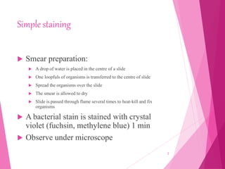
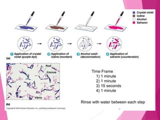
staining
BY- SANCHIT DHANKHAR
Simple staining
 Smear preparation:
 A drop of water is placed in the centre of a slide
 One loopfuls of organisms is transferred to the centre of slide
 Spread the organisms over the slide
 The smear is allowed to dry
 Slide is passed through flame several times to heat-kill and fix
organisms
 A bacterial stain is stained with crystal
violet (fuchsin, methylene blue) 1 min
 Observe under microscope
2
 Basic Dyes
 Methylene Blue
 Crystal Violet
 Carbol Fuchsin
 Safranin
 Malachite Green
 Acidic Dyes
 Picric Acid
 Nigrosin
 India Ink
 Eosin
3
DIFFERENCIAL STAINING
 Two or more reagents Distinguish
 Bacterial groups
 Specific Structures
 Example
 Gram stain
 Acid Fast Stain
4
GRAM STAINING
 1884 Christian Gram
 Staining technique that separates bacteria into two groups:
 Gram-positive bacteria
 Gram-negative bacteria
 It is based on the composition of their cell wall. Gram staining uses crystal violet to stain cell walls ,iodine as a
mordant, and a fuchsin or safranin counterstain to mark all bacteria. Gram-positive bacteria stain dark blue or
violet. Their cell wall is typically rich with peptidoglycan and lacks the secondary membrane
and lipopolysaccharide layer found in Gram-negative bacteria.
 Gram-negative organisms appear red or pink because they are counterstained. Because of presence of higher
lipid content, after alcohol-treatment, the porosity of the cell wall increases, hence the CVI complex (crystal
violet – iodine) can pass through. Thus, the primary stain is not retained. Also, in contrast to most Gram-
positive bacteria, Gram-negative bacteria have only a few layers of peptidoglycan and a secondary cell
membrane made primarily of lipopolysaccharide
5
Time Frame
1) 1 minute
2) 1 minute
3) 15 seconds
4) 1 minute
Rinse with water between each step
6
Ziehl-Neelsen Acid-Fast Stain
 - Acid fast retain red color in cell walls
 It is a special bacteriological stain used to identify acid-
fast organisms, mainly Mycobacteria. Mycobacterium
tuberculosis is the most important of this group because it is
responsible for tuberculosis (TB)
7
 The slide is flooded with Carbol Fuchsin,
 heated over steam bath at 90°C for 4min.
 The slide is then flooded with a mild solution of hydrochloric acid in isopropyl
alcohol to destain the Carbol Fuchsin, thus removing the stain from cells that
are unprotected by a waxy lipid layer.
Thereafter, the cells are stained in methylene blue and viewed on a
microscope.
8
ENDOSPORE STAINING
 Genera of bacteria that produce endospores
 Bacillus & Clostridium
 Other genera: Desulfotomaculum, Sporosarcina, Sporolactobacillus, Oscillospira,
Thermoactinomyces
 Schaeffer-Fulton Method for Endospore Staining
 Preparation of a Bacterial Smear-Air dry
 Application of Primary Dye (Malachite Green) w/ concurrent application of heat
 Allow slide to cool
 Rinse with water
 Application of Counterstain (Safranin) 9
10
11
 NEGATIVE STAINING
 Acid Dye
 (-) chromogen
 Repelled by (-) cell wall
 Cells
 Colorless
 Seen against dark background
12
Counting of bacteria
13
Turbidimetric method
14
THANKYOU
15

Staining- Pharmaceutical MIcrobiology

  • 1.
  • 2.
    Simple staining  Smearpreparation:  A drop of water is placed in the centre of a slide  One loopfuls of organisms is transferred to the centre of slide  Spread the organisms over the slide  The smear is allowed to dry  Slide is passed through flame several times to heat-kill and fix organisms  A bacterial stain is stained with crystal violet (fuchsin, methylene blue) 1 min  Observe under microscope 2
  • 3.
     Basic Dyes Methylene Blue  Crystal Violet  Carbol Fuchsin  Safranin  Malachite Green  Acidic Dyes  Picric Acid  Nigrosin  India Ink  Eosin 3
  • 4.
    DIFFERENCIAL STAINING  Twoor more reagents Distinguish  Bacterial groups  Specific Structures  Example  Gram stain  Acid Fast Stain 4
  • 5.
    GRAM STAINING  1884Christian Gram  Staining technique that separates bacteria into two groups:  Gram-positive bacteria  Gram-negative bacteria  It is based on the composition of their cell wall. Gram staining uses crystal violet to stain cell walls ,iodine as a mordant, and a fuchsin or safranin counterstain to mark all bacteria. Gram-positive bacteria stain dark blue or violet. Their cell wall is typically rich with peptidoglycan and lacks the secondary membrane and lipopolysaccharide layer found in Gram-negative bacteria.  Gram-negative organisms appear red or pink because they are counterstained. Because of presence of higher lipid content, after alcohol-treatment, the porosity of the cell wall increases, hence the CVI complex (crystal violet – iodine) can pass through. Thus, the primary stain is not retained. Also, in contrast to most Gram- positive bacteria, Gram-negative bacteria have only a few layers of peptidoglycan and a secondary cell membrane made primarily of lipopolysaccharide 5
  • 6.
    Time Frame 1) 1minute 2) 1 minute 3) 15 seconds 4) 1 minute Rinse with water between each step 6
  • 7.
    Ziehl-Neelsen Acid-Fast Stain - Acid fast retain red color in cell walls  It is a special bacteriological stain used to identify acid- fast organisms, mainly Mycobacteria. Mycobacterium tuberculosis is the most important of this group because it is responsible for tuberculosis (TB) 7
  • 8.
     The slideis flooded with Carbol Fuchsin,  heated over steam bath at 90°C for 4min.  The slide is then flooded with a mild solution of hydrochloric acid in isopropyl alcohol to destain the Carbol Fuchsin, thus removing the stain from cells that are unprotected by a waxy lipid layer. Thereafter, the cells are stained in methylene blue and viewed on a microscope. 8
  • 9.
    ENDOSPORE STAINING  Generaof bacteria that produce endospores  Bacillus & Clostridium  Other genera: Desulfotomaculum, Sporosarcina, Sporolactobacillus, Oscillospira, Thermoactinomyces  Schaeffer-Fulton Method for Endospore Staining  Preparation of a Bacterial Smear-Air dry  Application of Primary Dye (Malachite Green) w/ concurrent application of heat  Allow slide to cool  Rinse with water  Application of Counterstain (Safranin) 9
  • 10.
  • 11.
  • 12.
     NEGATIVE STAINING Acid Dye  (-) chromogen  Repelled by (-) cell wall  Cells  Colorless  Seen against dark background 12
  • 13.
  • 14.
  • 15.