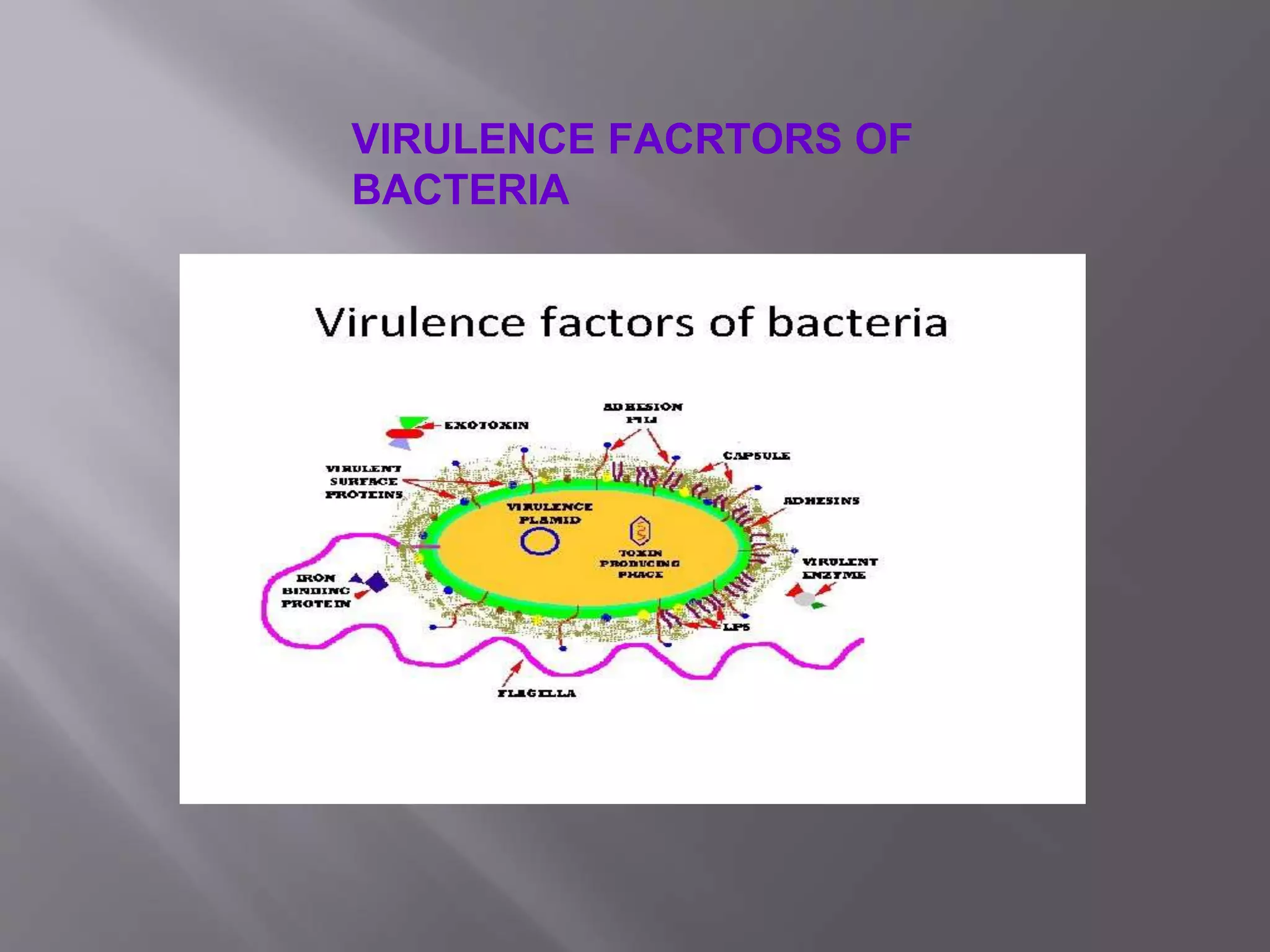
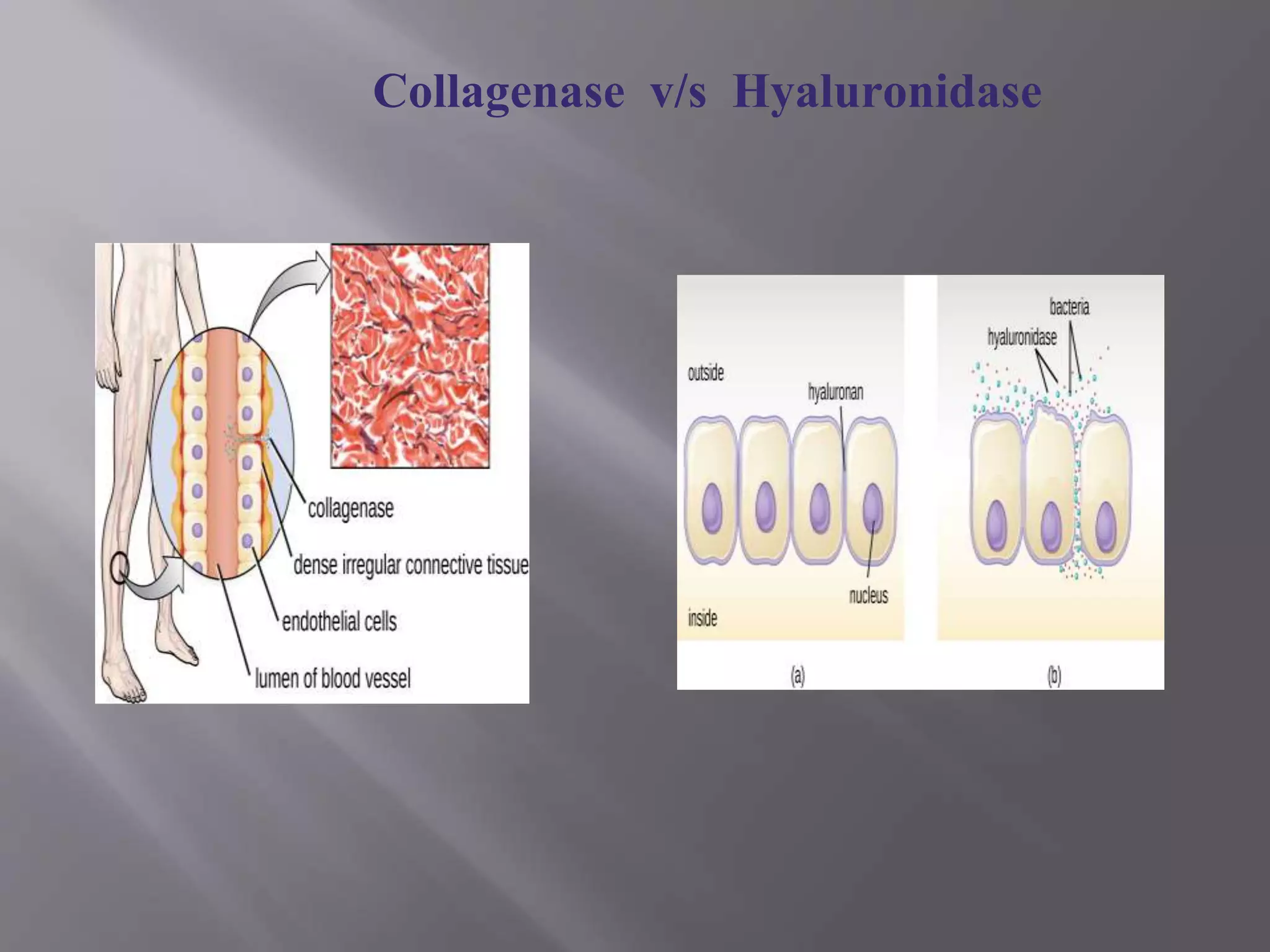
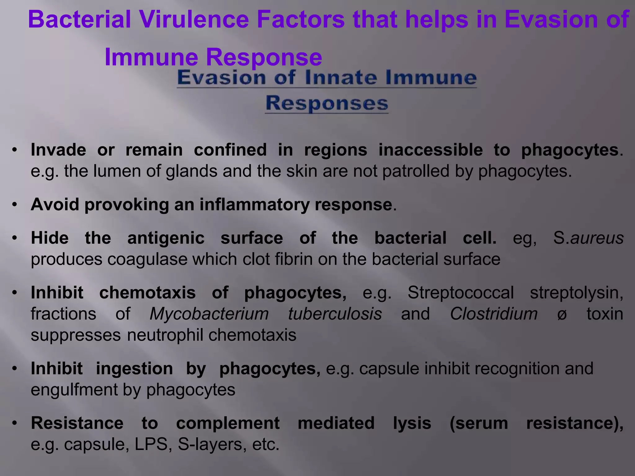
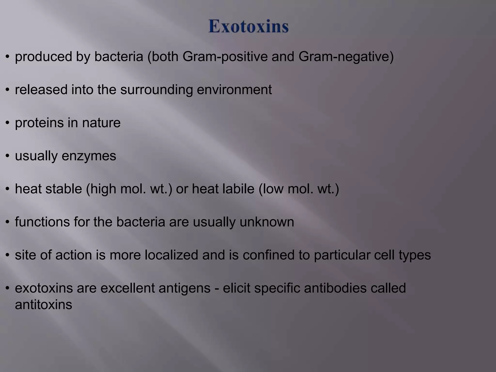
SRI PARAMAKALYANI COLLEGE
(REACCREDITED WITH B GRADE WITH A CGPA OF 2.71 IN THE II CYCLE BY NACC
AFFILIATED TO MANONMANIAM SUNDARANAR UNIVERSITY,THIRUNELVELI)
ALWARKURICHI -627 412 TAMILNADU INDIA.
POST GRADUATE& RESEARCHCENTER– DEPARTMENTOF MICROBIOLOGY
(GOVERMENTAIDED)
II SEM - CORE – IMMUNOLOGY
UNIT – I
VIRULENCE FACTORS
SUBMITTED TO ,
THE GUIDE
DR.S.VISWANATHAN,PH.D
HEAD OF THE DEPARTMENT
SRI PARAMAKALYANI COLLEGE
ALWARKURICHI.
SUBMITTED BY
PETCHIAMMAL.G
REG.NO : 20211231516118
I M.SC, MICROBIOLOGY
DATE: 28.04.2022
Introduction
Mechanism of Microbial pathogencity
Microbes and host interaction
Terminologies
virulence factor
Bacterial toxins
Autoimmune / Hpersensitivity Responses
Conclusion
References
OUTLINE
• To understand the virulence factors promote colonization to the
host.
• To know the bacterial virulence factor to damage the host.
• To differentiate the endotoxins and exotoxins.
• To identify bacterial virulance factors induce autoimmune disease.
OBJECTIVES
INTRODUCTIO
N
Ability of organism to cause disease or to interfere with a metabolic or physiologic
function of its host.
The word came from Latin,Virulentus (or) ‘full of poison’.
Virulence is the measure of the pathogenicity of an organism to cause disease;
usually used to describe the difference in disease causing capability between two
different strains of the same species.
Virulence can be expressed as
LD50 (lethal dose for 50% of the inoculated hosts) or
ID50 (infectious dose for 50% of the inoculated hosts).
 Saprophytism – Living on dead or decaying organic matter.
 Parasitism – Living on or within another living organism - there are different types
of host-parasite relationships.
 Commensalism – parasite lives on/in the host without causing any
 Opportunistic pathogen – The organism i s enerally harmless, but can
cause disease when it gains access to other sites or tissues.
 Obligate pathogen – parasite always causes disease .
 Infectivity – It is the capacity of the organism to penetrate the tissues of host, to
survive the host defenses, and to multiply and disseminate in host.
 Pathogenicity – It is the capacity of the microbial species to produce disease.
Pathogenic bacteria can be grouped in the three categories on the
basis of their invasive properties for eukaryotic cell;
• Facultative intracellular
• Obligate intracellular
• Extracellular
TERMINOLOGIES
Bacteremia - presence of bacteria in the blood.
Pyemia - Bacteremia involving pyogens ( pus-forming bacteria).
Viremia - presence of virus in the blood.
Toxemia - Toxin present in the blood.
Septicemia - Presence of actively multiplying bacteria in the blood.
Related to the presence of pathogen in the blood stream :
VIRULENCE FACRTORS OF
BACTERIA
• The factors produced by a microorganism and induce pathology in a host
are called virulence factors.
• These factors help pathogen to
 invade the host
 cause disease and
 evade host defenses
Virulence factors are classified into two categories
1. Virulence factors that promote bacterial colonization of the
host:
Adherence Factors
Invasion and/or Spreading Factors
Compete for iron and other nutrients;
Evasion of host immune responses
2. Virulence factors that damage the host.
Exotoxins
Endotoxins
 Bacterial motility - e.g.,non-motile mutants of Vibrio cholerae
are less virulent than the motile wild types.
 Bacterial enzymes - e.g., Streptococcus pyogenes produces
streptokinase that facilitate spread of the bacterium by liquefying
the fibrin clot.
Bacterial Virulence Factors that Promote
Colonization in the Host
Adhesins are proteins found on the cell wall of various bacteria that bind to
specific receptor molecules on the surface of host cells and enable the
bacterium to adhere intimately to that cell in order to colonize and resist
physical removal, e.g. common fimbriae, capsule, biofilm, liptotechoic acid,
Fibronectin binding protein (FBP), etc.
fibronectin
"Spreading Factors" are a family of bacterial enzymes that affect the
physical properties of tissue matrices and intercellular spaces, thereby
promoting the spread of the pathogen.
• Hyaluronidase - depolymerize hyaluronic acid, the interstitial cement
substance of connective’ tissue; produced by streptococci, staphylococci, and
clostridia.
• Collagenase - breaks down collagen; produced by Clostridium histolyticum
and Clostridium perfringens.
• Neuraminidase - degrades neuraminic acid (also called sialic acid) present
on epithelial cells of the mucosa; produced by Vibrio cholerae, Shigella
dysenteriae, P.multocida, and M.haemolytica.
• Streptokinase and Staphylokinase – convert inactive plasminogen to plasmin which
digests fibrin.
• Edema Factor of B.anthracis - adenylate cyclase activity promote bacterial
invasion.
Collagenase v/s Hyaluronidase
• Some bacteria have mechanisms by virtue of which they initiate phagocytosis
in non-phagocytic cells for invasion by:
 binding to some receptor on cell, eg. Yersinia pestis.
 injecting invasins, such as Type III secretion system in bacterial cytoplasm,
eg. Salmonella.
• In either case changes in host cell cytoskeleton cause the bacteria to be ingested.
• Some pathogens can utilize actin fibres intracellularly to move
through host cells (transcytosis), eg. Listeria monocytogenes
• Invasins may also mediate uptake of bacteria into professional phagocytic
cells in a way that bypass normal phagosome formation
The bacteria having the type III secretion system on contact with cells, delivers
proteins into the cells which cause polymerization and depolymerization of
actin filaments resulting in cytoskeletal rearrangement. Thus the invasins is
able to trick the non-phagocytic cell into behaving like a phagocyte and engulf
the bacterium into phagosome like vacuole. The bacteria then cause the
vacuole membrane to rupture and escape into the cytoplasm
Bacteria compete for nutrients by synthesizing specific transport
systems or cell wall components capable of binding limiting substrates
and transporting them into the cell.
• Siderophores—low MW compounds that chelate iron with very high
affinity, eg. E.coli.
• Direct binding of host transferrin, lactoferrin, ferritin, or heme by
bacterial surface receptors, eg. Yersinia species.
• Exotoxins that lyse host cells (can be used to obtain other
nutrients as well), eg. Haemolysins.
• Invade or remain confined in regions inaccessible to phagocytes.
e.g. the lumen of glands and the skin are not patrolled by phagocytes.
• Avoid provoking an inflammatory response.
• Hide the antigenic surface of the bacterial cell. eg, S.aureus
produces coagulase which clot fibrin on the bacterial surface
• Inhibit chemotaxis of phagocytes, e.g. Streptococcal streptolysin,
fractions of Mycobacterium tuberculosis and Clostridium ø toxin
suppresses neutrophil chemotaxis
• Inhibit ingestion by phagocytes, e.g. capsule inhibit recognition and
engulfment by phagocytes
• Resistance to complement mediated lysis (serum resistance),
e.g. capsule, LPS, S-layers, etc.
Bacterial Virulence Factors that helps in Evasion of
Immune Response
Resistance to opsonization/phagocytosis
• LPS O polysaccharide
• S-layer
• Extracellular products: enzymes that inactivate C5a
chemoattractant (S. pyogenes), toxins that kill phagocytes
(leukotoxins) (Mannheimia haemolytica),inhibit migration, or reduce
oxidative burst.
Strategies for surviving phagocytosis
I. Escape from phagosome before fusion with lysosome (example: Listeria
monocytogenes, mediated by listeriolysin)
II. Prevent phagosome-lysosome fusion – eg, Salmonella, Mycobacterium
Legionella and Chlamydia.
III. Express factors that allow survival in phagolysosome
Virulence factors that damage the host include
•The cell wall components that bind to host cells causing them to
synthesize chemokines.
• secrete pro-inflammatory cytokines and
•Toxins.
• Induce autoimmune responses.
Bacterial Virulence Factors that
damage the Host
Bacterial Cell Wall Components that Promote Synthesis and
Secretion of Inflammatory Cytokines and Chemokines.
• LPS of Gram-positive bacteria, and teichoic acids and glycopeptides of
Gram-positive bacteria induces cytokine production and secretion
• These cytokines, such as TNF-alpha, IL-1, interleukin-6 IL-6, IL-8, and
platelet-activating factor (PAF) promote inflammation and lead to activation of
the complement pathways and the coagulation pathway.
• At moderate levels, inflammation, products of the complement pathways, and
products of the coagulation pathway are essential for body defense.
However, these when excessive produced in c y t o k i n e amounts cause
exaggerated inflammatory response which leads to MOSF.
• In some bacteria, lipoproteins in the outer membrane may also play a role in
leading to excessive cytokine production.
I. Endotoxin
II. Exotoxin
.
•Toxins are of two types
• Endotoxins are the lipopolysaccharides component of the outer
membrane of cell wall of the Gram negative bacterial cell.
• The endotoxins are released into the medium only following the death
or the lysis of cells which occurs during late growth stages of culture.
• The toxic effects of endotoxins are observed only after they are released
in to the medium.
• All endotoxins exhibit similar pharmacological effects.
• They cause pyrexia, blood changes and shock.
• Inflammation
• Fever production
• Tissue destruction
• Respiratory distress
• Capillary damage (leading to petechial rash , capillary leakage, and
hypovolemia)
• Intravascular coagulation
• Hypotension
• Decreased cardiac output
• Irreversible shock
• Wasting of the body
• Diarrhea (from endotoxin in intestines)
• Allow bacteria to cross the blood-brain barrier
• produced by bacteria (both Gram-positive and Gram-negative)
• released into the surrounding environment
• proteins in nature
• usually enzymes
• heat stable (high mol. wt.) or heat labile (low mol. wt.)
• functions for the bacteria are usually unknown
• site of action is more localized and is confined to particular cell types
• exotoxins are excellent antigens - elicit specific antibodies called
antitoxins
On the basis of mode of action:
• super antigens, e.g. Toxic shock syndrome toxin-1 produced by some
strains of Staphylococcus aureus.
• toxins that act on the extracellular matrix of connective tissue,
e.g.Clostridium perfringens collagenase.
• A-B toxins, e.g. botulinum toxin.
• exotoxins that damage host cell membranes, e.g. botulinum toxin.
On the basis of site of action:
• cytotoxins, e.g.diphtheria toxin and erythrogenic toxins.
• neurotoxins, e.g.botulinum toxin and tetanus toxin.
• enterotoxins, e.g. cholera toxin and staphylococcal enterotoxin.
 Super antigens bind directly to the outside of MHC-II
molecules .
 TCRs and activate many T4-lymphocytes.
 A specific TCR is not required for activation.
 break down of host macromolecules , such as collagen,
hyaluronic acid, proteins including immunoglobulins, etc.
 play an important role in disease development by providing nutrients
and/or helping in dissemination deeper in body tissues
 cause extensive tissue damage.
 Examples:
 Hyaluronidase–aid in the spread of bacteria by
degrading extracellular matrix.
 Collagenase –aids in dissemination
 DNase–reduces viscosity of debris from dead cells
 Proteases – tissue damage
 consist of two parts: A (active) - enzymatic component, and B – binding
component
 determines the host cell specificity of the toxin .
A – B Toxins
a) Diphtheria toxin: ADP-ribosylation of host EF-2; host cells are killed
due to the blocking of translation of mRNA into polypeptides.
b) Cholera toxin: ADP-ribosylation of a cAMP regulatory protein,
which causes loss of ion regulation, water loss, diarrhea.
c) Shiga toxin: cleaves host rRNA, which blocks translation and kills
the host cell.
d) Clostridium botulinum: large subunit targets neurons, small
subunit cleave snare proteins inhibiting neurotransmitter release from
neurons-causes paralysis
 Cause damage or disruption of plasma membranes, which leads to osmotic
lysisand cell death.
 Three types of membrane disrupting toxins:
 Enzymes that hydrolyze phospholipids: phospholipase,
sphingomyelinase
 Toxins with detergent-like surfactant activity that disrupt
membrane by lipid solubilization
 Pore forming toxins (the most common): proteins that insert in
the host membrane and form a hydrophilic pore.
•C. perfringens phospholipase or alpha toxin (lecithinase)
•Leukocidin - Staphylococcus aureus and Streptococcus pyogenes
•Elastase - Pseudomonas aeruginosa
•Haemolysins
EXAMPLES
ENDOTOXIN /
EXOTOXIN
• Producing cross-reacting antibodies or auto reactive cytotoxic T-
lymphocytes made in response to bacterial antigens that accidentally
cross-react with epitopes on host cells destroying the host cells to which
they have bound and/or activate the classical complement pathway
that stimulates the inflammatory response resulting in more tissue damage,
e.g. rheumatic fever triggered by some strains of Streptococcus pyogenes
• Stimulating the production of immune complexes that activate the
complement pathway resulting in inflammatory response, which destroys
tissues, e.g. acute glomerulonephritis following infection by Streptococcus
pyogenes.
CONCLUSION
•The virulence factors encoded by the microbial system are very interesting
and significant.
•Because the elucidation of the virulence mechanisms at molecular and
cellular level could be useful to develop strategies against microbe-mediated
pathogenesis.
REFERENCES
• https://www.ncbi.nlm.nih.gov/pmc/articles/P
MC5243249/.
• https://www.sciencedirect.com/topics/immun
ology-and microbiology/virulence-factor.
• https://www.news-
medical.net/amp/health/What-are-Virulence.
Factors.aspx.
• https://en.m.wikipedia.org/wiki/Virulence_fact
or
BENEFITS TO GAINED KNOWLEDGE
Communication skill
Gained subject knowledge
Personal development
Time management
THANK YOU
•The chairmen
•The secretory
•The principle Management committee
•The principle,sri paramakalyani college
•The Head – Department of Microbiology
•The staff members – Department of
microbiology
THANK YOU

VIRULENCE FACTORS (1).pptx

  • 1.
    SRI PARAMAKALYANI COLLEGE (REACCREDITEDWITH B GRADE WITH A CGPA OF 2.71 IN THE II CYCLE BY NACC AFFILIATED TO MANONMANIAM SUNDARANAR UNIVERSITY,THIRUNELVELI) ALWARKURICHI -627 412 TAMILNADU INDIA. POST GRADUATE& RESEARCHCENTER– DEPARTMENTOF MICROBIOLOGY (GOVERMENTAIDED) II SEM - CORE – IMMUNOLOGY UNIT – I VIRULENCE FACTORS SUBMITTED TO , THE GUIDE DR.S.VISWANATHAN,PH.D HEAD OF THE DEPARTMENT SRI PARAMAKALYANI COLLEGE ALWARKURICHI. SUBMITTED BY PETCHIAMMAL.G REG.NO : 20211231516118 I M.SC, MICROBIOLOGY DATE: 28.04.2022
  • 2.
    Introduction Mechanism of Microbialpathogencity Microbes and host interaction Terminologies virulence factor Bacterial toxins Autoimmune / Hpersensitivity Responses Conclusion References OUTLINE
  • 3.
    • To understandthe virulence factors promote colonization to the host. • To know the bacterial virulence factor to damage the host. • To differentiate the endotoxins and exotoxins. • To identify bacterial virulance factors induce autoimmune disease. OBJECTIVES
  • 4.
    INTRODUCTIO N Ability of organismto cause disease or to interfere with a metabolic or physiologic function of its host. The word came from Latin,Virulentus (or) ‘full of poison’. Virulence is the measure of the pathogenicity of an organism to cause disease; usually used to describe the difference in disease causing capability between two different strains of the same species. Virulence can be expressed as LD50 (lethal dose for 50% of the inoculated hosts) or ID50 (infectious dose for 50% of the inoculated hosts).
  • 5.
     Saprophytism –Living on dead or decaying organic matter.  Parasitism – Living on or within another living organism - there are different types of host-parasite relationships.  Commensalism – parasite lives on/in the host without causing any  Opportunistic pathogen – The organism i s enerally harmless, but can cause disease when it gains access to other sites or tissues.  Obligate pathogen – parasite always causes disease .  Infectivity – It is the capacity of the organism to penetrate the tissues of host, to survive the host defenses, and to multiply and disseminate in host.  Pathogenicity – It is the capacity of the microbial species to produce disease.
  • 7.
    Pathogenic bacteria canbe grouped in the three categories on the basis of their invasive properties for eukaryotic cell; • Facultative intracellular • Obligate intracellular • Extracellular
  • 8.
    TERMINOLOGIES Bacteremia - presenceof bacteria in the blood. Pyemia - Bacteremia involving pyogens ( pus-forming bacteria). Viremia - presence of virus in the blood. Toxemia - Toxin present in the blood. Septicemia - Presence of actively multiplying bacteria in the blood. Related to the presence of pathogen in the blood stream :
  • 9.
  • 10.
    • The factorsproduced by a microorganism and induce pathology in a host are called virulence factors. • These factors help pathogen to  invade the host  cause disease and  evade host defenses
  • 11.
    Virulence factors areclassified into two categories 1. Virulence factors that promote bacterial colonization of the host: Adherence Factors Invasion and/or Spreading Factors Compete for iron and other nutrients; Evasion of host immune responses 2. Virulence factors that damage the host. Exotoxins Endotoxins
  • 13.
     Bacterial motility- e.g.,non-motile mutants of Vibrio cholerae are less virulent than the motile wild types.  Bacterial enzymes - e.g., Streptococcus pyogenes produces streptokinase that facilitate spread of the bacterium by liquefying the fibrin clot. Bacterial Virulence Factors that Promote Colonization in the Host
  • 14.
    Adhesins are proteinsfound on the cell wall of various bacteria that bind to specific receptor molecules on the surface of host cells and enable the bacterium to adhere intimately to that cell in order to colonize and resist physical removal, e.g. common fimbriae, capsule, biofilm, liptotechoic acid, Fibronectin binding protein (FBP), etc.
  • 17.
  • 18.
    "Spreading Factors" area family of bacterial enzymes that affect the physical properties of tissue matrices and intercellular spaces, thereby promoting the spread of the pathogen. • Hyaluronidase - depolymerize hyaluronic acid, the interstitial cement substance of connective’ tissue; produced by streptococci, staphylococci, and clostridia. • Collagenase - breaks down collagen; produced by Clostridium histolyticum and Clostridium perfringens. • Neuraminidase - degrades neuraminic acid (also called sialic acid) present on epithelial cells of the mucosa; produced by Vibrio cholerae, Shigella dysenteriae, P.multocida, and M.haemolytica. • Streptokinase and Staphylokinase – convert inactive plasminogen to plasmin which digests fibrin. • Edema Factor of B.anthracis - adenylate cyclase activity promote bacterial invasion.
  • 19.
  • 20.
    • Some bacteriahave mechanisms by virtue of which they initiate phagocytosis in non-phagocytic cells for invasion by:  binding to some receptor on cell, eg. Yersinia pestis.  injecting invasins, such as Type III secretion system in bacterial cytoplasm, eg. Salmonella. • In either case changes in host cell cytoskeleton cause the bacteria to be ingested. • Some pathogens can utilize actin fibres intracellularly to move through host cells (transcytosis), eg. Listeria monocytogenes • Invasins may also mediate uptake of bacteria into professional phagocytic cells in a way that bypass normal phagosome formation
  • 21.
    The bacteria havingthe type III secretion system on contact with cells, delivers proteins into the cells which cause polymerization and depolymerization of actin filaments resulting in cytoskeletal rearrangement. Thus the invasins is able to trick the non-phagocytic cell into behaving like a phagocyte and engulf the bacterium into phagosome like vacuole. The bacteria then cause the vacuole membrane to rupture and escape into the cytoplasm
  • 23.
    Bacteria compete fornutrients by synthesizing specific transport systems or cell wall components capable of binding limiting substrates and transporting them into the cell. • Siderophores—low MW compounds that chelate iron with very high affinity, eg. E.coli. • Direct binding of host transferrin, lactoferrin, ferritin, or heme by bacterial surface receptors, eg. Yersinia species. • Exotoxins that lyse host cells (can be used to obtain other nutrients as well), eg. Haemolysins.
  • 24.
    • Invade orremain confined in regions inaccessible to phagocytes. e.g. the lumen of glands and the skin are not patrolled by phagocytes. • Avoid provoking an inflammatory response. • Hide the antigenic surface of the bacterial cell. eg, S.aureus produces coagulase which clot fibrin on the bacterial surface • Inhibit chemotaxis of phagocytes, e.g. Streptococcal streptolysin, fractions of Mycobacterium tuberculosis and Clostridium ø toxin suppresses neutrophil chemotaxis • Inhibit ingestion by phagocytes, e.g. capsule inhibit recognition and engulfment by phagocytes • Resistance to complement mediated lysis (serum resistance), e.g. capsule, LPS, S-layers, etc. Bacterial Virulence Factors that helps in Evasion of Immune Response
  • 26.
    Resistance to opsonization/phagocytosis •LPS O polysaccharide • S-layer • Extracellular products: enzymes that inactivate C5a chemoattractant (S. pyogenes), toxins that kill phagocytes (leukotoxins) (Mannheimia haemolytica),inhibit migration, or reduce oxidative burst.
  • 27.
    Strategies for survivingphagocytosis I. Escape from phagosome before fusion with lysosome (example: Listeria monocytogenes, mediated by listeriolysin) II. Prevent phagosome-lysosome fusion – eg, Salmonella, Mycobacterium Legionella and Chlamydia. III. Express factors that allow survival in phagolysosome
  • 28.
    Virulence factors thatdamage the host include •The cell wall components that bind to host cells causing them to synthesize chemokines. • secrete pro-inflammatory cytokines and •Toxins. • Induce autoimmune responses. Bacterial Virulence Factors that damage the Host
  • 29.
    Bacterial Cell WallComponents that Promote Synthesis and Secretion of Inflammatory Cytokines and Chemokines. • LPS of Gram-positive bacteria, and teichoic acids and glycopeptides of Gram-positive bacteria induces cytokine production and secretion • These cytokines, such as TNF-alpha, IL-1, interleukin-6 IL-6, IL-8, and platelet-activating factor (PAF) promote inflammation and lead to activation of the complement pathways and the coagulation pathway. • At moderate levels, inflammation, products of the complement pathways, and products of the coagulation pathway are essential for body defense. However, these when excessive produced in c y t o k i n e amounts cause exaggerated inflammatory response which leads to MOSF. • In some bacteria, lipoproteins in the outer membrane may also play a role in leading to excessive cytokine production.
  • 30.
  • 31.
    • Endotoxins arethe lipopolysaccharides component of the outer membrane of cell wall of the Gram negative bacterial cell. • The endotoxins are released into the medium only following the death or the lysis of cells which occurs during late growth stages of culture. • The toxic effects of endotoxins are observed only after they are released in to the medium. • All endotoxins exhibit similar pharmacological effects. • They cause pyrexia, blood changes and shock.
  • 33.
    • Inflammation • Feverproduction • Tissue destruction • Respiratory distress • Capillary damage (leading to petechial rash , capillary leakage, and hypovolemia) • Intravascular coagulation • Hypotension • Decreased cardiac output • Irreversible shock • Wasting of the body • Diarrhea (from endotoxin in intestines) • Allow bacteria to cross the blood-brain barrier
  • 34.
    • produced bybacteria (both Gram-positive and Gram-negative) • released into the surrounding environment • proteins in nature • usually enzymes • heat stable (high mol. wt.) or heat labile (low mol. wt.) • functions for the bacteria are usually unknown • site of action is more localized and is confined to particular cell types • exotoxins are excellent antigens - elicit specific antibodies called antitoxins
  • 35.
    On the basisof mode of action: • super antigens, e.g. Toxic shock syndrome toxin-1 produced by some strains of Staphylococcus aureus. • toxins that act on the extracellular matrix of connective tissue, e.g.Clostridium perfringens collagenase. • A-B toxins, e.g. botulinum toxin. • exotoxins that damage host cell membranes, e.g. botulinum toxin. On the basis of site of action: • cytotoxins, e.g.diphtheria toxin and erythrogenic toxins. • neurotoxins, e.g.botulinum toxin and tetanus toxin. • enterotoxins, e.g. cholera toxin and staphylococcal enterotoxin.
  • 36.
     Super antigensbind directly to the outside of MHC-II molecules .  TCRs and activate many T4-lymphocytes.  A specific TCR is not required for activation.
  • 37.
     break downof host macromolecules , such as collagen, hyaluronic acid, proteins including immunoglobulins, etc.  play an important role in disease development by providing nutrients and/or helping in dissemination deeper in body tissues  cause extensive tissue damage.  Examples:  Hyaluronidase–aid in the spread of bacteria by degrading extracellular matrix.  Collagenase –aids in dissemination  DNase–reduces viscosity of debris from dead cells  Proteases – tissue damage
  • 38.
     consist oftwo parts: A (active) - enzymatic component, and B – binding component  determines the host cell specificity of the toxin . A – B Toxins
  • 39.
    a) Diphtheria toxin:ADP-ribosylation of host EF-2; host cells are killed due to the blocking of translation of mRNA into polypeptides. b) Cholera toxin: ADP-ribosylation of a cAMP regulatory protein, which causes loss of ion regulation, water loss, diarrhea. c) Shiga toxin: cleaves host rRNA, which blocks translation and kills the host cell. d) Clostridium botulinum: large subunit targets neurons, small subunit cleave snare proteins inhibiting neurotransmitter release from neurons-causes paralysis
  • 41.
     Cause damageor disruption of plasma membranes, which leads to osmotic lysisand cell death.  Three types of membrane disrupting toxins:  Enzymes that hydrolyze phospholipids: phospholipase, sphingomyelinase  Toxins with detergent-like surfactant activity that disrupt membrane by lipid solubilization  Pore forming toxins (the most common): proteins that insert in the host membrane and form a hydrophilic pore. •C. perfringens phospholipase or alpha toxin (lecithinase) •Leukocidin - Staphylococcus aureus and Streptococcus pyogenes •Elastase - Pseudomonas aeruginosa •Haemolysins EXAMPLES
  • 42.
  • 43.
    • Producing cross-reactingantibodies or auto reactive cytotoxic T- lymphocytes made in response to bacterial antigens that accidentally cross-react with epitopes on host cells destroying the host cells to which they have bound and/or activate the classical complement pathway that stimulates the inflammatory response resulting in more tissue damage, e.g. rheumatic fever triggered by some strains of Streptococcus pyogenes • Stimulating the production of immune complexes that activate the complement pathway resulting in inflammatory response, which destroys tissues, e.g. acute glomerulonephritis following infection by Streptococcus pyogenes.
  • 44.
    CONCLUSION •The virulence factorsencoded by the microbial system are very interesting and significant. •Because the elucidation of the virulence mechanisms at molecular and cellular level could be useful to develop strategies against microbe-mediated pathogenesis.
  • 45.
    REFERENCES • https://www.ncbi.nlm.nih.gov/pmc/articles/P MC5243249/. • https://www.sciencedirect.com/topics/immun ology-andmicrobiology/virulence-factor. • https://www.news- medical.net/amp/health/What-are-Virulence. Factors.aspx. • https://en.m.wikipedia.org/wiki/Virulence_fact or
  • 46.
    BENEFITS TO GAINEDKNOWLEDGE Communication skill Gained subject knowledge Personal development Time management
  • 48.
    THANK YOU •The chairmen •Thesecretory •The principle Management committee •The principle,sri paramakalyani college •The Head – Department of Microbiology •The staff members – Department of microbiology THANK YOU