The document discusses microbial pathogenicity and the progression of infection and disease. It provides details on:
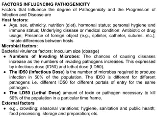
1) The factors that influence a microbe's pathogenicity, including host factors like age and immune status, and microbial factors like virulence factors and inoculum size.
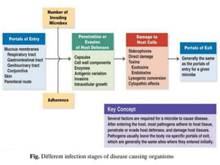
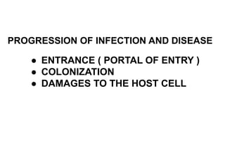
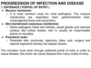
2) The steps in pathogenesis which include a microbe gaining access to the host, adhering to tissues, penetrating defenses, and damaging the host directly or through toxins.
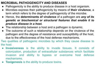
3) The two qualities that allow microbes to cause disease - invasiveness and toxigenesis. It also discusses bacterial adherence, biofilm formation, and how pathogens prevent host defenses.


![Lecture 6- Bacteria- Phathogenesis [Autosaved].pptx](https://cdn.slidesharecdn.com/ss_thumbnails/lecture6-bacteria-phathogenesisautosaved-220830041321-bf3b2198-thumbnail.jpg?width=640&height=640&fit=bounds)


