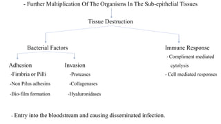
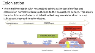
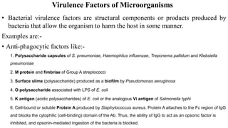
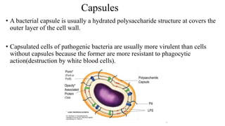
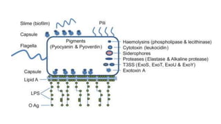
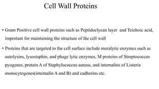
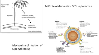
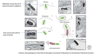
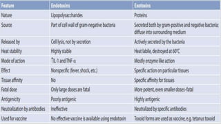
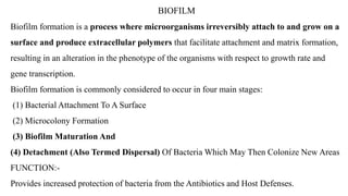
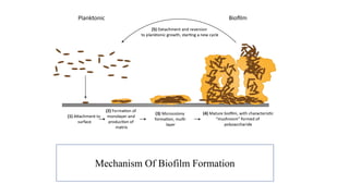
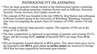
SEMINAR
An activity in which a group of persons engaged in
advanced study of a subject meet in a general
direction of an expert staff member for a discussion
leading to in depth study of the subject/problem.
BACTERIAL PATHOGENICITY AND
VIRULENCE FACTORS
INDEX
โ€ข INTRODUCTION
โ€ข TYPES OF BACTERIAL PATHOGENS
โ€ข REQUIREMENTS OF PATHOGENICITY
โ€ข VIRULENCE FACTORS
โ€ข BIOFILM
โ€ข PATHOGENICITY ISLAND
โ€ข QUORUM SENSING
โ€ข BACTERIAL SECRETION
PATHOGENICITY AND VIRULENCE
โ€ข Pathogenesis โ€“ Ability of a microbial species to produce a disease.
โ€ข Virulence โ€“ its is the relative degree of pathogenesis(tissue damage), which may vary
between different strains of the same organism depending upon their expression of the
virulence factors.
Virulence of a strain may undergo spontaneous or induced variation.
๏‚ง Exaltation โ€“ Enhancement of virulence, which can be induced experimentally by serial
passage into susceptible host.
๏‚ง Attenuation โ€“ Reduction of virulence, achieved by passage through unfavourable
hosts, repeated cultures in artificial media, growth in high temperature or in the
presence of weak antiseptics, desiccation or prolonged storage in culture.
Types of bacterial pathogens
โ€ข Primary Pathogens- capable of establishing infection and causing disease in
previously healthy individuals with intact immunological defenses.
โ€ข Opportunistic Pathogens- rarely cause disease in individuals with intact
immunological defenses. When the immunity gets compromised then only they
cause pathogenicity. For eg
- Coagulase negative Staphylococcus
- Escherichia coli
INFECTION VS. COLONISATION
โ€ข Infection โ€“ pathogen enters ๏ƒ  establishes itself ๏ƒ  multiplies๏ƒ  invades
normal anatomical barrier of host ๏ƒ  disease ๏ƒ  clinical manifestation
โ€ข Colonization โ€“ Pathogenic organism enters ๏ƒ does not invade, don't cause
disease and don`t elicit immune response ๏ƒ host is asymptomatic carrier.
Microorganism enters the host and initiates infection.
Attachment to the surface of the host
Interaction of bacterial adhesins with cell-surface receptors.
Invasion of the cell and Attachment without invasion
deeper tissues occurs via:-
Toxins
Enzymes
Requirements for Pathogenicity
- Further Multiplication Of The Organisms In The Sub-epithelial Tissues
Tissue Destruction
Bacterial Factors Immune Response
- Compliment mediated
Adhesion Invasion cytolysis
-Fimbria or Pilli -Proteases - Cell mediated responses
-Non Pilus adhesins -Collagenases
-Bio-film formation -Hyaluronidases
- Entry into the bloodstream and causing disseminated infection.
VIRULENCE FACTORS MECHANISM
CAPSULE Prevents the phagocytosis of bacteria by preventing the
phagocytes from adhering to the organism
CELL WALL PROTEINS
- Protein A
- M Protein
Binds to IgG & prevents the activation of complement
Associated with Rheumatic Fever
CYTOTOXINS
- Hemolysins
- Leukocidins
Lyse and damage the RBCs and WBCs
FIMBRIA or PILLI Bind to sugar residues on the host cells
NON-PILUS ADHESINS
- M Protein
- Lipoteichoic acid
- Cell Surface Lectin
Exacerbates Rheumatic fever
Helps in adhesion by binding to CD14 and Toll like
receptors
Binds to specific carbohydrate structures on the cell
BIOFILM Composed of multiple bacteria to form a slime layer to
evade the host immunity
PROTEASES Breakdown of protein in the cell
COLLAGENASES Breakdown the collagen peptide bonds
HYALURONIDASES Degrade the host cell hyaluronic acid
Transmission
Refers to the spread of the micro-organism from the reservoir or source to a susceptible
host in different ways.
โ€ข Contact:
Direct Contact โ€“ By skin/mucosa of an infected person.
Organisms โ€“ HIV, Neisseria gonorrhoea, Chlamydia trachomatis and Treponema pallidum etc.
Indirect Contact โ€“ Through Fomites( in-animate objects) such as clothing, toys etc.
Eg. Face towels used by different persons which can lead to the spread of Trachoma.
โ€ข Inhalation: Agents causing respiratory infection, which are spread into the environment
through secretions from the nose or throat during sneezing, coughing or speaking in the
form of droplets. Eg. Influenza virus, Corona virus etc.
โ€ข Air-Borne Transmission: Droplet nuclei <10um in size can remain suspended in
the air for long periods and can act as a source of infection.
โ€ข Droplet( Dust) Transmission: Larger droplets > 10 um in size
โ€ข Air Borne Droplet Nuclei: Small droplets <10um in size which can travel
farther distances.
โ€ข Ingestion: Infections like cholera, dysentery, food poisoning etc and most of
parasitic infections which can be acquired through contaminated food or water.
โ€ข Inoculation โ€“ Animal bite๏ƒ  rabies virus
Inoculated directly in tissues๏ƒ  spores of Clostridium tetani present in
soil, gets deposited in host tissue, leads to severe infections
โ€ข Transmission of Blood Borne Infections ๏ƒ  Needle pricks, sharp injuries, blood
transfusions, etc.
โ€ข Vector borne - Mechanical vectors ๏ƒ carry microbes and transmit them to
eatables.
โ€ข Biological vectors ๏ƒ  pathogen multiply in body of vectors, eg โ€“
Anopheles mosquito in malaria, culex mosquito in filariarsis.
Colonization
โ€ข The initial interaction with host tissues occurs at a mucosal surface and
colonization normally requires adhesion to the mucosal cell surface. This allows
the establishment of a focus of infection that may remain localized or may
subsequently spread to other tissues.
Adhesion
โ€ข It is an essential preliminary to colonization and then penetration through tissues.
Successful colonization also requires that bacteria are able to acquire essential
nutrientsโ€”in particular ironโ€”for growth. At the molecular level, adhesion
involves surface interactions between specific receptors on the mammalian cell
membrane (usually carbohydrates) and ligands (usually proteins) on the bacterial
surface.
Aggressins or Virulence Determinants
โ€ข In order to survive and multiply within the host, many organisms produce a
variety of substances that allow them to avoid host defence mechanisms. These
substances, termed aggressins, include capsules and extracellular slime
substances, surface proteins and carbohydrates, enzymes, toxins, and other small
molecules.
โ€ข For example, although 6 separate serotypes of encapsulated Haemophilus
influenzae are recognized, serious infection is almost exclusively associated with
isolates of serotype b (hence Hib vaccine).
Invasion
โ€ข Invasion is penetration of host cells and tissues (beyond the skin and mucous
surfaces), and is mediated by a complex array of molecules, often described as
โ€˜invasinsโ€™ . these can be in the form of bacterial surface or secreted proteins
which target host cell molecules (receptors).
โ€ข Invasins are a class of proteins associated with the penetration of pathogens
into host cells. Invasins play a role in promoting entry during the initial stage of
infection.
- For eg. Coagulase, staphylokinase and leukocidins in Staphylococcus spp. And
Hyaluronic acid and lipoteichoic acid in Streptococcus spp. etc
โ€ข Coagulase- Coats to fibrin in the blood which protects the bacterium from
phagocytosis
โ€ข Staphylokinase- It turns human plasminogen into plasmin which digests the
components of the extracellular matrix and causes extensive proteolysis.
โ€ข Leucocidins- It damages the membranes of the host cells and targets the
phagocytes, natural killer cells and T-lymphocytes, thus affecting both innate and
adaptive immunity.
โ€ข Hyaluronic acid- Constitutes the polysaccharide capsule which protects the
streptococci from phagocytosis by counteracting against the PMNs and they can
also bind to CD44 receptors and causing epithelial disruption.
โ€ข Lipoteichoic acid- Help in anchoring the streptococci to the cells of the host and
tightly adhere to them thus initiating the first step in the process of adhesion
Virulence Factors of Microorganisms
โ€ข Bacterial virulence factors are structural components or products produced by
bacteria that allow the organism to harm the host in some manner.
Examples are:-
โ€ข Anti-phagocytic factors like:-
1. Polysaccharide capsules of S. pneumoniae, Haemophilus influenzae, Treponema pallidum and Klebsiella
pneumoniae
2. M protein and fimbriae of Group A streptococci
3. Surface slime (polysaccharide) produced as a biofilm by Pseudomonas aeruginosa
4. O polysaccharide associated with LPS of E. coli
5. K antigen (acidic polysaccharides) of E. coli or the analogous Vi antigen of Salmonella typhi
6. Cell-bound or soluble Protein A produced by Staphylococcus aureus. Protein A attaches to the Fc region of IgG
and blocks the cytophilic (cell-binding) domain of the Ab. Thus, the ability of IgG to act as an opsonic factor is
inhibited, and opsonin-mediated ingestion of the bacteria is blocked.
Capsules
โ€ข A bacterial capsule is usually a hydrated polysaccharide structure at covers the
outer layer of the cell wall.
โ€ข Capsulated cells of pathogenic bacteria are usually more virulent than cells
without capsules because the former are more resistant to phagocytic
action(destruction by white blood cells).
Cell Wall Proteins
โ€ข Gram Positive cell wall proteins such as Peptidoclycan layer and Teichoic acid,
important for maintaining the structure of the cell wall
โ€ข Proteins that are targeted to the cell surface include muralytic enzymes such as
autolysins, lysostaphin, and phage lytic enzymes, M proteins of Streptococcus
pyogenes, protein A of Staphylococcus aureus, and internalins of Listeria
monocytogenes(internalin A and B) and cadherins etc.
M Protein Mechanism Of Streptococcus
Mechanism of Invasion of
Staphylococcus
Listeria monocytogenes Internalin B Activates Junctional Endocytosis to Accelerate Intestinal Invasion
Adhesion occurs by Int A
And Int B and E Cadherin
Lysis occurs by Listeria
Lysin O (LLO)
CYTOTOXINS
Exotoxins
Exotoxins, in contrast to endotoxin, are diffusible proteins secreted into the external
medium by the pathogen. Most pathogens secrete various protein molecules that
facilitate Adhesion to, or Invasion of, the host. Many others cause damage to host
cells. The damage may be
โ€ข Physiological - CHOLERA TOXIN which Promotes Electrolyte (And Fluid)
Excretion From Enterocytes Without Killing The Cells
โ€ข Pathological - DIPHTHERIA TOXIN which Inhibits Protein Synthesis And
Induces Cell Death
โ€ข Type I (membrane acting) toxins bind surface receptors and stimulate transmembrane signals, and
include the super-antigenic toxins.
โ€ข Type II (membrane damaging) toxins directly affect membranes, forming pores or disrupting lipid
bilayers.
โ€ข Type III (intracellular effector) toxins translocate an active enzymatic component into the cell and
modify an intracellular target molecule.
Endotoxins
Endotoxins are the lipid A portion of lipopolysaccharide (LPS). They are present
as an integral part of the cell wall of gram-negative bacteria. They are released
from the bacterial surface by natural lysis of the bacteria and are responsible for
various biological effects in the host.
Mechanism:-
1. Macrophage Activation โ€“ It binds to specific receptor on macrophages and stimulate
the release of acute phase cytokines e.g. IL-1, TNF-a, IL-6, Nitric oxide and
prostaglandins.
2. Compliment Activation โ€“ Activation of alternative pathway of complement and release
of C3a and C5a further promoting Inflammatory reaction
3. Endothelial Activation โ€“ Increased vascular permeability
4. Coagulation Pathway Activation - Hageman factor and other coagulation factors
leading to thrombosis and DIC.
5. Platelet Activation & Mast cell Activation
BIOFILM
Biofilm formation is a process where microorganisms irreversibly attach to and grow on a
surface and produce extracellular polymers that facilitate attachment and matrix formation,
resulting in an alteration in the phenotype of the organisms with respect to growth rate and
gene transcription.
Biofilm formation is commonly considered to occur in four main stages:
(1) Bacterial Attachment To A Surface
(2) Microcolony Formation
(3) Biofilm Maturation And
(4) Detachment (Also Termed Dispersal) Of Bacteria Which May Then Colonize New Areas
FUNCTION:-
Provides increased protection of bacteria from the Antibiotics and Host Defenses.
Mechanism Of Biofilm Formation
PATHOGENICITY ISLANDS(PAI)
โ€ข They are large genomic islands located in the chromosomal regions containing
sets of genes encoding numerous virulence factors such as Adhesins, Secretory
Systems, Toxins, Invasins, Iron Uptake system etc.
โ€ข The concept of PAI was founded in the late 1980s by Jรถrg Hacker and colleagues
in Werner Goebel's group at the University of Wรผrzburg, Wรผrzburg, Germany,
who were investigating the genetic basis of virulence of UPEC strains 536 and
J96.
โ€ข PAI occupy relatively large genomic regions. The majority of PAI are in the
range of 10 to 200 kb.
โ€ข The base composition is expressed as percentage of guanine and cytosine (G+C)
bases, and the average G+C content of bacterial DNA can range from 25 to
75%.
โ€ข PAI are frequently located adjacent to tRNA genes. This observation gave rise to
the hypothesis that tRNA genes serve as anchor points for insertion of foreign
DNA that has been acquired by horizontal gene transfer.
DR(Direct Repeats) are defined as DNA
sequences of 16 to 20 bp (up to 130 bp)
with a perfect or nearly perfect sequence
repetition
-Recognition sequences for enzymes
involved in excision of mobile genetic
elements, thus contributing to the
instability of a PAI.
Virulence genes (V1 to V4) and are
frequently interspersed with other mobility
elements like inertion sequences.
IS(Insertion sequences) IS elements can
also result in the mobilization of larger
portions of DNA. PAI can also represent
integrated plasmids, conjugative
transposons, bacteriophages or parts of
these elements
- Isc And Isd stand for complete and
defective(remnant of insertion
sequence) insertion sequences.
Int(Integrases) are responsible for transfer pathogenic elements to other
species of bacteria.
BACTERIAL SECRETORY SYSTEMS
๏ถBacterial secretion systems are protein complexes present on the cell
membranes of bacteria for secretion of substances. Specifically, they are the
cellular devices used by pathogenic bacteria to secrete their virulence factors
(mainly of proteins) to invade the host cells.
๏ฑType I
โ€ข Type I secretion system (T1SS or TOSS) is found in Gram-negative bacteria.
โ€ข It depends on chaperone activity using Hly and Tol proteins.
HlyA binds to HlyB HlyAB complex HlyD
Uncoiling and it moves to the outer membrane HlyA gets secreted
out.
ABC Transporter Activates
โ€ข Type II (T2SS) secretion system depends on the Sec or Tat system for initial
secretion inside the gram negative bacteria.
โ€ข Sec โ€“ Transmembrane translocation of proteins and Tat- Twin arginine
translocation of proteins in their folded state.
โ€ข Made up of General Secretory Proteins(GSPs)
Exoproteins are
Transported across the
inner membrane and
into the periplasm
Pre-pseudopilins are also
transported from the
cytoplasm into the periplasm
Cleavage by Pre-pilin peptidase
GspO And converted into mature
pseudopilins
Secretion ATPase GspE
Bind and hydrolyze ATP to produce
energy
Exoproteins, in the periplasm are
able to enter the secretion machinery
Pseudopilus then forces the
exoproteins out through the secretin
GspD and into the extracellular
matrix and get pushed out of the cell.
โ€ข Type III Secretory system: Helps to inject bacterial effector proteins directly into
the host cell cytoplasm, bypassing the extracellular matrix.
โ€ข Eg. Yesinia pestis can directly inject toxins from its cytoplasm into the hostโ€™s cytoplasm
Direct interactions with filamentous (F-actin)
or globular (G-actin) actin.
Activated by guanine nucleotide exchange factors
Down-regulated by GTPase-activated proteins
Activated Rho GTPases recruit proteins
Anchor the actin cytoskeleton to the plasma
membrane and activate GEFs
(guanine nucleotide exchange factors)
Type IV Secretory System
โ€ข Secrete proteinaceous effectors in order to manipulate and often kill rival bacterial
and eukaryotic cells.
โ€ข Eg. Legionella pneumophila that causes legionellosis (Legionnaires' disease) has a
T4SS called ICM/DOT (intracellular multiplication/defect in organelle
trafficking genes) that transport many bacterial proteins into its eukaryotic host.
โ€ข It can transport single proteins, as well as protein-protein and DNA-protein
complexes. Secretion is transferred directly from the recipient cell through the cell
membranes.
Type V Secretory System
โ€ข Type V secretion systems (T5SS) are different from other secretion systems is that
they secrete themselves and only involves the outer cell membrane.
โ€ข For secreted protein to pass through the inner cell membrane, T5SS depends on
Sec system. They have a ฮฒ-barrel domain, which inserts into the outer cell
membrane
โ€ข Forms a channel that can transport secreted
protein along with it.
โ€ข They are also called the autotransporter
systems
Type VI Secretory System
๏ถThe tubules consist of repeating units
of the proteins TssA and TssB (VipA/VipB)
arranged as a sheath around
a tube built from stacked hexameric rings of the
haemolysin co-regulated protein (Hcp)
๏ถThe baseplate and phage tail-like complex
interact in the bacterial cytoplasm,
and then are recruited to the cell envelope
by the membrane complex.
๏ถMembers of the second class of substrates
are targeted for secretion via interaction with
the phage tail spike-like protein VgrG.
TWO COMPONENT SIGNAL TRANSDUCTION
- Regulatory protein pair that senses change in external environment and produce
adaptive response.
- Occurs by transfer of phosphate molecule from sensor to effector proteins
- Phosphorylation of effector proteins regulate strength and duration of response by
binding to promoters in nucleic acid that allow for expression of gene that codes for
virulence factors.
- Eg- enteric gram negative bacteria, B. fragilis, B. pertussis, M. tuberculosis, Vibrio
species and Clostridium perfringens, etc.
QUORUM SENSING
Definition:
Cell to cell communication process involving production, detection and response
to extracellular signalling molecule k/a Autoinducers.
Components โ€“
1) Signalling molecule/autoinducers
2) Receptor that can specifically detect signalling molecule
3) Universal autoinducer AI-2.
In GNB`s โ€“ N-Acylhomoserine lactone (commonest)
In GPB`s โ€“ Oligopeptides (8-10 amino acid long)
โ€ข Mechanism of QUORUM SENSING :
Microbes secrete โ€˜Autoinducersโ€™ (AHLs) {AHL is plasma membrane diffusible}
Less AIs donot produce the response as threshold level is not reached.
As bacteria multiplies AHL increases extracellularly and start moving inside the bacteria.
Activation of genes responsible for various function
THANK YOU

BACTERIAL PATHOGENICITY AND VIRULENCE FACTORS.pptx

  • 1.
    SEMINAR An activity inwhich a group of persons engaged in advanced study of a subject meet in a general direction of an expert staff member for a discussion leading to in depth study of the subject/problem.
  • 2.
  • 3.
    INDEX โ€ข INTRODUCTION โ€ข TYPESOF BACTERIAL PATHOGENS โ€ข REQUIREMENTS OF PATHOGENICITY โ€ข VIRULENCE FACTORS โ€ข BIOFILM โ€ข PATHOGENICITY ISLAND โ€ข QUORUM SENSING โ€ข BACTERIAL SECRETION
  • 4.
    PATHOGENICITY AND VIRULENCE โ€ขPathogenesis โ€“ Ability of a microbial species to produce a disease. โ€ข Virulence โ€“ its is the relative degree of pathogenesis(tissue damage), which may vary between different strains of the same organism depending upon their expression of the virulence factors. Virulence of a strain may undergo spontaneous or induced variation. ๏‚ง Exaltation โ€“ Enhancement of virulence, which can be induced experimentally by serial passage into susceptible host. ๏‚ง Attenuation โ€“ Reduction of virulence, achieved by passage through unfavourable hosts, repeated cultures in artificial media, growth in high temperature or in the presence of weak antiseptics, desiccation or prolonged storage in culture.
  • 5.
    Types of bacterialpathogens โ€ข Primary Pathogens- capable of establishing infection and causing disease in previously healthy individuals with intact immunological defenses. โ€ข Opportunistic Pathogens- rarely cause disease in individuals with intact immunological defenses. When the immunity gets compromised then only they cause pathogenicity. For eg - Coagulase negative Staphylococcus - Escherichia coli
  • 6.
    INFECTION VS. COLONISATION โ€ขInfection โ€“ pathogen enters ๏ƒ  establishes itself ๏ƒ  multiplies๏ƒ  invades normal anatomical barrier of host ๏ƒ  disease ๏ƒ  clinical manifestation โ€ข Colonization โ€“ Pathogenic organism enters ๏ƒ does not invade, don't cause disease and don`t elicit immune response ๏ƒ host is asymptomatic carrier.
  • 7.
    Microorganism enters thehost and initiates infection. Attachment to the surface of the host Interaction of bacterial adhesins with cell-surface receptors. Invasion of the cell and Attachment without invasion deeper tissues occurs via:- Toxins Enzymes Requirements for Pathogenicity
  • 8.
    - Further MultiplicationOf The Organisms In The Sub-epithelial Tissues Tissue Destruction Bacterial Factors Immune Response - Compliment mediated Adhesion Invasion cytolysis -Fimbria or Pilli -Proteases - Cell mediated responses -Non Pilus adhesins -Collagenases -Bio-film formation -Hyaluronidases - Entry into the bloodstream and causing disseminated infection.
  • 9.
    VIRULENCE FACTORS MECHANISM CAPSULEPrevents the phagocytosis of bacteria by preventing the phagocytes from adhering to the organism CELL WALL PROTEINS - Protein A - M Protein Binds to IgG & prevents the activation of complement Associated with Rheumatic Fever CYTOTOXINS - Hemolysins - Leukocidins Lyse and damage the RBCs and WBCs FIMBRIA or PILLI Bind to sugar residues on the host cells NON-PILUS ADHESINS - M Protein - Lipoteichoic acid - Cell Surface Lectin Exacerbates Rheumatic fever Helps in adhesion by binding to CD14 and Toll like receptors Binds to specific carbohydrate structures on the cell BIOFILM Composed of multiple bacteria to form a slime layer to evade the host immunity PROTEASES Breakdown of protein in the cell COLLAGENASES Breakdown the collagen peptide bonds HYALURONIDASES Degrade the host cell hyaluronic acid
  • 10.
    Transmission Refers to thespread of the micro-organism from the reservoir or source to a susceptible host in different ways. โ€ข Contact: Direct Contact โ€“ By skin/mucosa of an infected person. Organisms โ€“ HIV, Neisseria gonorrhoea, Chlamydia trachomatis and Treponema pallidum etc. Indirect Contact โ€“ Through Fomites( in-animate objects) such as clothing, toys etc. Eg. Face towels used by different persons which can lead to the spread of Trachoma. โ€ข Inhalation: Agents causing respiratory infection, which are spread into the environment through secretions from the nose or throat during sneezing, coughing or speaking in the form of droplets. Eg. Influenza virus, Corona virus etc.
  • 11.
    โ€ข Air-Borne Transmission:Droplet nuclei <10um in size can remain suspended in the air for long periods and can act as a source of infection. โ€ข Droplet( Dust) Transmission: Larger droplets > 10 um in size โ€ข Air Borne Droplet Nuclei: Small droplets <10um in size which can travel farther distances. โ€ข Ingestion: Infections like cholera, dysentery, food poisoning etc and most of parasitic infections which can be acquired through contaminated food or water. โ€ข Inoculation โ€“ Animal bite๏ƒ  rabies virus Inoculated directly in tissues๏ƒ  spores of Clostridium tetani present in soil, gets deposited in host tissue, leads to severe infections
  • 12.
    โ€ข Transmission ofBlood Borne Infections ๏ƒ  Needle pricks, sharp injuries, blood transfusions, etc. โ€ข Vector borne - Mechanical vectors ๏ƒ carry microbes and transmit them to eatables. โ€ข Biological vectors ๏ƒ  pathogen multiply in body of vectors, eg โ€“ Anopheles mosquito in malaria, culex mosquito in filariarsis.
  • 13.
    Colonization โ€ข The initialinteraction with host tissues occurs at a mucosal surface and colonization normally requires adhesion to the mucosal cell surface. This allows the establishment of a focus of infection that may remain localized or may subsequently spread to other tissues.
  • 14.
    Adhesion โ€ข It isan essential preliminary to colonization and then penetration through tissues. Successful colonization also requires that bacteria are able to acquire essential nutrientsโ€”in particular ironโ€”for growth. At the molecular level, adhesion involves surface interactions between specific receptors on the mammalian cell membrane (usually carbohydrates) and ligands (usually proteins) on the bacterial surface.
  • 15.
    Aggressins or VirulenceDeterminants โ€ข In order to survive and multiply within the host, many organisms produce a variety of substances that allow them to avoid host defence mechanisms. These substances, termed aggressins, include capsules and extracellular slime substances, surface proteins and carbohydrates, enzymes, toxins, and other small molecules. โ€ข For example, although 6 separate serotypes of encapsulated Haemophilus influenzae are recognized, serious infection is almost exclusively associated with isolates of serotype b (hence Hib vaccine).
  • 16.
    Invasion โ€ข Invasion ispenetration of host cells and tissues (beyond the skin and mucous surfaces), and is mediated by a complex array of molecules, often described as โ€˜invasinsโ€™ . these can be in the form of bacterial surface or secreted proteins which target host cell molecules (receptors). โ€ข Invasins are a class of proteins associated with the penetration of pathogens into host cells. Invasins play a role in promoting entry during the initial stage of infection. - For eg. Coagulase, staphylokinase and leukocidins in Staphylococcus spp. And Hyaluronic acid and lipoteichoic acid in Streptococcus spp. etc
  • 17.
    โ€ข Coagulase- Coatsto fibrin in the blood which protects the bacterium from phagocytosis โ€ข Staphylokinase- It turns human plasminogen into plasmin which digests the components of the extracellular matrix and causes extensive proteolysis. โ€ข Leucocidins- It damages the membranes of the host cells and targets the phagocytes, natural killer cells and T-lymphocytes, thus affecting both innate and adaptive immunity. โ€ข Hyaluronic acid- Constitutes the polysaccharide capsule which protects the streptococci from phagocytosis by counteracting against the PMNs and they can also bind to CD44 receptors and causing epithelial disruption. โ€ข Lipoteichoic acid- Help in anchoring the streptococci to the cells of the host and tightly adhere to them thus initiating the first step in the process of adhesion
  • 19.
    Virulence Factors ofMicroorganisms โ€ข Bacterial virulence factors are structural components or products produced by bacteria that allow the organism to harm the host in some manner. Examples are:- โ€ข Anti-phagocytic factors like:- 1. Polysaccharide capsules of S. pneumoniae, Haemophilus influenzae, Treponema pallidum and Klebsiella pneumoniae 2. M protein and fimbriae of Group A streptococci 3. Surface slime (polysaccharide) produced as a biofilm by Pseudomonas aeruginosa 4. O polysaccharide associated with LPS of E. coli 5. K antigen (acidic polysaccharides) of E. coli or the analogous Vi antigen of Salmonella typhi 6. Cell-bound or soluble Protein A produced by Staphylococcus aureus. Protein A attaches to the Fc region of IgG and blocks the cytophilic (cell-binding) domain of the Ab. Thus, the ability of IgG to act as an opsonic factor is inhibited, and opsonin-mediated ingestion of the bacteria is blocked.
  • 20.
    Capsules โ€ข A bacterialcapsule is usually a hydrated polysaccharide structure at covers the outer layer of the cell wall. โ€ข Capsulated cells of pathogenic bacteria are usually more virulent than cells without capsules because the former are more resistant to phagocytic action(destruction by white blood cells).
  • 22.
    Cell Wall Proteins โ€ขGram Positive cell wall proteins such as Peptidoclycan layer and Teichoic acid, important for maintaining the structure of the cell wall โ€ข Proteins that are targeted to the cell surface include muralytic enzymes such as autolysins, lysostaphin, and phage lytic enzymes, M proteins of Streptococcus pyogenes, protein A of Staphylococcus aureus, and internalins of Listeria monocytogenes(internalin A and B) and cadherins etc.
  • 23.
    M Protein MechanismOf Streptococcus Mechanism of Invasion of Staphylococcus
  • 24.
    Listeria monocytogenes InternalinB Activates Junctional Endocytosis to Accelerate Intestinal Invasion Adhesion occurs by Int A And Int B and E Cadherin Lysis occurs by Listeria Lysin O (LLO)
  • 25.
    CYTOTOXINS Exotoxins Exotoxins, in contrastto endotoxin, are diffusible proteins secreted into the external medium by the pathogen. Most pathogens secrete various protein molecules that facilitate Adhesion to, or Invasion of, the host. Many others cause damage to host cells. The damage may be โ€ข Physiological - CHOLERA TOXIN which Promotes Electrolyte (And Fluid) Excretion From Enterocytes Without Killing The Cells โ€ข Pathological - DIPHTHERIA TOXIN which Inhibits Protein Synthesis And Induces Cell Death
  • 26.
    โ€ข Type I(membrane acting) toxins bind surface receptors and stimulate transmembrane signals, and include the super-antigenic toxins. โ€ข Type II (membrane damaging) toxins directly affect membranes, forming pores or disrupting lipid bilayers. โ€ข Type III (intracellular effector) toxins translocate an active enzymatic component into the cell and modify an intracellular target molecule.
  • 28.
    Endotoxins Endotoxins are thelipid A portion of lipopolysaccharide (LPS). They are present as an integral part of the cell wall of gram-negative bacteria. They are released from the bacterial surface by natural lysis of the bacteria and are responsible for various biological effects in the host. Mechanism:- 1. Macrophage Activation โ€“ It binds to specific receptor on macrophages and stimulate the release of acute phase cytokines e.g. IL-1, TNF-a, IL-6, Nitric oxide and prostaglandins. 2. Compliment Activation โ€“ Activation of alternative pathway of complement and release of C3a and C5a further promoting Inflammatory reaction 3. Endothelial Activation โ€“ Increased vascular permeability 4. Coagulation Pathway Activation - Hageman factor and other coagulation factors leading to thrombosis and DIC. 5. Platelet Activation & Mast cell Activation
  • 30.
    BIOFILM Biofilm formation isa process where microorganisms irreversibly attach to and grow on a surface and produce extracellular polymers that facilitate attachment and matrix formation, resulting in an alteration in the phenotype of the organisms with respect to growth rate and gene transcription. Biofilm formation is commonly considered to occur in four main stages: (1) Bacterial Attachment To A Surface (2) Microcolony Formation (3) Biofilm Maturation And (4) Detachment (Also Termed Dispersal) Of Bacteria Which May Then Colonize New Areas FUNCTION:- Provides increased protection of bacteria from the Antibiotics and Host Defenses.
  • 31.
  • 32.
    PATHOGENICITY ISLANDS(PAI) โ€ข Theyare large genomic islands located in the chromosomal regions containing sets of genes encoding numerous virulence factors such as Adhesins, Secretory Systems, Toxins, Invasins, Iron Uptake system etc. โ€ข The concept of PAI was founded in the late 1980s by Jรถrg Hacker and colleagues in Werner Goebel's group at the University of Wรผrzburg, Wรผrzburg, Germany, who were investigating the genetic basis of virulence of UPEC strains 536 and J96. โ€ข PAI occupy relatively large genomic regions. The majority of PAI are in the range of 10 to 200 kb. โ€ข The base composition is expressed as percentage of guanine and cytosine (G+C) bases, and the average G+C content of bacterial DNA can range from 25 to 75%. โ€ข PAI are frequently located adjacent to tRNA genes. This observation gave rise to the hypothesis that tRNA genes serve as anchor points for insertion of foreign DNA that has been acquired by horizontal gene transfer.
  • 33.
    DR(Direct Repeats) aredefined as DNA sequences of 16 to 20 bp (up to 130 bp) with a perfect or nearly perfect sequence repetition -Recognition sequences for enzymes involved in excision of mobile genetic elements, thus contributing to the instability of a PAI. Virulence genes (V1 to V4) and are frequently interspersed with other mobility elements like inertion sequences. IS(Insertion sequences) IS elements can also result in the mobilization of larger portions of DNA. PAI can also represent integrated plasmids, conjugative transposons, bacteriophages or parts of these elements - Isc And Isd stand for complete and defective(remnant of insertion sequence) insertion sequences. Int(Integrases) are responsible for transfer pathogenic elements to other species of bacteria.
  • 34.
    BACTERIAL SECRETORY SYSTEMS ๏ถBacterialsecretion systems are protein complexes present on the cell membranes of bacteria for secretion of substances. Specifically, they are the cellular devices used by pathogenic bacteria to secrete their virulence factors (mainly of proteins) to invade the host cells. ๏ฑType I โ€ข Type I secretion system (T1SS or TOSS) is found in Gram-negative bacteria. โ€ข It depends on chaperone activity using Hly and Tol proteins. HlyA binds to HlyB HlyAB complex HlyD Uncoiling and it moves to the outer membrane HlyA gets secreted out. ABC Transporter Activates
  • 35.
    โ€ข Type II(T2SS) secretion system depends on the Sec or Tat system for initial secretion inside the gram negative bacteria. โ€ข Sec โ€“ Transmembrane translocation of proteins and Tat- Twin arginine translocation of proteins in their folded state. โ€ข Made up of General Secretory Proteins(GSPs) Exoproteins are Transported across the inner membrane and into the periplasm Pre-pseudopilins are also transported from the cytoplasm into the periplasm Cleavage by Pre-pilin peptidase GspO And converted into mature pseudopilins Secretion ATPase GspE Bind and hydrolyze ATP to produce energy Exoproteins, in the periplasm are able to enter the secretion machinery Pseudopilus then forces the exoproteins out through the secretin GspD and into the extracellular matrix and get pushed out of the cell.
  • 36.
    โ€ข Type IIISecretory system: Helps to inject bacterial effector proteins directly into the host cell cytoplasm, bypassing the extracellular matrix. โ€ข Eg. Yesinia pestis can directly inject toxins from its cytoplasm into the hostโ€™s cytoplasm Direct interactions with filamentous (F-actin) or globular (G-actin) actin. Activated by guanine nucleotide exchange factors Down-regulated by GTPase-activated proteins Activated Rho GTPases recruit proteins Anchor the actin cytoskeleton to the plasma membrane and activate GEFs (guanine nucleotide exchange factors)
  • 37.
    Type IV SecretorySystem โ€ข Secrete proteinaceous effectors in order to manipulate and often kill rival bacterial and eukaryotic cells. โ€ข Eg. Legionella pneumophila that causes legionellosis (Legionnaires' disease) has a T4SS called ICM/DOT (intracellular multiplication/defect in organelle trafficking genes) that transport many bacterial proteins into its eukaryotic host. โ€ข It can transport single proteins, as well as protein-protein and DNA-protein complexes. Secretion is transferred directly from the recipient cell through the cell membranes.
  • 38.
    Type V SecretorySystem โ€ข Type V secretion systems (T5SS) are different from other secretion systems is that they secrete themselves and only involves the outer cell membrane. โ€ข For secreted protein to pass through the inner cell membrane, T5SS depends on Sec system. They have a ฮฒ-barrel domain, which inserts into the outer cell membrane โ€ข Forms a channel that can transport secreted protein along with it. โ€ข They are also called the autotransporter systems
  • 39.
    Type VI SecretorySystem ๏ถThe tubules consist of repeating units of the proteins TssA and TssB (VipA/VipB) arranged as a sheath around a tube built from stacked hexameric rings of the haemolysin co-regulated protein (Hcp) ๏ถThe baseplate and phage tail-like complex interact in the bacterial cytoplasm, and then are recruited to the cell envelope by the membrane complex. ๏ถMembers of the second class of substrates are targeted for secretion via interaction with the phage tail spike-like protein VgrG.
  • 40.
    TWO COMPONENT SIGNALTRANSDUCTION - Regulatory protein pair that senses change in external environment and produce adaptive response. - Occurs by transfer of phosphate molecule from sensor to effector proteins - Phosphorylation of effector proteins regulate strength and duration of response by binding to promoters in nucleic acid that allow for expression of gene that codes for virulence factors. - Eg- enteric gram negative bacteria, B. fragilis, B. pertussis, M. tuberculosis, Vibrio species and Clostridium perfringens, etc.
  • 41.
    QUORUM SENSING Definition: Cell tocell communication process involving production, detection and response to extracellular signalling molecule k/a Autoinducers. Components โ€“ 1) Signalling molecule/autoinducers 2) Receptor that can specifically detect signalling molecule 3) Universal autoinducer AI-2. In GNB`s โ€“ N-Acylhomoserine lactone (commonest) In GPB`s โ€“ Oligopeptides (8-10 amino acid long)
  • 42.
    โ€ข Mechanism ofQUORUM SENSING : Microbes secrete โ€˜Autoinducersโ€™ (AHLs) {AHL is plasma membrane diffusible} Less AIs donot produce the response as threshold level is not reached. As bacteria multiplies AHL increases extracellularly and start moving inside the bacteria. Activation of genes responsible for various function
  • 43.