Downloaded 384 times
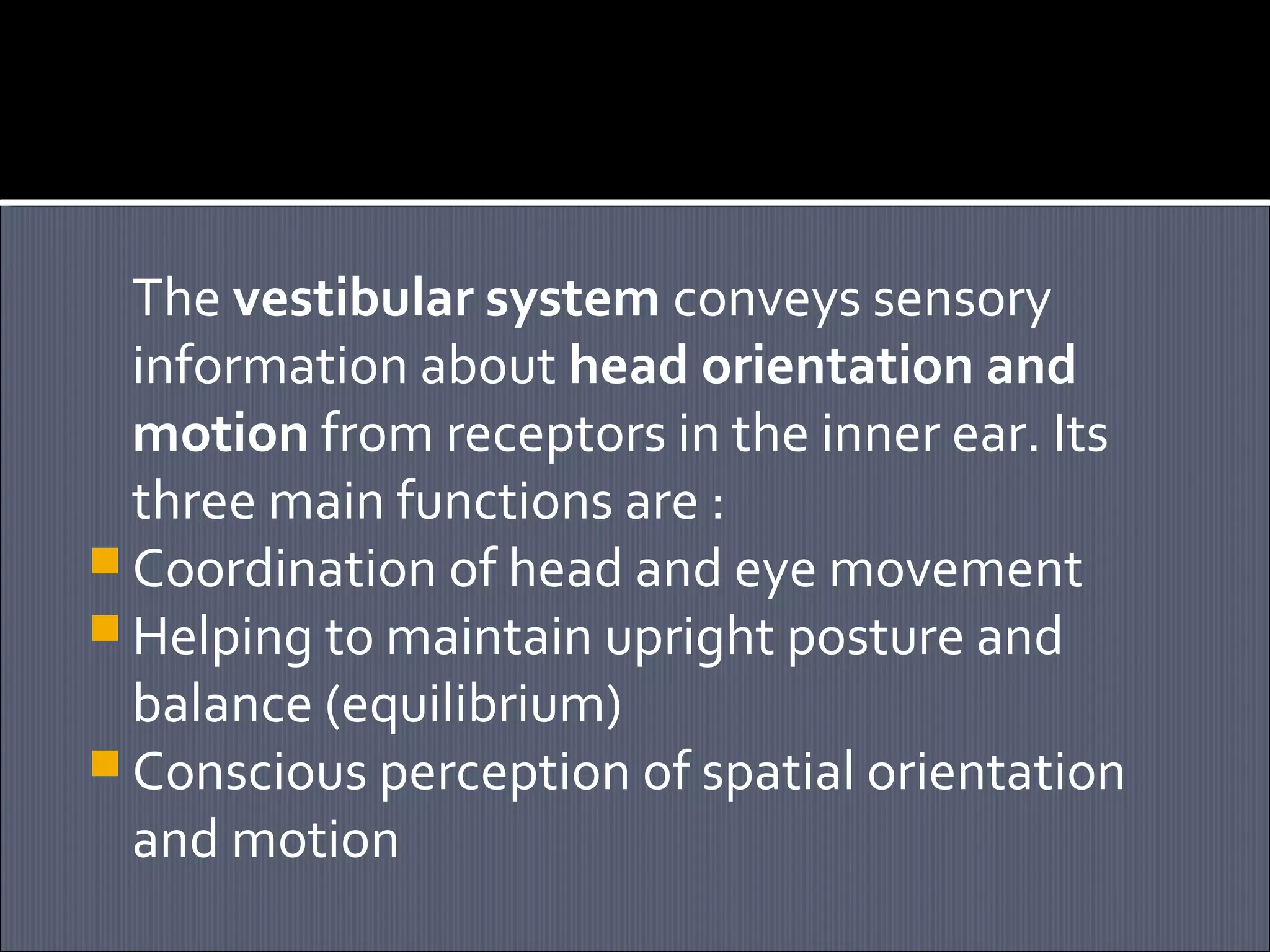
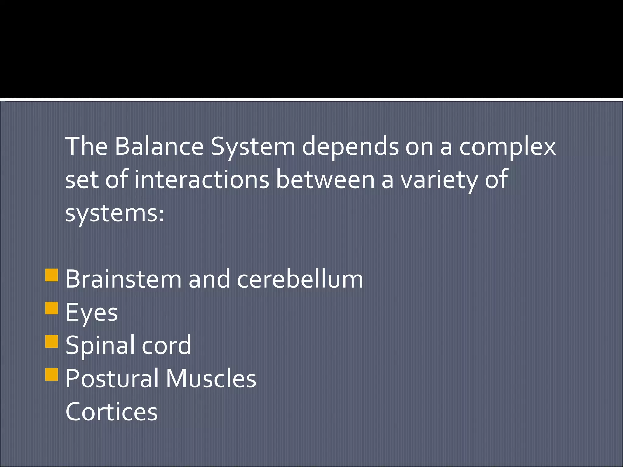

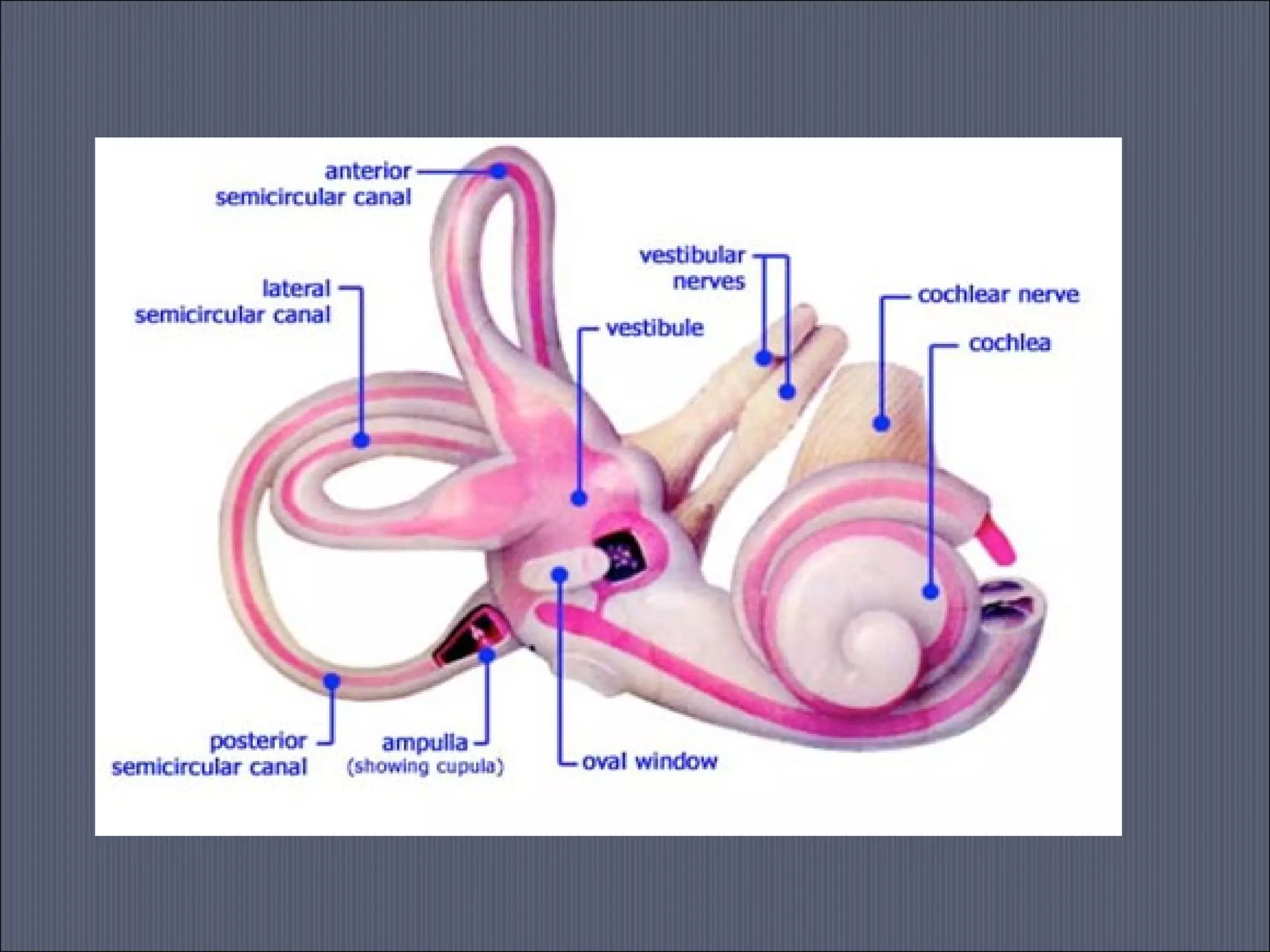


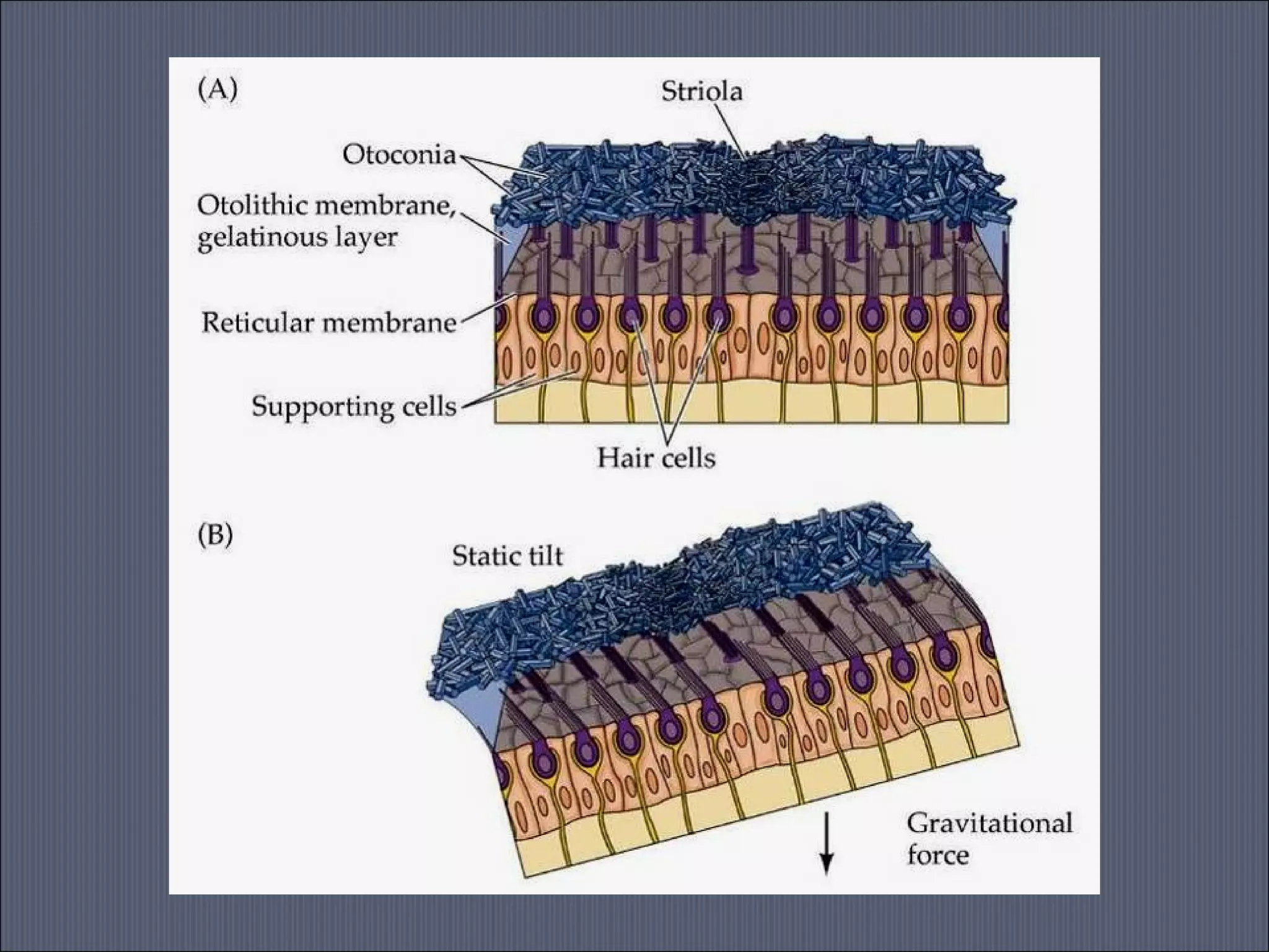

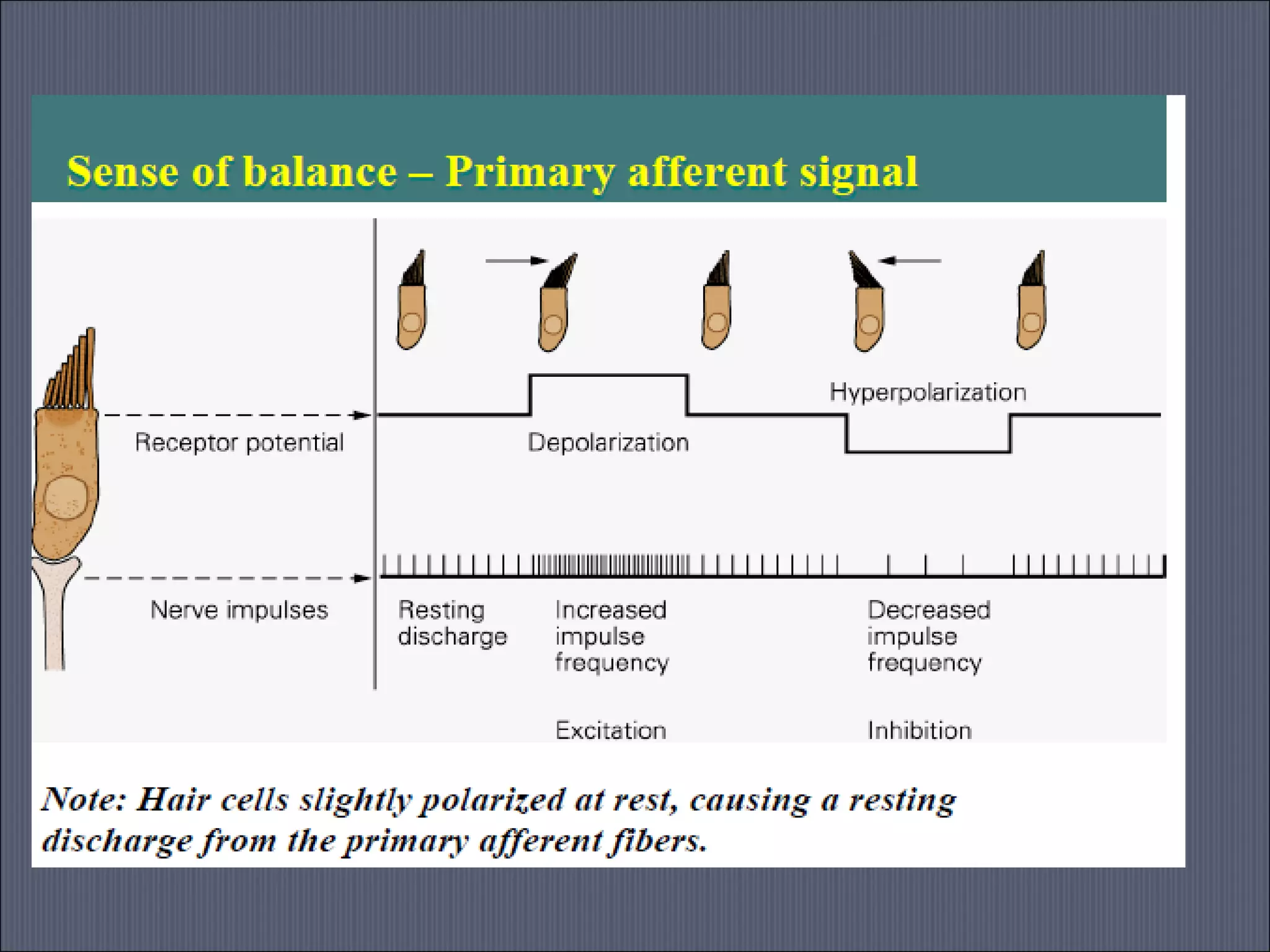
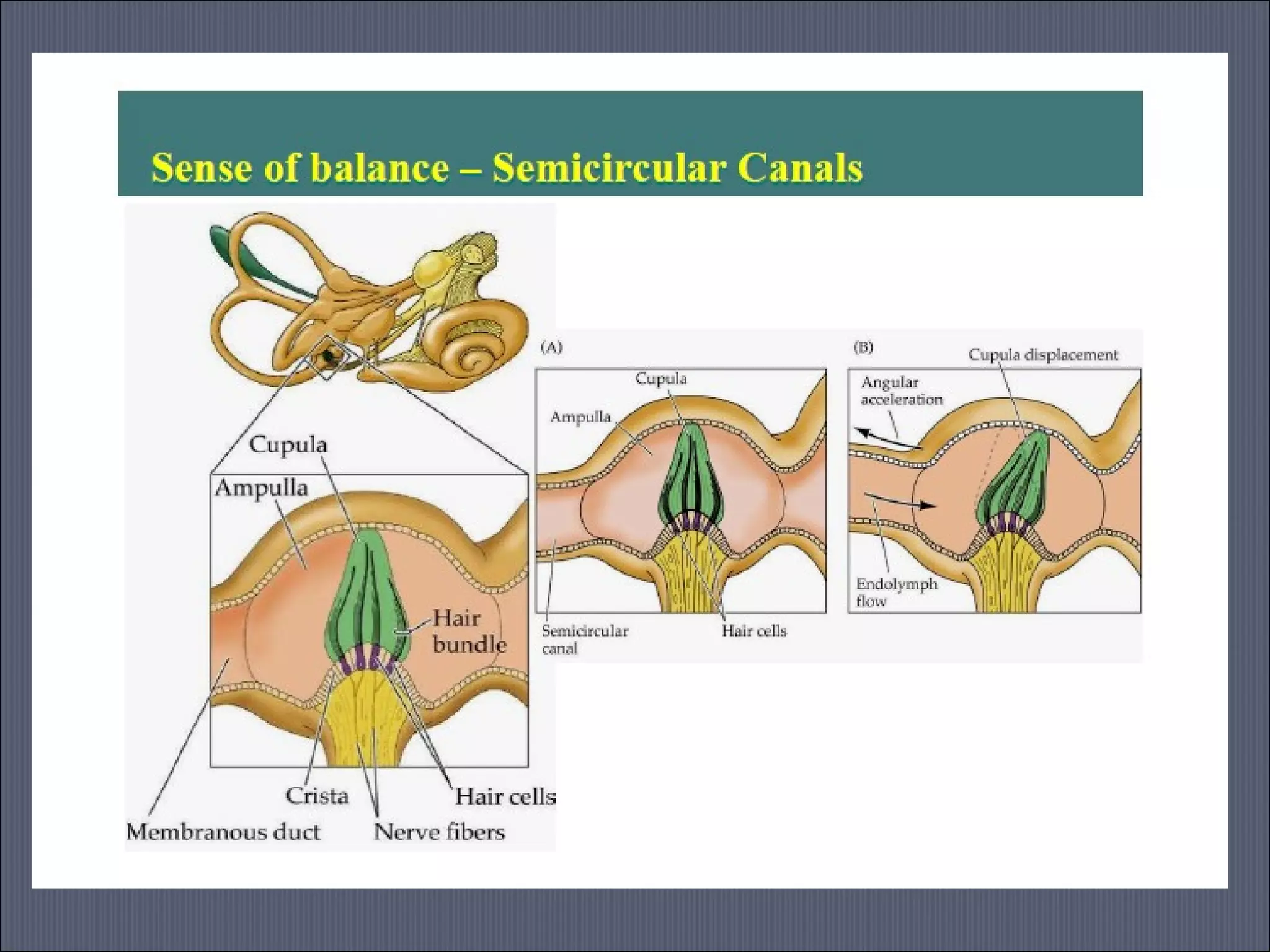


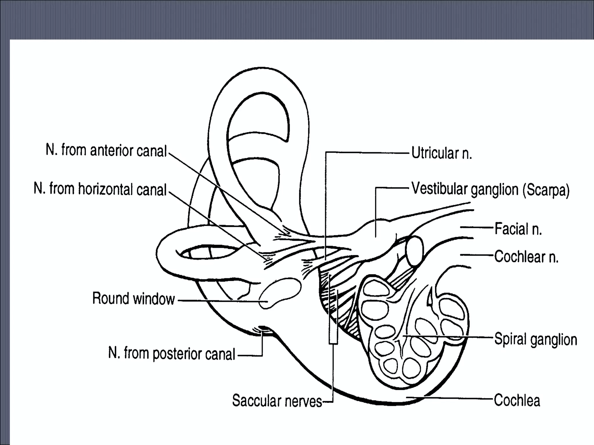
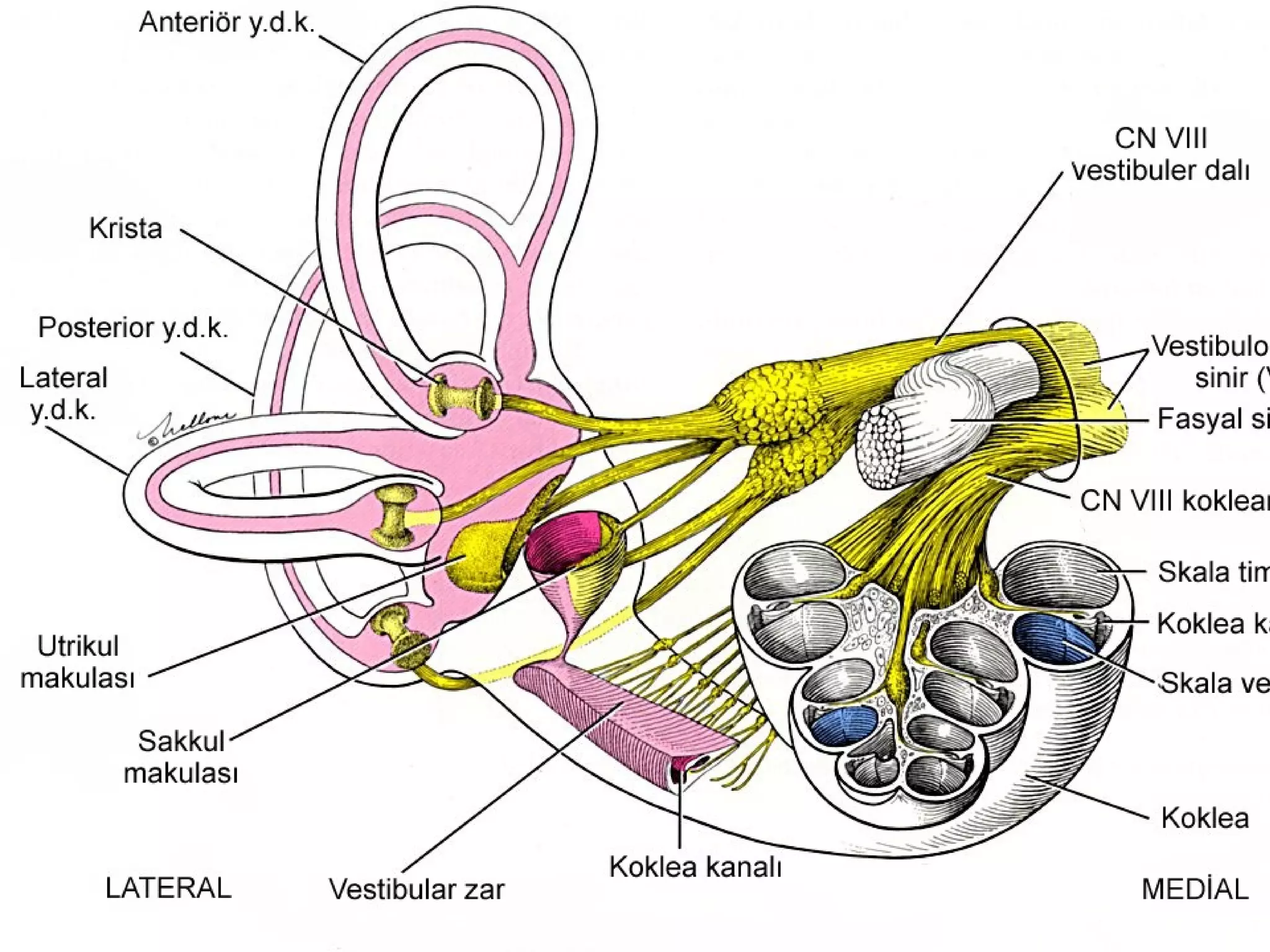
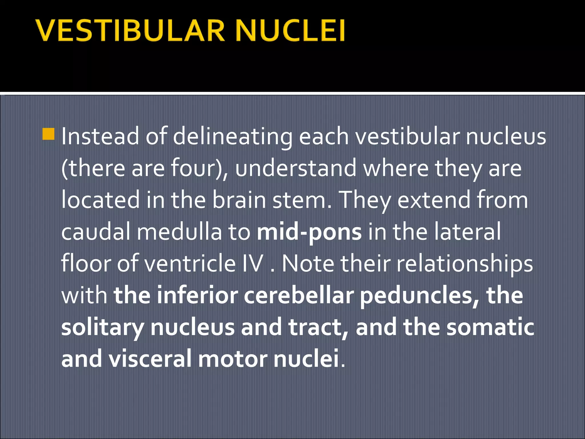


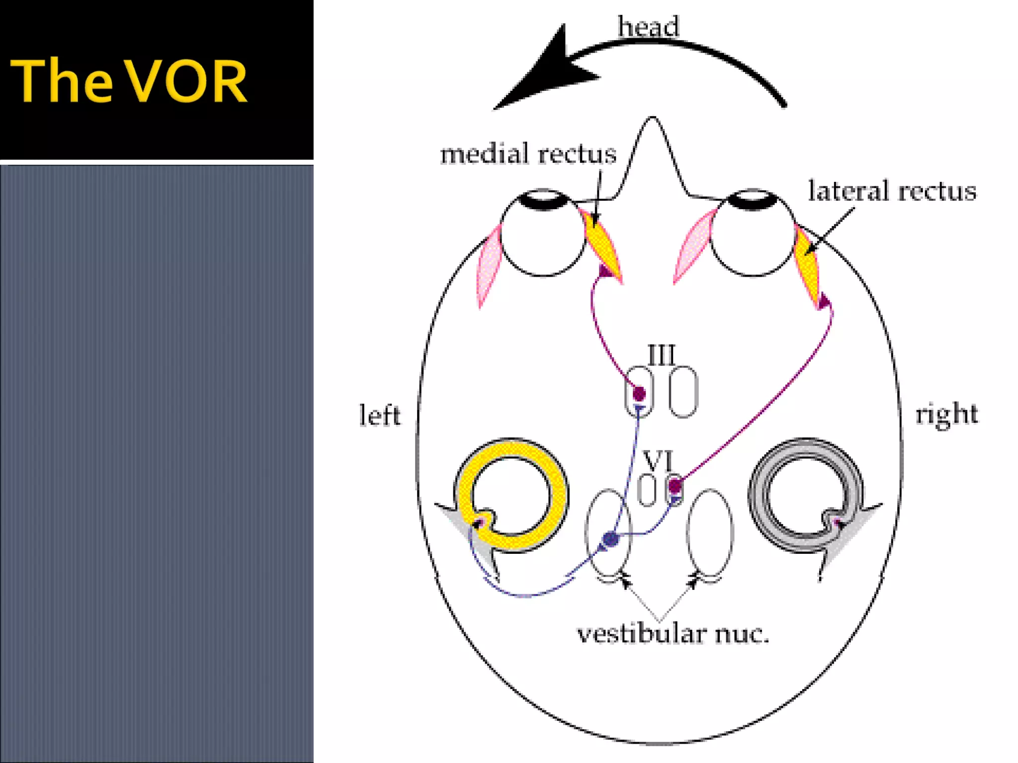
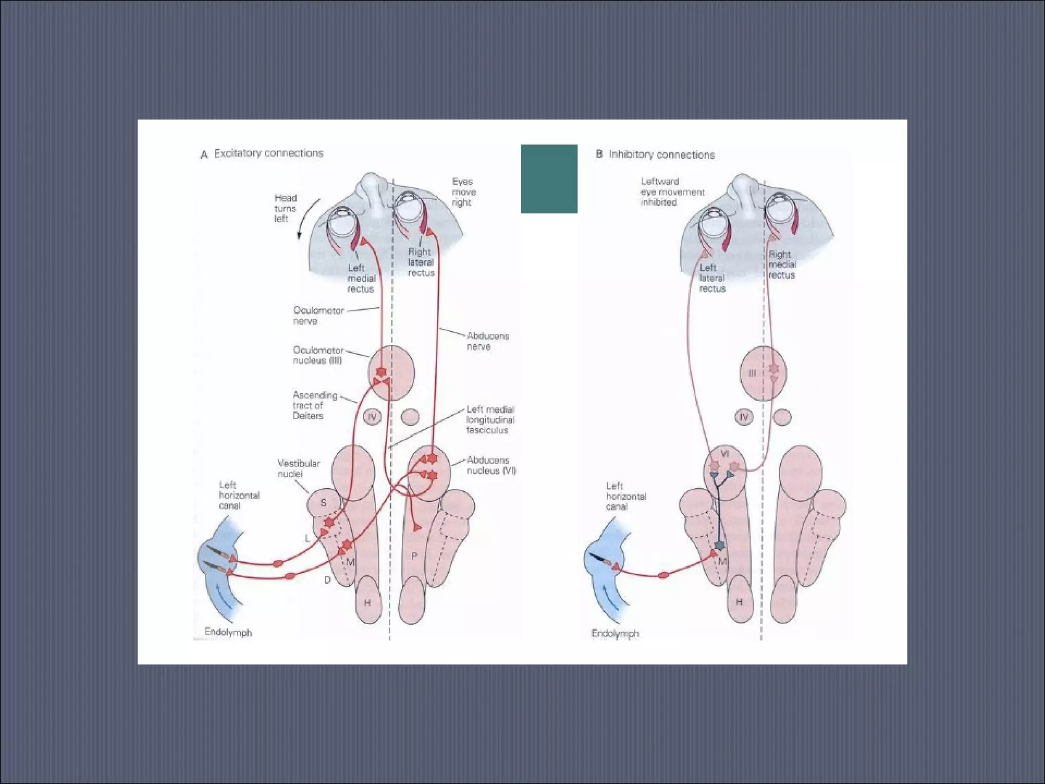

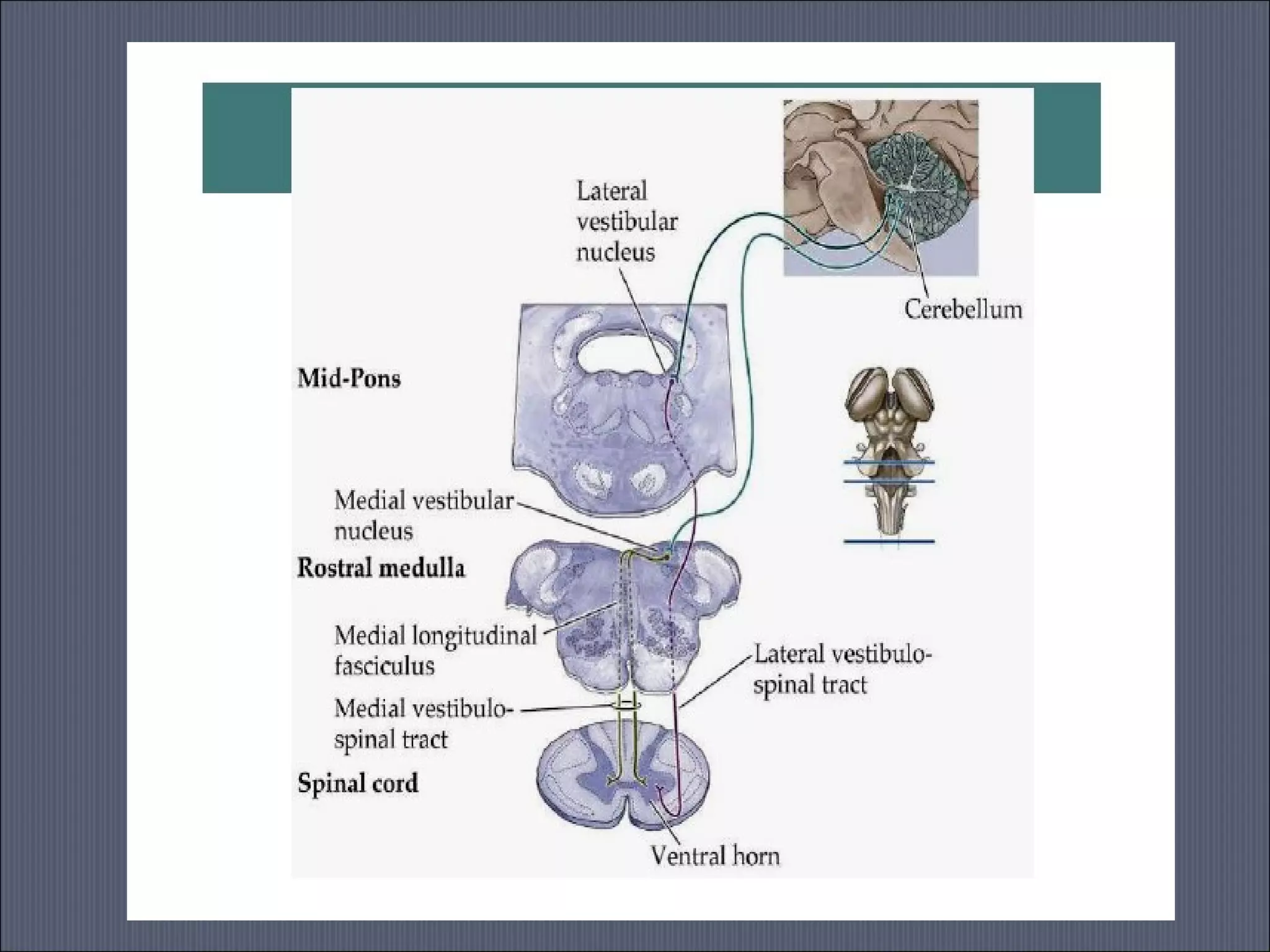
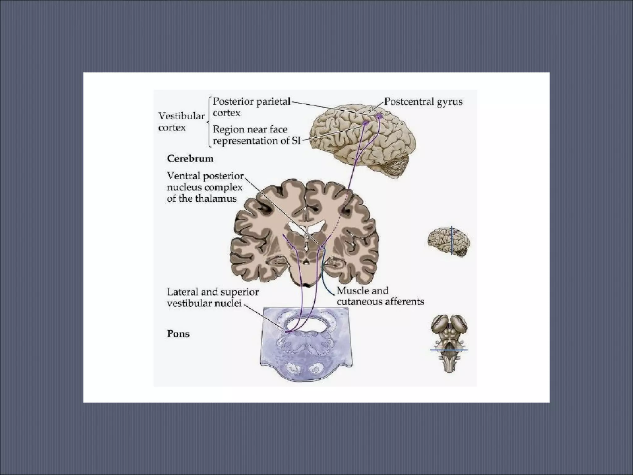
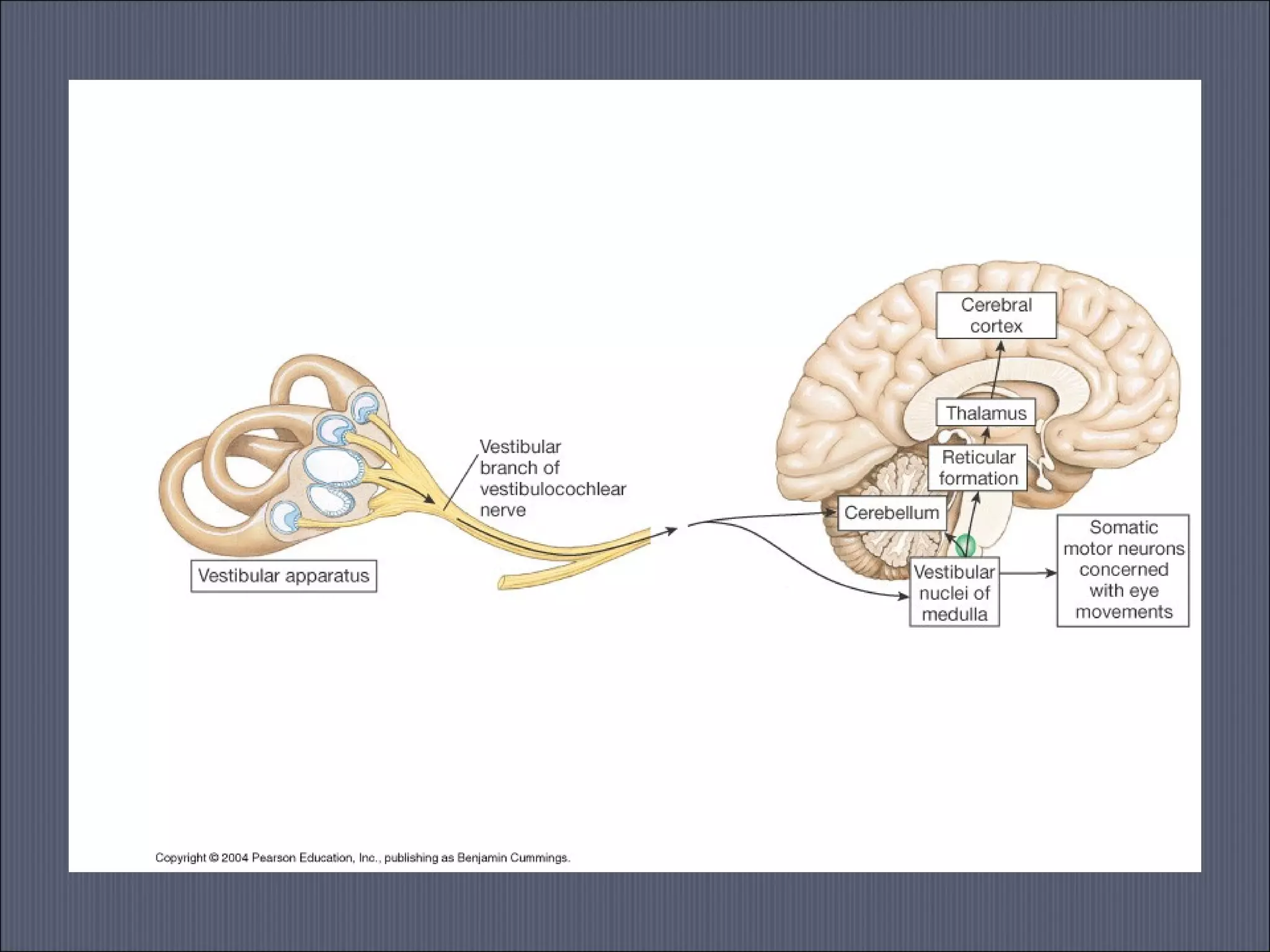
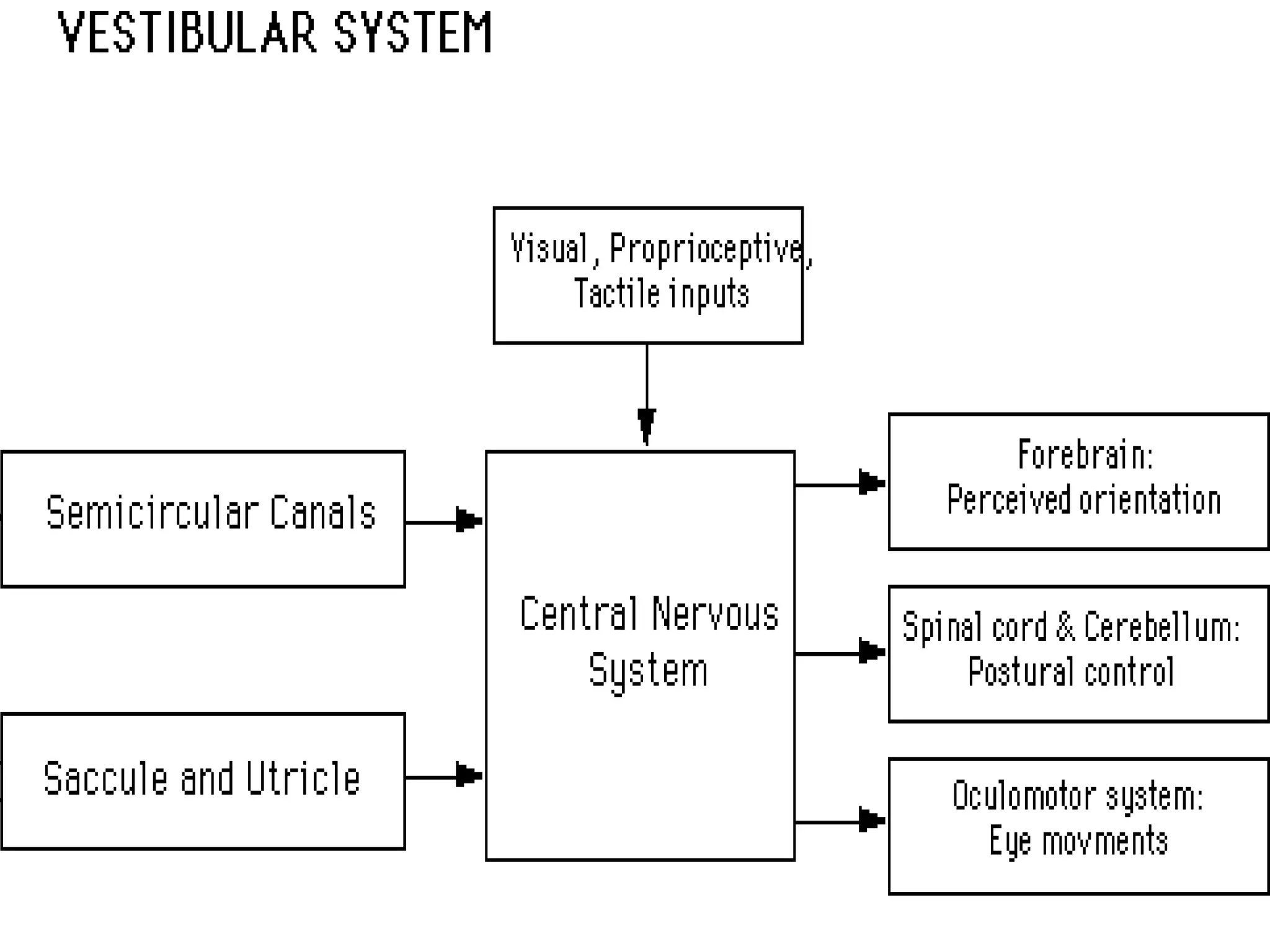


The vestibular system detects head motion and orientation using receptors in the inner ear. It has three main functions: coordinating eye and head movements, maintaining balance and posture, and perceiving spatial orientation. The vestibular system interacts with the brainstem, cerebellum, eyes, spinal cord and muscles. Sensory information travels to the brainstem via the vestibular nerve and projects to areas controlling eye movements, posture and spatial awareness. Damage can cause issues with balance, coordination and perception.


![Coded Agents – with UiPath SDK + LangGraph [Virtual Hands-on Workshop]](https://cdn.slidesharecdn.com/ss_thumbnails/codedagentsdeck-251215155422-5497c599-thumbnail.jpg?width=640&height=640&fit=bounds)


