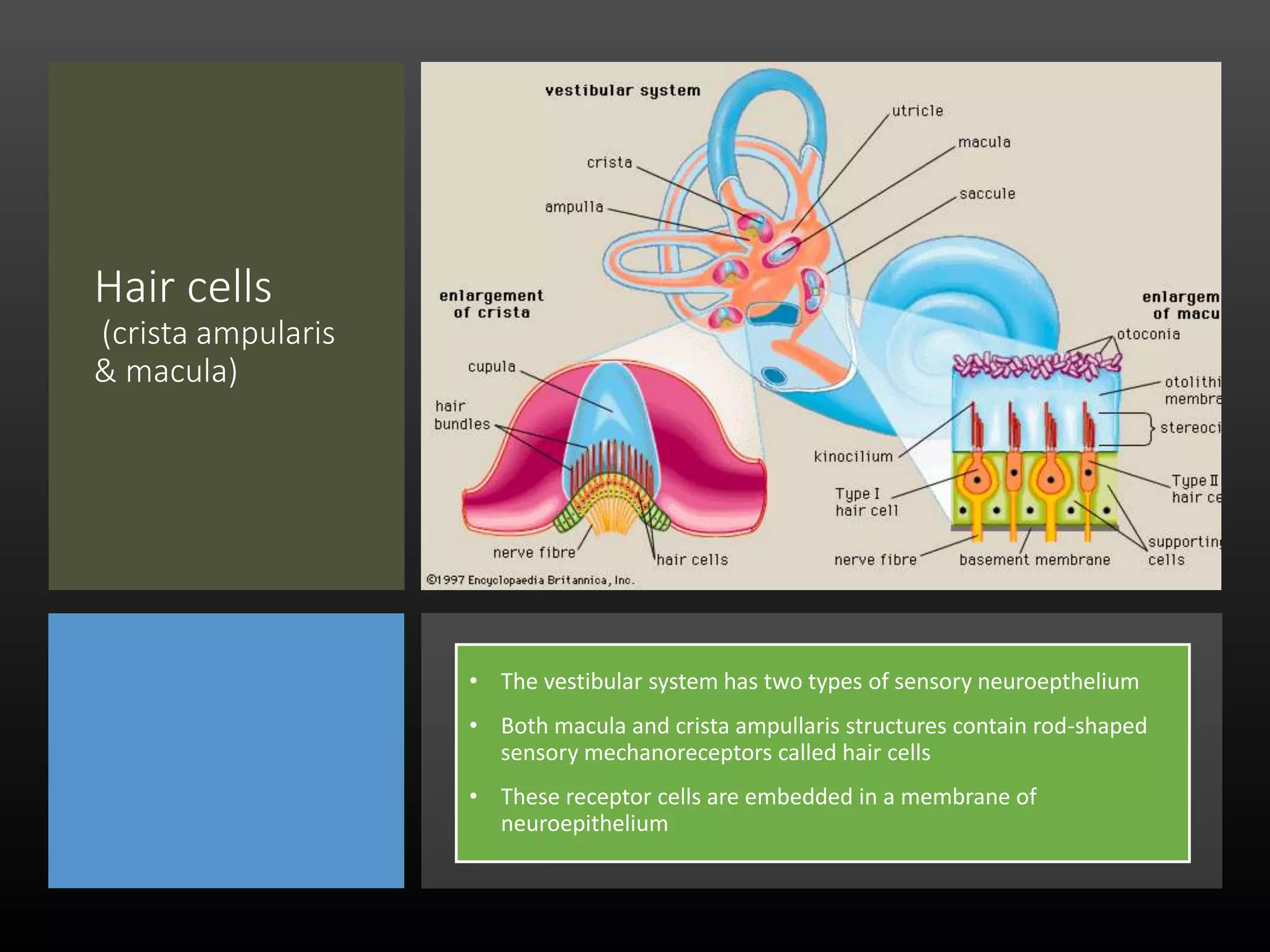
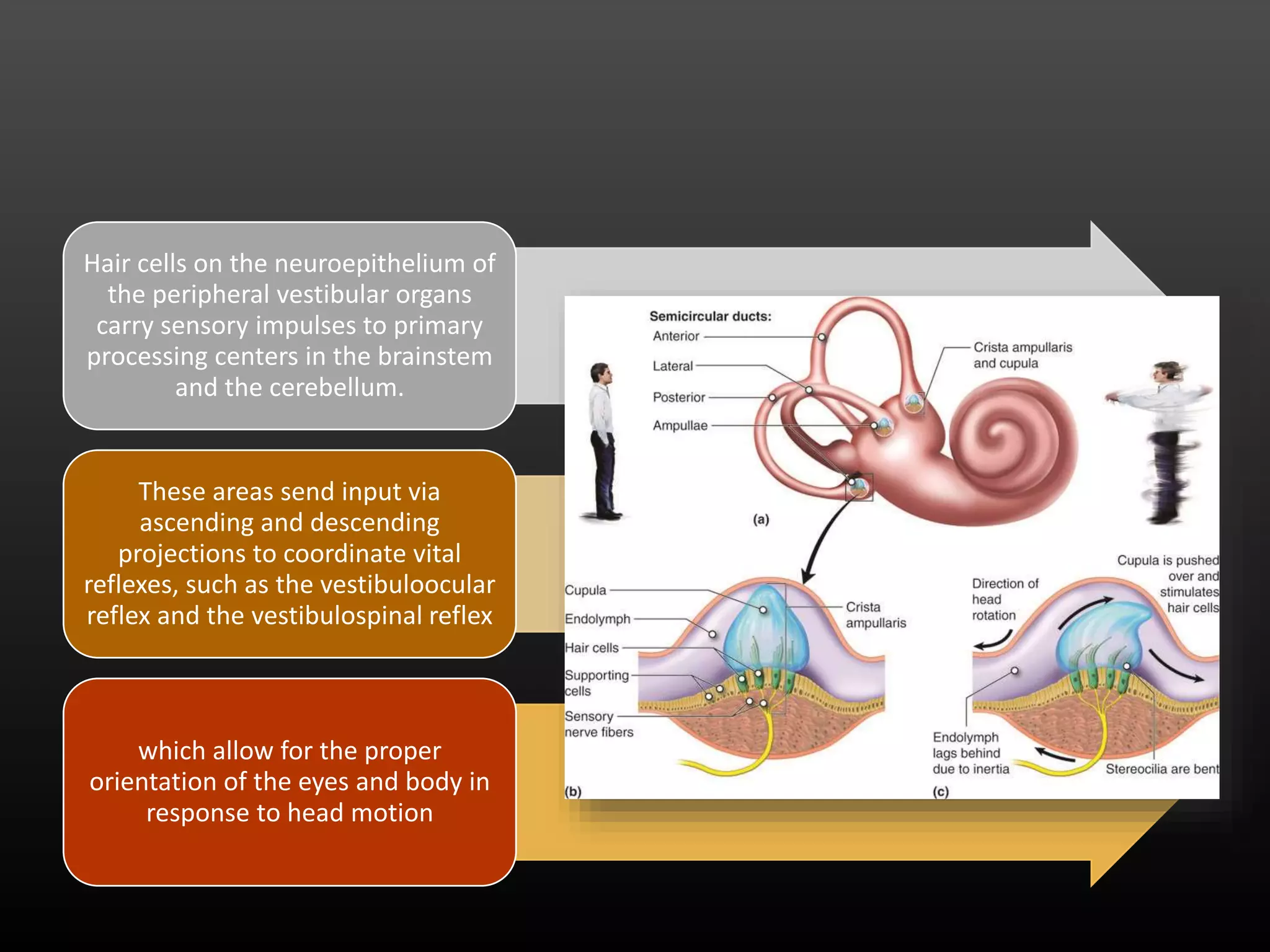
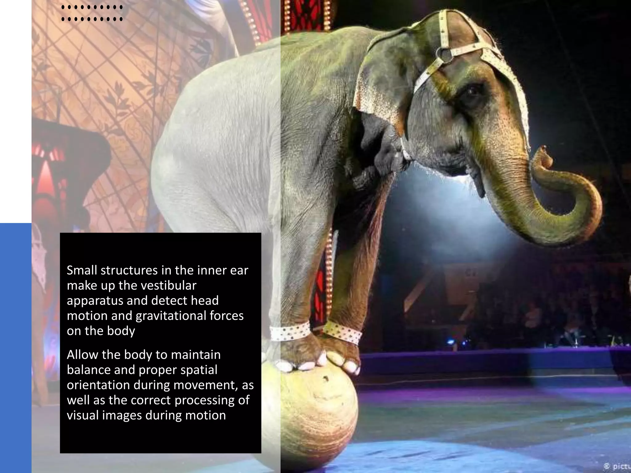
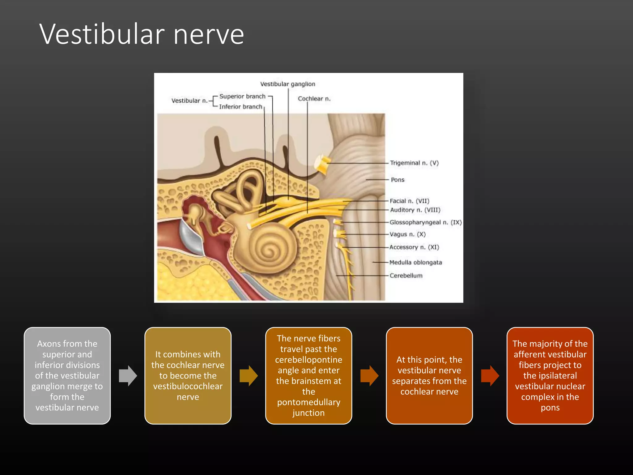
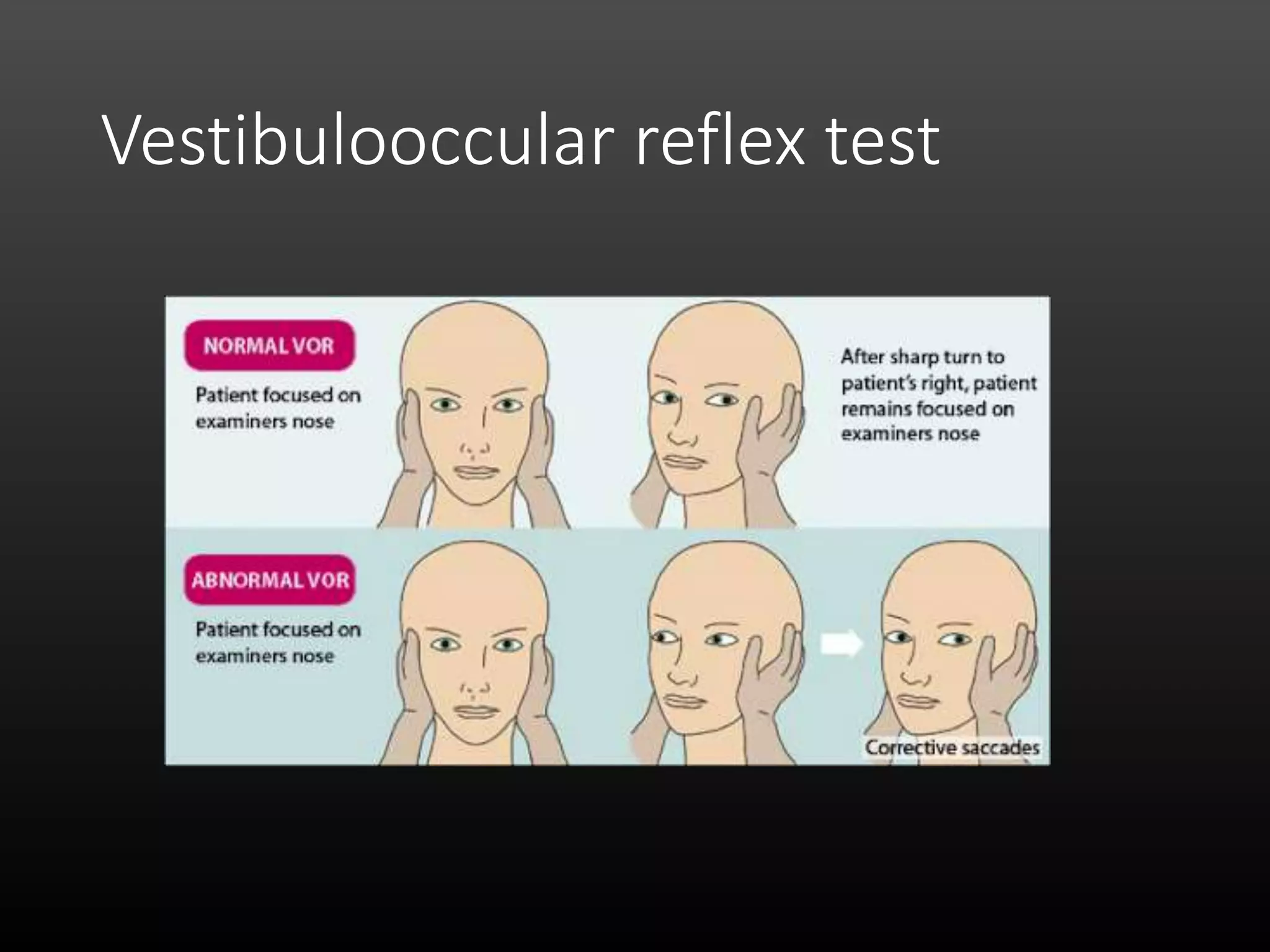
VESTIBULAR
SYSTEM
Aditya
Johan
Romadhon
introduction
A sense of proper sensory processing of head motion and
the coordination of visual and postural movements to
maintain equilibrium
Posture is a subsconcious adjustment of tone in different
muscle so as to maintain balance during displacement of
the body caused by gravity or acceleration
Balance is the ability to maintain the body center of mass
over its base support
The vestibular system is an intricate organization that
involves multiple levels of sensory processing to achieve
this goal
Vestibular structures The five major
vestibular
structures are
located in the
inner ear
the utricle
the saccule
and the
lateral,
superior, and
posterior
semicircular
canals
Hair cells
(crista ampularis
& macula)
• The vestibular system has two types of sensory neuroepthelium
• Both macula and crista ampullaris structures contain rod-shaped
sensory mechanoreceptors called hair cells
• These receptor cells are embedded in a membrane of
neuroepithelium
Hair cells on the neuroepithelium of
the peripheral vestibular organs
carry sensory impulses to primary
processing centers in the brainstem
and the cerebellum.
These areas send input via
ascending and descending
projections to coordinate vital
reflexes, such as the vestibuloocular
reflex and the vestibulospinal reflex
which allow for the proper
orientation of the eyes and body in
response to head motion
Tip Link function
(sodium-
potassium pump)
• Hair Cell “tip link” function. When head motion results in
tilting of the stereocilia toward the kinocilium, shifting of the
“tip links” causes mechanical opening of the transduction
channels resulting in an influx of K and depolarization of the
hair cells.
• The bending of the stereocilia away from the kinocilium
decreases the “tip link” tension and results in mechanical
closure of the channel causing hyperpolarization of the hair
cell
Dynamic changes of interstereociliary
Utricle & saccule
The utricle and
saccule are
structures of the
static labyrinth
that sense the
orientation of
the head in
space
They respond to
linear
acceleration,
gravitational
forces, and tilting
of the head
Each contains a
sensory
neuroepithelium
called the macula
The macula of
the utricle senses
motion in the
horizontal plane
The macula of
the saccule
senses motion in
the vertical plane
Semicircular canal
• The semicircular ducts
have the same basic
structure as the bony
semicircular canals in
which they are contained
• They make up the kinetic
labyrinth that senses
angular acceleration or
rotation of the head and
are oriented at right angles
to one another
Small structures in the inner ear
make up the vestibular
apparatus and detect head
motion and gravitational forces
on the body
Allow the body to maintain
balance and proper spatial
orientation during movement, as
well as the correct processing of
visual images during motion
Vestibular nerve
Axons from the
superior and
inferior divisions
of the vestibular
ganglion merge to
form the
vestibular nerve
It combines with
the cochlear nerve
to become the
vestibulocochlear
nerve
The nerve fibers
travel past the
cerebellopontine
angle and enter
the brainstem at
the
pontomedullary
junction
At this point, the
vestibular nerve
separates from the
cochlear nerve
The majority of the
afferent vestibular
fibers project to
the ipsilateral
vestibular nuclear
complex in the
pons
Vestibular nuclear
complex
Some of the nerve fibers project to
the flocculo-nodular lobe of the
cerebellum and the adjacent
vermian cortex
• This complex is the primary
processor of vestibular input and
consists of four major nuclei:
medial, superior, lateral, and
inferior
• The medial vestibular nucleus is
the largest and makes up the
medial column
• The lateral column consists of the
superior, lateral, and inferior
vestibular nuclei
The medial vestibular nucleus has bilateral descending
projections in the medial vestibulospinal tract to the
cervical spinal cord to allow for the coordination of
head and neck motion
The lateral vestibular nucleus form the lateral
vestibular tract in the ipsilateral spinal cord, functions
in the vestibular spinal reflex by coordinating reflexive
tone in the trunk muscles and proximal extensors of
the limbs to maintain posture and balance
The superior vestibular nucleus, it sends ascending
efferent fibers to the extraocular muscles via the
medial longitudinal fasciculus in order to coordinate
the vestibuloocular reflex
The inferior vestibular nucleus has projections that go
to the other three vestibular nuclei and to the
cerebellum
Cerebellum
Vestibuloocular
reflex
The vestibuloocular reflex
coordinates eye movement in order
to stabilize retinal images during
head rotation
Vestibulooccular reflex test
Vestibulospinal reflex
This reflex involves many complex connections which
integrate input from the macula, crista ampullaris,
visual system, and axial and limb muscles by the
brainstem, and cerebellum in order for the
maintenance of posture and balance.
It involves both the lateral and medial vestibular spinal
tracts
SEKIAN
&
TERIMAKASIH

Vestibular system

  • 1.
  • 2.
    introduction A sense ofproper sensory processing of head motion and the coordination of visual and postural movements to maintain equilibrium Posture is a subsconcious adjustment of tone in different muscle so as to maintain balance during displacement of the body caused by gravity or acceleration Balance is the ability to maintain the body center of mass over its base support The vestibular system is an intricate organization that involves multiple levels of sensory processing to achieve this goal
  • 3.
    Vestibular structures Thefive major vestibular structures are located in the inner ear the utricle the saccule and the lateral, superior, and posterior semicircular canals
  • 4.
    Hair cells (crista ampularis &macula) • The vestibular system has two types of sensory neuroepthelium • Both macula and crista ampullaris structures contain rod-shaped sensory mechanoreceptors called hair cells • These receptor cells are embedded in a membrane of neuroepithelium
  • 5.
    Hair cells onthe neuroepithelium of the peripheral vestibular organs carry sensory impulses to primary processing centers in the brainstem and the cerebellum. These areas send input via ascending and descending projections to coordinate vital reflexes, such as the vestibuloocular reflex and the vestibulospinal reflex which allow for the proper orientation of the eyes and body in response to head motion
  • 6.
    Tip Link function (sodium- potassiumpump) • Hair Cell “tip link” function. When head motion results in tilting of the stereocilia toward the kinocilium, shifting of the “tip links” causes mechanical opening of the transduction channels resulting in an influx of K and depolarization of the hair cells. • The bending of the stereocilia away from the kinocilium decreases the “tip link” tension and results in mechanical closure of the channel causing hyperpolarization of the hair cell
  • 7.
    Dynamic changes ofinterstereociliary
  • 8.
    Utricle & saccule Theutricle and saccule are structures of the static labyrinth that sense the orientation of the head in space They respond to linear acceleration, gravitational forces, and tilting of the head Each contains a sensory neuroepithelium called the macula The macula of the utricle senses motion in the horizontal plane The macula of the saccule senses motion in the vertical plane
  • 9.
    Semicircular canal • Thesemicircular ducts have the same basic structure as the bony semicircular canals in which they are contained • They make up the kinetic labyrinth that senses angular acceleration or rotation of the head and are oriented at right angles to one another
  • 10.
    Small structures inthe inner ear make up the vestibular apparatus and detect head motion and gravitational forces on the body Allow the body to maintain balance and proper spatial orientation during movement, as well as the correct processing of visual images during motion
  • 11.
    Vestibular nerve Axons fromthe superior and inferior divisions of the vestibular ganglion merge to form the vestibular nerve It combines with the cochlear nerve to become the vestibulocochlear nerve The nerve fibers travel past the cerebellopontine angle and enter the brainstem at the pontomedullary junction At this point, the vestibular nerve separates from the cochlear nerve The majority of the afferent vestibular fibers project to the ipsilateral vestibular nuclear complex in the pons
  • 12.
    Vestibular nuclear complex Some ofthe nerve fibers project to the flocculo-nodular lobe of the cerebellum and the adjacent vermian cortex
  • 13.
    • This complexis the primary processor of vestibular input and consists of four major nuclei: medial, superior, lateral, and inferior • The medial vestibular nucleus is the largest and makes up the medial column • The lateral column consists of the superior, lateral, and inferior vestibular nuclei
  • 14.
    The medial vestibularnucleus has bilateral descending projections in the medial vestibulospinal tract to the cervical spinal cord to allow for the coordination of head and neck motion The lateral vestibular nucleus form the lateral vestibular tract in the ipsilateral spinal cord, functions in the vestibular spinal reflex by coordinating reflexive tone in the trunk muscles and proximal extensors of the limbs to maintain posture and balance The superior vestibular nucleus, it sends ascending efferent fibers to the extraocular muscles via the medial longitudinal fasciculus in order to coordinate the vestibuloocular reflex The inferior vestibular nucleus has projections that go to the other three vestibular nuclei and to the cerebellum
  • 15.
  • 16.
    Vestibuloocular reflex The vestibuloocular reflex coordinateseye movement in order to stabilize retinal images during head rotation
  • 17.
  • 18.
    Vestibulospinal reflex This reflexinvolves many complex connections which integrate input from the macula, crista ampullaris, visual system, and axial and limb muscles by the brainstem, and cerebellum in order for the maintenance of posture and balance. It involves both the lateral and medial vestibular spinal tracts
  • 19.