Downloaded 203 times


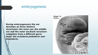
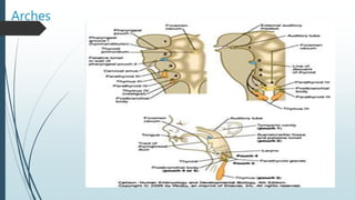
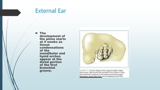
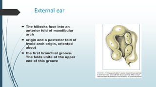
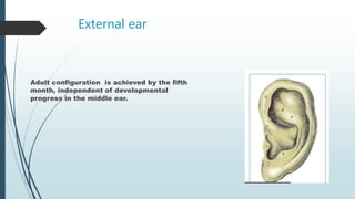


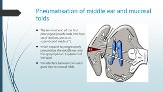

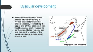

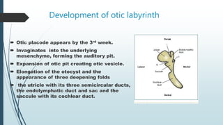
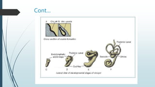
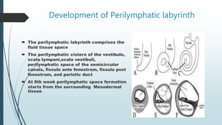


The ear develops from three germ layers into three main structures - the inner, middle, and outer ear. The outer ear develops from hillocks in the mandibular and hyoid arches, which fuse to form the pinna. The external auditory canal develops from the first branchial groove. The middle ear cavities develop from outpouchings of the first and second pharyngeal pouches. Ossicles develop from the first and second branchial arches. The inner ear develops from the otic placode, forming the fluid-filled cochlea and vestibular system. The facial and acoustic nerves also develop during this period to innervate the ear structures.


