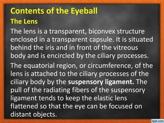
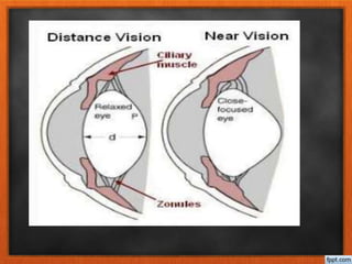
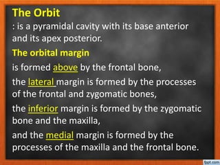
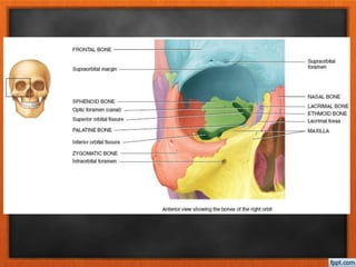
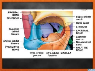
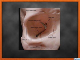
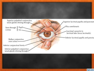
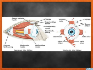
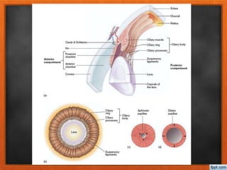
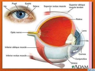
Orbital Region
Dr. Haydar Muneer Salih
The Orbital Region
The orbits are a pair of bony cavities that
contain the eyeballs; their associated
muscles, nerves, vessels, and fat; and most
of the lacrimal apparatus. The orbital
opening is guarded by two thin, movable
folds, the eyelids.
The Orbit
: is a pyramidal cavity with its base anterior
and its apex posterior.
The orbital margin
is formed above by the frontal bone,
the lateral margin is formed by the processes
of the frontal and zygomatic bones,
the inferior margin is formed by the zygomatic
bone and the maxilla,
and the medial margin is formed by the
processes of the maxilla and the frontal bone.
Roof: Formed by the orbital plate of the frontal
bone, which separates the orbital cavity from
the anterior cranial fossa
Lateral wall: Formed by the zygomatic bone and
the greater wing of the sphenoid
Floor: Formed by the orbital plate of the
maxilla, which separates the orbital cavity from
the maxillary sinus
Medial wall: Formed from by the frontal
process of the maxilla, the lacrimal bone, the
orbital plate of the ethmoid and the body of the
sphenoid Openings into the Orbital Cavity
Orbital opening:
About one sixth of the eye is exposed; the
remainder is protected by the walls of the orbit.
Supraorbital notch (Foramen): situated on the
superior orbital margin.
Infraorbital groove and canal: Situated on the
floor of the orbit in the orbital plate of the
maxilla;
Nasolacrimal canal: Located anteriorly on the
medial wall; it communicates with the inferior
meatus of the nose. It transmits the
nasolacrimal duct.
Inferior orbital fissure: Located posteriorly
between the maxilla and the greater wing of the
sphenoid; it communicates with the
pterygopalatine fossa. It transmits the maxillary
nerve and its zygomatic branch, the inferior
ophthalmic vein, and sympathetic nerves.
Superior orbital fissure: Located posteriorly
between the greater and lesser wings of the
sphenoid; it communicates with the middle
cranial fossa. It transmits the trigeminal V1, the
trochlear nerve, the oculomotor nerve, the
abducent nerve, the superior ophthalmic vein.
Optic canal: Located posteriorly in the
lesser wing of the sphenoid ; it
communicates with the middle cranial
fossa. It transmits the optic nerve and the
ophthalmic artery.
Nerves of the Orbit
1. Optic Nerve
2. Lacrimal Nerve
3. Frontal Nerve
4. Trochlear Nerve
5. Oculomotor Nerve
6. Nasociliary Nerve
7. Abducent Nerve
Blood Vessels and Lymph Vessels of the Orbit
Ophthalmic Artery
The ophthalmic artery is a branch of the
internal carotid artery after that vessel
emerges from the cavernous sinus. It enters
the orbit through the optic canal with the
optic nerve. It runs forward and crosses the
optic nerve to reach the medial wall of the
orbit. It gives off numerous branches, which
accompany the nerves in the orbital cavity.
Ophthalmic Veins
The superior ophthalmic vein communicates in
front with the facial vein.
The inferior ophthalmic vein communicates
through the inferior orbital fissure with the
pterygoid venous plexus. Both veins pass
backward through the superior orbital fissure
and drain into the cavernous sinus.
Lymph Vessels
No lymph vessels or nodes are present in the
orbital cavity.
Fascial Sheath of the Eyeball
• It separates the eyeball from the orbital fat
and provides it with a socket for free
movement.
• The lower part of the fascial sheath, which
passes beneath the eyeball and connects
the check ligaments, is thickened and serves
to suspend the eyeball; it is called the
suspensory ligament of the eye.
By this means, the eye is suspended from the
medial and lateral walls of the orbit, as if in a
hammock.
The Eyelid
• The eyelids protect the eye from injury and
excessive light by their closure. The upper
eyelid is larger and more mobile than the
lower, and they meet each other at the
medial and lateral angles
• The superficial surface of the eyelids is
covered by skin, and the deep surface is
covered by a mucous membrane called the
conjunctiva.
The conjunctiva is a thin mucous
membrane that lines the eyelids and is
reflected at the superior and inferior
fornices onto the anterior surface of the
eyeball. Its epithelium is continuous with
that of the cornea. The upper lateral part
of the superior fornix is pierced by the
ducts of the lacrimal gland. The
conjunctiva thus forms a potential space,
the conjunctival sac, which is open at the
palpebral fissure.
• The framework of the eyelids is formed by a
fibrous sheet, the orbital septum. This is
attached to the periosteum at the orbital
margins. The orbital septum is thickened at
the margins of the lids to form the superior
and inferior tarsal plates.
• The lateral ends of the plates are attached
by a band, the lateral palpebral ligament,
to a bony tubercle just within the orbital
margin. The medial ends of the plates are
attached by a band, the medial palpebral
ligament, to the crest of the lacrimal bone.
Movements of the Eyelids
The position of the eyelids at rest depends on
the tone of the orbicularis oculi and the
levator palpebrae superioris muscles and the
position of the eyeball.
The eyelids are closed by the contraction of
the orbicularis oculi and the relaxation of the
levator palpebrae superioris muscles.
The eye is opened by the levator palpebrae
superioris raising the upper lid.
Muscles of orbital region
A. Muscles of eyeball
1. Extrinsic muscles:
Superior rectus
Inferior rectus
Medial rectus
Lateral rectus
Superior oblique
Inferior oblique
Muscles of orbital region
A. Muscles of eyeball
2. Intrinsic muscles:
Sphincter pupillae of iris
Dilator pupillae of iris
Ciliary muscle
B. Muscles of eyelid
Structure of the Eye
The eyeball is embedded in orbital fat but
is separated from it by the fascial sheath of
the eyeball. The eyeball consists of three
coats, which, from without inward, are the
fibrous coat, the vascular pigmented coat,
and the nervous coat.
1. Fibrous Coat
The Sclera
The opaque sclera is composed of dense fibrous
tissue and is white. Posteriorly, it is pierced by
the optic nerve and is fused with the dural
sheath of that nerve. At the lamina cribrosa
The sclera is also pierced by the ciliary arteries
and nerves and their associated veins, the
venae vorticosae. The sclera is directly
continuous in front with the cornea at the
corneoscleral junction, or limbus.
1. Fibrous Coat
The Cornea
The transparent cornea is largely responsible
for the refraction of the light entering the eye.
It is in contact posteriorly with the aqueous
humor.
The cornea is avascular and alymphatic. It is
nourished by diffusion from the aqueous
humor and from the capillaries at its edge.
Nerve Supply Long ciliary nerves from the
ophthalmic division of the trigeminal nerve
Function of the Cornea
The cornea is the most important refractive
medium of the eye.
2. Vascular Pigmented Coat
The Choroid
The choroid is composed of an outer
pigmented layer and an inner, highly vascular
layer.
The Ciliary Body
is continuous posteriorly with the choroid, and
anteriorly it lies behind the peripheral margin
of the iris. It is composed of the ciliary ring,
the ciliary processes, and the ciliary muscle.
2. Vascular Pigmented Coat
The Iris and Pupil
The iris is a thin, contractile, pigmented
diaphragm with a central aperture, the pupil.
It is suspended in the aqueous humor
between the cornea and the lens. The
periphery of the iris is attached to the anterior
surface of the ciliary body. It divides the space
between the lens and the cornea into an
anterior and a posterior chamber.
3. Nervous Coat: The Retina
The retina consists of an outer pigmented
layer and an inner nervous layer. Its outer
surface is in contact with the choroid, and its
inner surface is in contact with the vitreous
body.
The optic nerve leaves the retina about 3 mm
to the medial side of the macula lutea by the
optic disc. The optic disc is slightly depressed
at its center, where it is pierced by the central
artery of the retina.
Contents of the Eyeball
Aqueous Humor
• The aqueous humor is a clear fluid that fills
the anterior and posterior chambers of the
eyeball. It is believed to be a secretion from
the ciliary processes, Obstruction to the
draining of the aqueous humor results in a
rise in intraocular pressure called glaucoma.
• The function of the aqueous humor is to
support the wall of the eyeball by exerting
internal pressure and thus maintaining its
optical shape
Contents of the Eyeball
Vitreous Body
The vitreous body fills the eyeball behind
the lens and is a transparent gel.
The function of the vitreous body is to
contribute slightly to the magnifying
power of the eye. It supports the posterior
surface of the lens and assists in holding
the neural part of the retina against the
pigmented part of the retina.
Contents of the Eyeball
The Lens
The lens is a transparent, biconvex structure
enclosed in a transparent capsule. It is situated
behind the iris and in front of the vitreous
body and is encircled by the ciliary processes.
The equatorial region, or circumference, of the
lens is attached to the ciliary processes of the
ciliary body by the suspensory ligament. The
pull of the radiating fibers of the suspensory
ligament tends to keep the elastic lens
flattened so that the eye can be focused on
distant objects.
Orbital region
Orbital region
Orbital region
Orbital region

Orbital region

  • 1.
  • 2.
    The Orbital Region Theorbits are a pair of bony cavities that contain the eyeballs; their associated muscles, nerves, vessels, and fat; and most of the lacrimal apparatus. The orbital opening is guarded by two thin, movable folds, the eyelids.
  • 4.
    The Orbit : isa pyramidal cavity with its base anterior and its apex posterior. The orbital margin is formed above by the frontal bone, the lateral margin is formed by the processes of the frontal and zygomatic bones, the inferior margin is formed by the zygomatic bone and the maxilla, and the medial margin is formed by the processes of the maxilla and the frontal bone.
  • 6.
    Roof: Formed bythe orbital plate of the frontal bone, which separates the orbital cavity from the anterior cranial fossa Lateral wall: Formed by the zygomatic bone and the greater wing of the sphenoid Floor: Formed by the orbital plate of the maxilla, which separates the orbital cavity from the maxillary sinus Medial wall: Formed from by the frontal process of the maxilla, the lacrimal bone, the orbital plate of the ethmoid and the body of the sphenoid Openings into the Orbital Cavity
  • 8.
    Orbital opening: About onesixth of the eye is exposed; the remainder is protected by the walls of the orbit. Supraorbital notch (Foramen): situated on the superior orbital margin. Infraorbital groove and canal: Situated on the floor of the orbit in the orbital plate of the maxilla; Nasolacrimal canal: Located anteriorly on the medial wall; it communicates with the inferior meatus of the nose. It transmits the nasolacrimal duct.
  • 10.
    Inferior orbital fissure:Located posteriorly between the maxilla and the greater wing of the sphenoid; it communicates with the pterygopalatine fossa. It transmits the maxillary nerve and its zygomatic branch, the inferior ophthalmic vein, and sympathetic nerves. Superior orbital fissure: Located posteriorly between the greater and lesser wings of the sphenoid; it communicates with the middle cranial fossa. It transmits the trigeminal V1, the trochlear nerve, the oculomotor nerve, the abducent nerve, the superior ophthalmic vein.
  • 12.
    Optic canal: Locatedposteriorly in the lesser wing of the sphenoid ; it communicates with the middle cranial fossa. It transmits the optic nerve and the ophthalmic artery.
  • 13.
    Nerves of theOrbit 1. Optic Nerve 2. Lacrimal Nerve 3. Frontal Nerve 4. Trochlear Nerve 5. Oculomotor Nerve 6. Nasociliary Nerve 7. Abducent Nerve
  • 15.
    Blood Vessels andLymph Vessels of the Orbit Ophthalmic Artery The ophthalmic artery is a branch of the internal carotid artery after that vessel emerges from the cavernous sinus. It enters the orbit through the optic canal with the optic nerve. It runs forward and crosses the optic nerve to reach the medial wall of the orbit. It gives off numerous branches, which accompany the nerves in the orbital cavity.
  • 18.
    Ophthalmic Veins The superiorophthalmic vein communicates in front with the facial vein. The inferior ophthalmic vein communicates through the inferior orbital fissure with the pterygoid venous plexus. Both veins pass backward through the superior orbital fissure and drain into the cavernous sinus. Lymph Vessels No lymph vessels or nodes are present in the orbital cavity.
  • 20.
    Fascial Sheath ofthe Eyeball • It separates the eyeball from the orbital fat and provides it with a socket for free movement. • The lower part of the fascial sheath, which passes beneath the eyeball and connects the check ligaments, is thickened and serves to suspend the eyeball; it is called the suspensory ligament of the eye. By this means, the eye is suspended from the medial and lateral walls of the orbit, as if in a hammock.
  • 22.
    The Eyelid • Theeyelids protect the eye from injury and excessive light by their closure. The upper eyelid is larger and more mobile than the lower, and they meet each other at the medial and lateral angles • The superficial surface of the eyelids is covered by skin, and the deep surface is covered by a mucous membrane called the conjunctiva.
  • 24.
    The conjunctiva isa thin mucous membrane that lines the eyelids and is reflected at the superior and inferior fornices onto the anterior surface of the eyeball. Its epithelium is continuous with that of the cornea. The upper lateral part of the superior fornix is pierced by the ducts of the lacrimal gland. The conjunctiva thus forms a potential space, the conjunctival sac, which is open at the palpebral fissure.
  • 26.
    • The frameworkof the eyelids is formed by a fibrous sheet, the orbital septum. This is attached to the periosteum at the orbital margins. The orbital septum is thickened at the margins of the lids to form the superior and inferior tarsal plates. • The lateral ends of the plates are attached by a band, the lateral palpebral ligament, to a bony tubercle just within the orbital margin. The medial ends of the plates are attached by a band, the medial palpebral ligament, to the crest of the lacrimal bone.
  • 28.
    Movements of theEyelids The position of the eyelids at rest depends on the tone of the orbicularis oculi and the levator palpebrae superioris muscles and the position of the eyeball. The eyelids are closed by the contraction of the orbicularis oculi and the relaxation of the levator palpebrae superioris muscles. The eye is opened by the levator palpebrae superioris raising the upper lid.
  • 30.
    Muscles of orbitalregion A. Muscles of eyeball 1. Extrinsic muscles: Superior rectus Inferior rectus Medial rectus Lateral rectus Superior oblique Inferior oblique
  • 33.
    Muscles of orbitalregion A. Muscles of eyeball 2. Intrinsic muscles: Sphincter pupillae of iris Dilator pupillae of iris Ciliary muscle B. Muscles of eyelid
  • 35.
    Structure of theEye The eyeball is embedded in orbital fat but is separated from it by the fascial sheath of the eyeball. The eyeball consists of three coats, which, from without inward, are the fibrous coat, the vascular pigmented coat, and the nervous coat.
  • 36.
    1. Fibrous Coat TheSclera The opaque sclera is composed of dense fibrous tissue and is white. Posteriorly, it is pierced by the optic nerve and is fused with the dural sheath of that nerve. At the lamina cribrosa The sclera is also pierced by the ciliary arteries and nerves and their associated veins, the venae vorticosae. The sclera is directly continuous in front with the cornea at the corneoscleral junction, or limbus.
  • 38.
    1. Fibrous Coat TheCornea The transparent cornea is largely responsible for the refraction of the light entering the eye. It is in contact posteriorly with the aqueous humor. The cornea is avascular and alymphatic. It is nourished by diffusion from the aqueous humor and from the capillaries at its edge. Nerve Supply Long ciliary nerves from the ophthalmic division of the trigeminal nerve Function of the Cornea The cornea is the most important refractive medium of the eye.
  • 40.
    2. Vascular PigmentedCoat The Choroid The choroid is composed of an outer pigmented layer and an inner, highly vascular layer. The Ciliary Body is continuous posteriorly with the choroid, and anteriorly it lies behind the peripheral margin of the iris. It is composed of the ciliary ring, the ciliary processes, and the ciliary muscle.
  • 42.
    2. Vascular PigmentedCoat The Iris and Pupil The iris is a thin, contractile, pigmented diaphragm with a central aperture, the pupil. It is suspended in the aqueous humor between the cornea and the lens. The periphery of the iris is attached to the anterior surface of the ciliary body. It divides the space between the lens and the cornea into an anterior and a posterior chamber.
  • 44.
    3. Nervous Coat:The Retina The retina consists of an outer pigmented layer and an inner nervous layer. Its outer surface is in contact with the choroid, and its inner surface is in contact with the vitreous body. The optic nerve leaves the retina about 3 mm to the medial side of the macula lutea by the optic disc. The optic disc is slightly depressed at its center, where it is pierced by the central artery of the retina.
  • 46.
    Contents of theEyeball Aqueous Humor • The aqueous humor is a clear fluid that fills the anterior and posterior chambers of the eyeball. It is believed to be a secretion from the ciliary processes, Obstruction to the draining of the aqueous humor results in a rise in intraocular pressure called glaucoma. • The function of the aqueous humor is to support the wall of the eyeball by exerting internal pressure and thus maintaining its optical shape
  • 48.
    Contents of theEyeball Vitreous Body The vitreous body fills the eyeball behind the lens and is a transparent gel. The function of the vitreous body is to contribute slightly to the magnifying power of the eye. It supports the posterior surface of the lens and assists in holding the neural part of the retina against the pigmented part of the retina.
  • 49.
    Contents of theEyeball The Lens The lens is a transparent, biconvex structure enclosed in a transparent capsule. It is situated behind the iris and in front of the vitreous body and is encircled by the ciliary processes. The equatorial region, or circumference, of the lens is attached to the ciliary processes of the ciliary body by the suspensory ligament. The pull of the radiating fibers of the suspensory ligament tends to keep the elastic lens flattened so that the eye can be focused on distant objects.