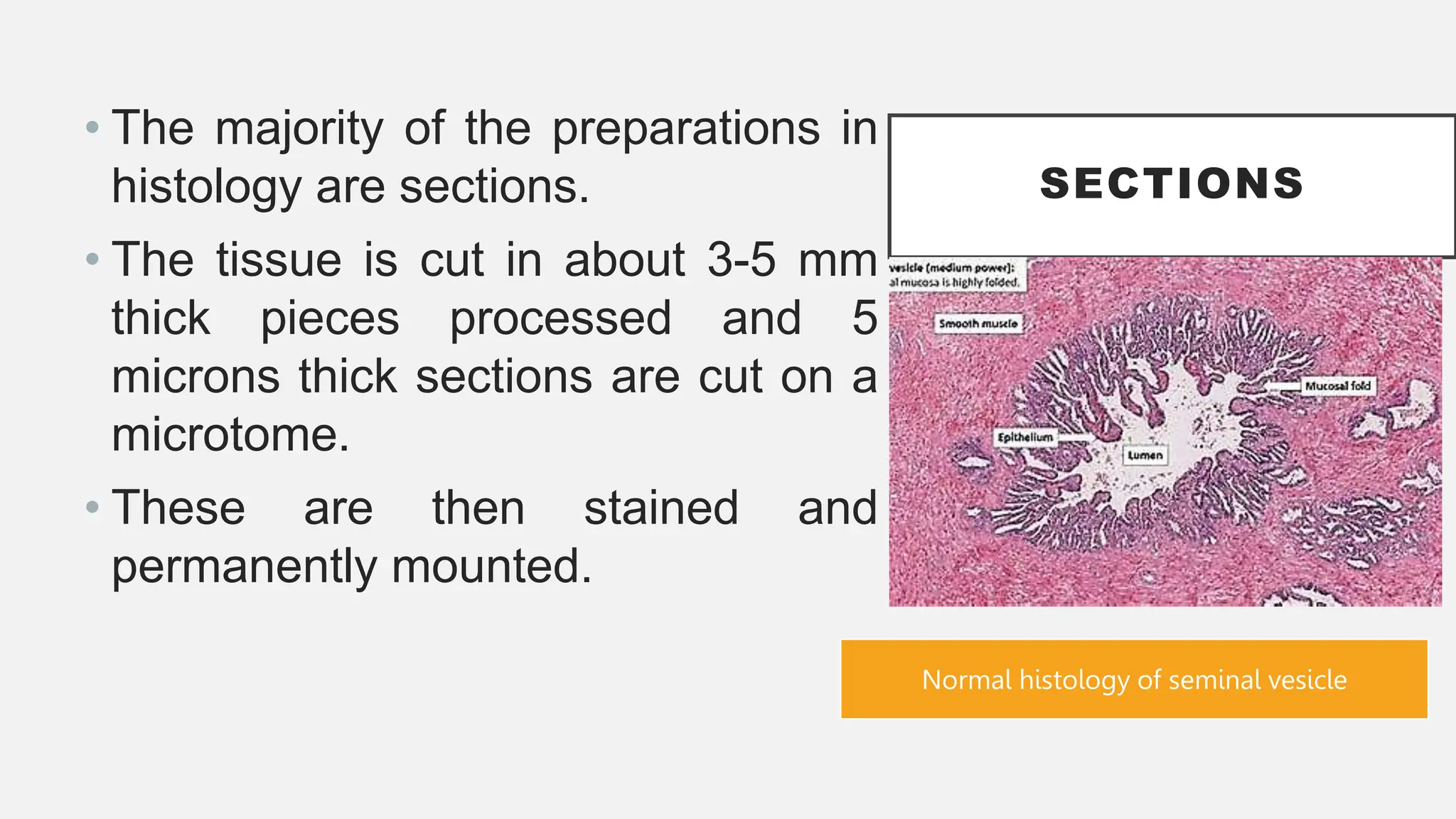
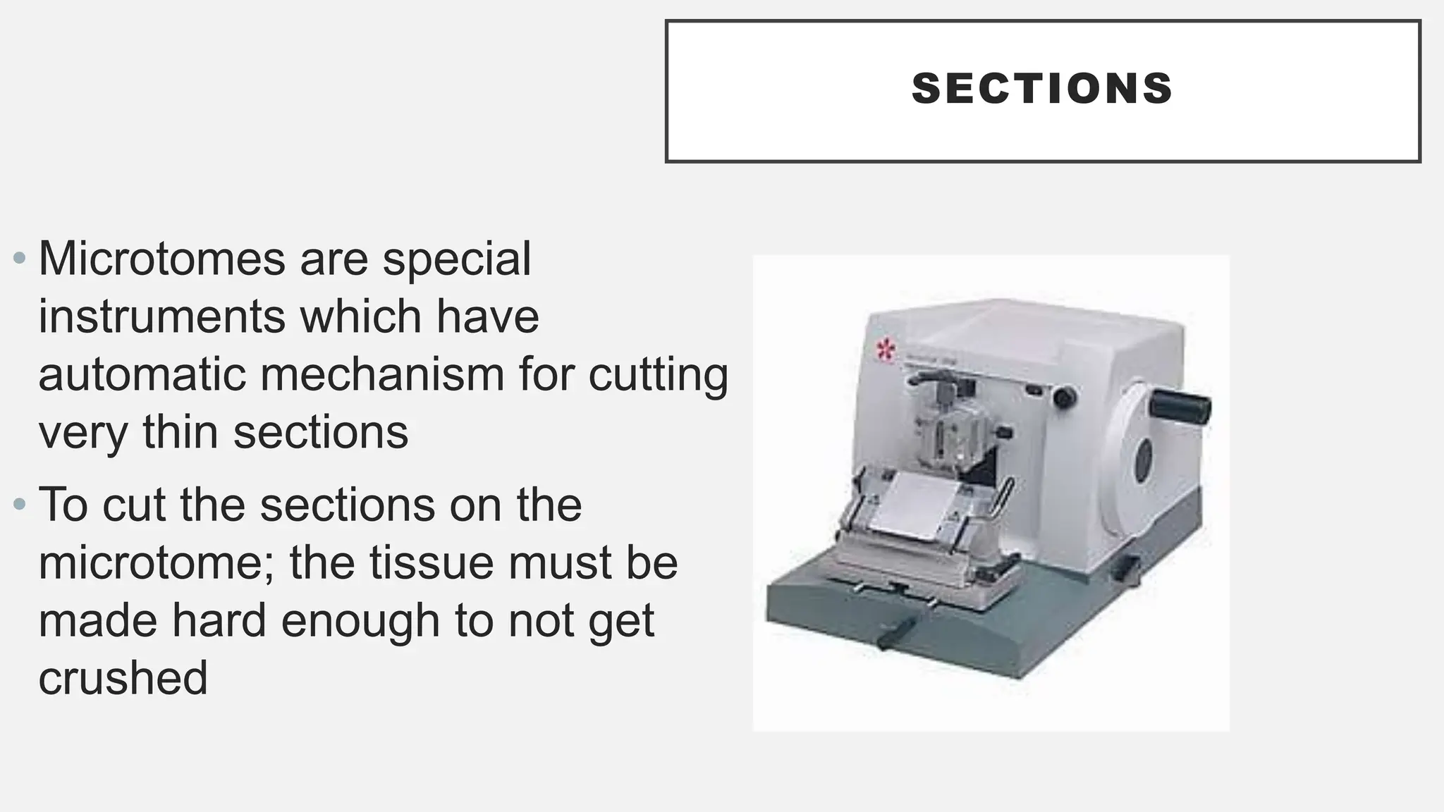
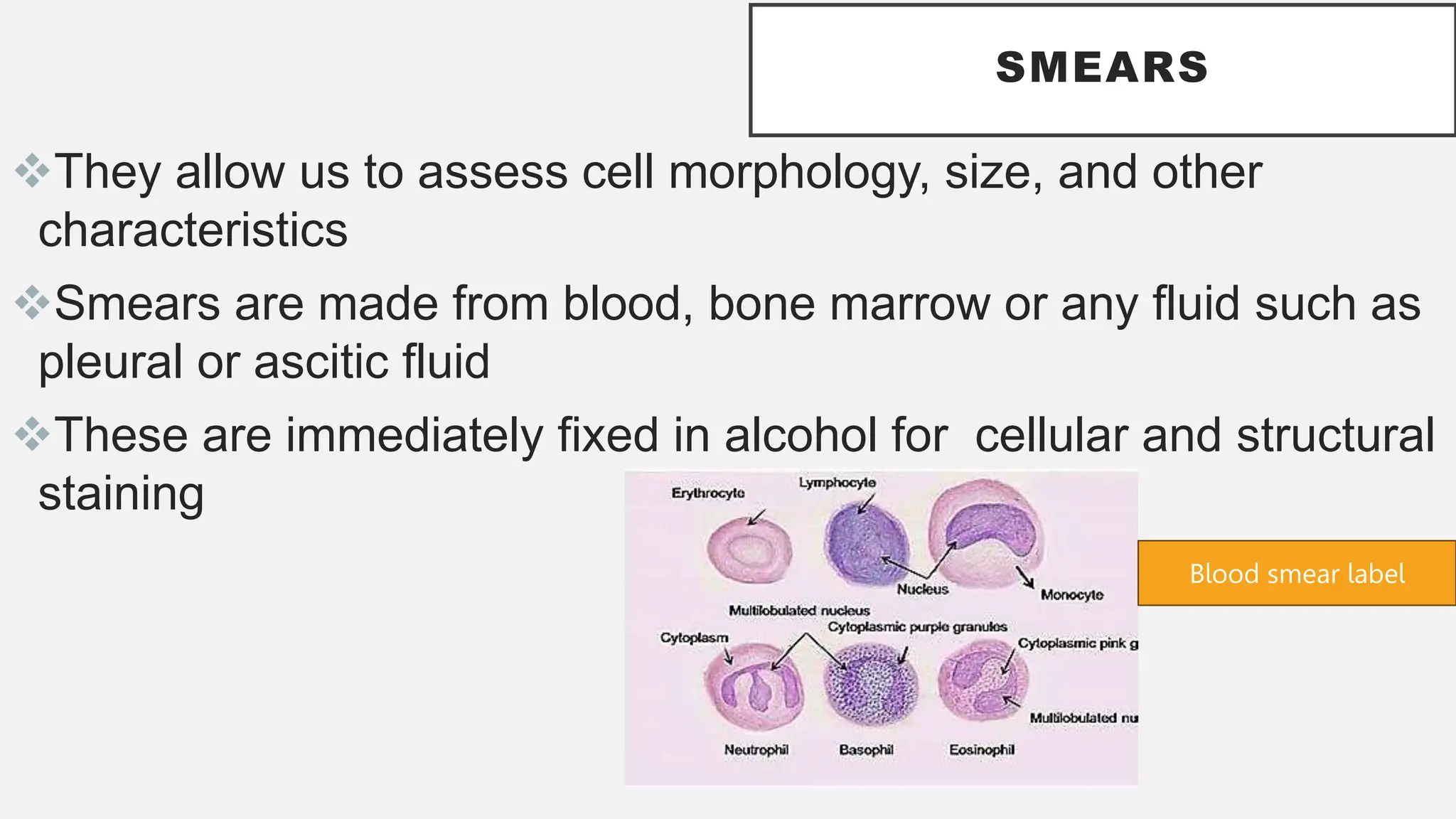
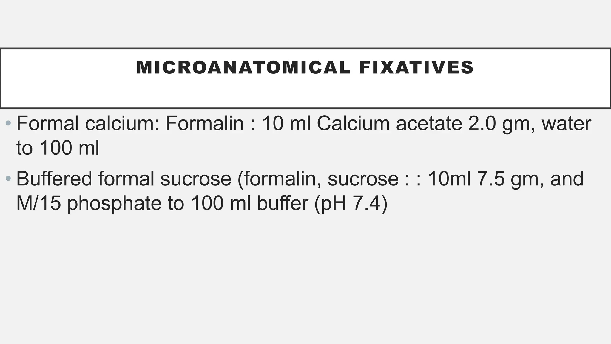
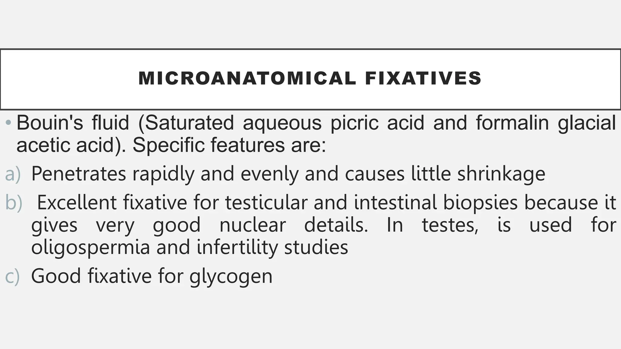
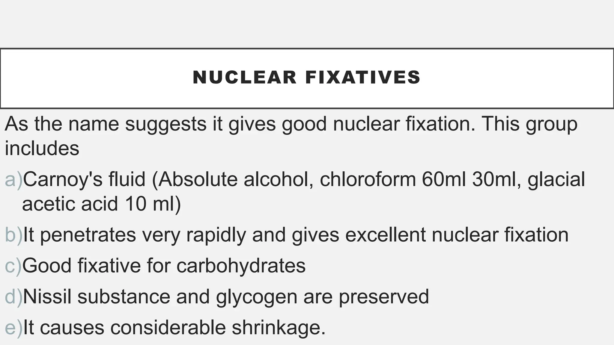
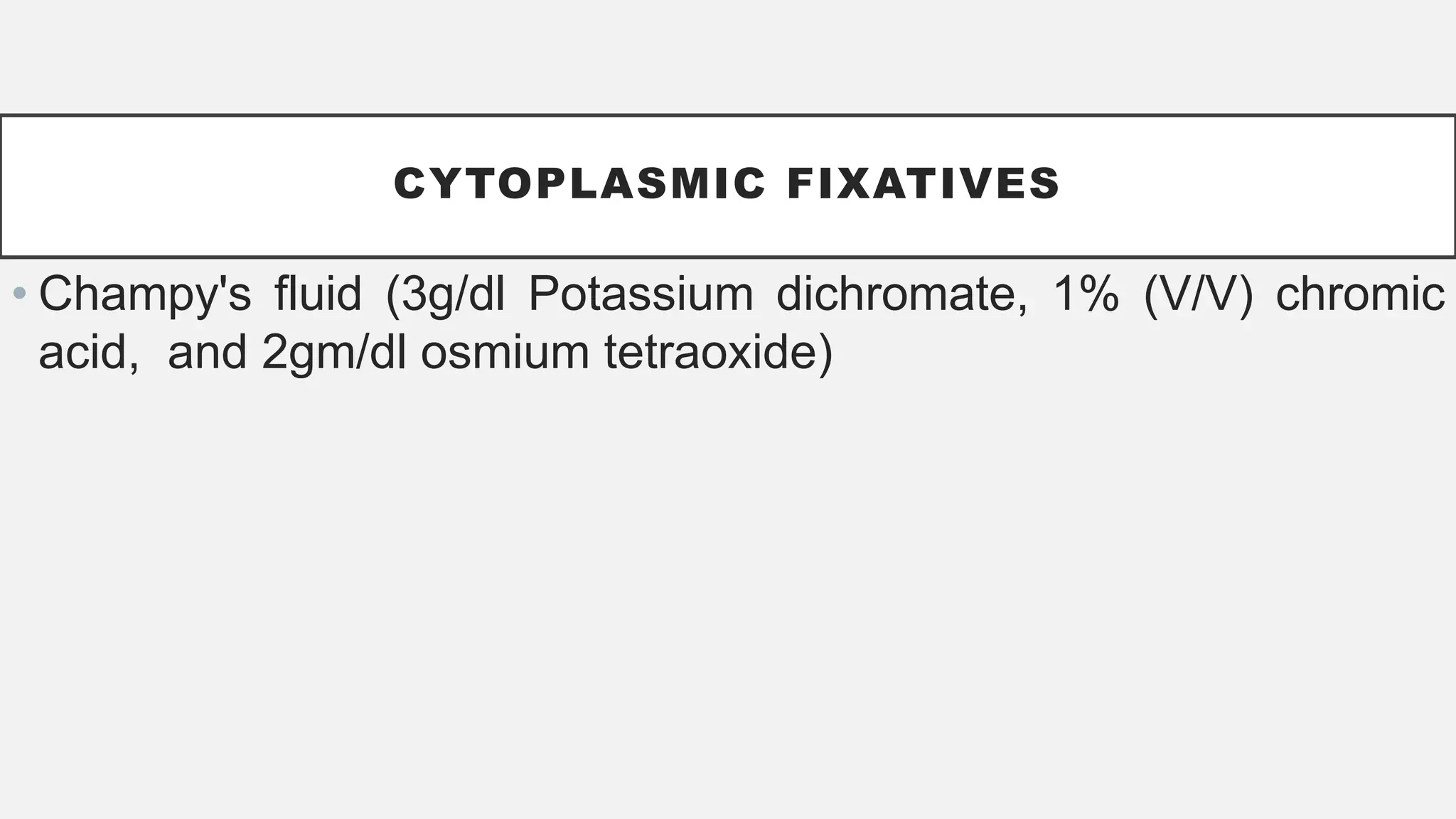
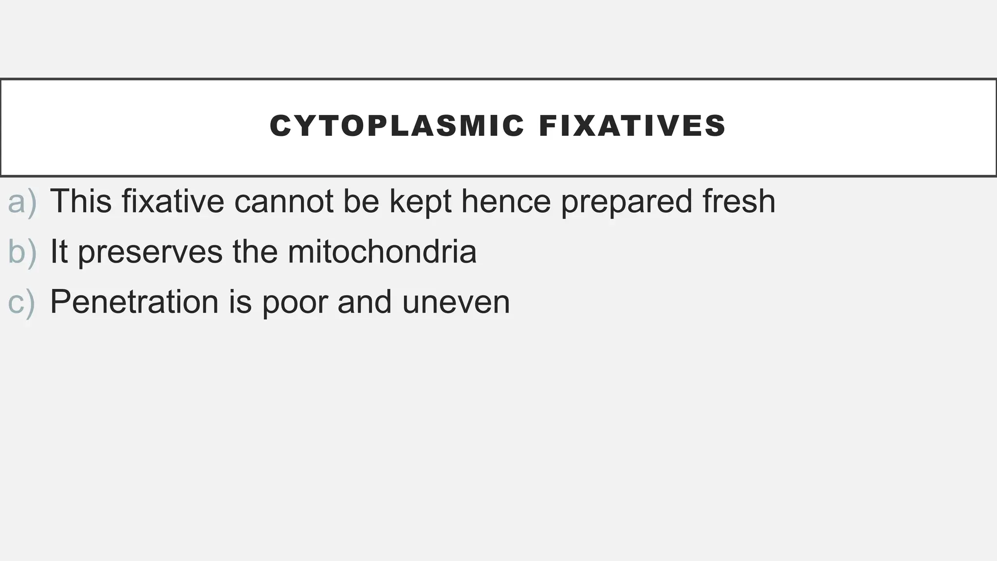
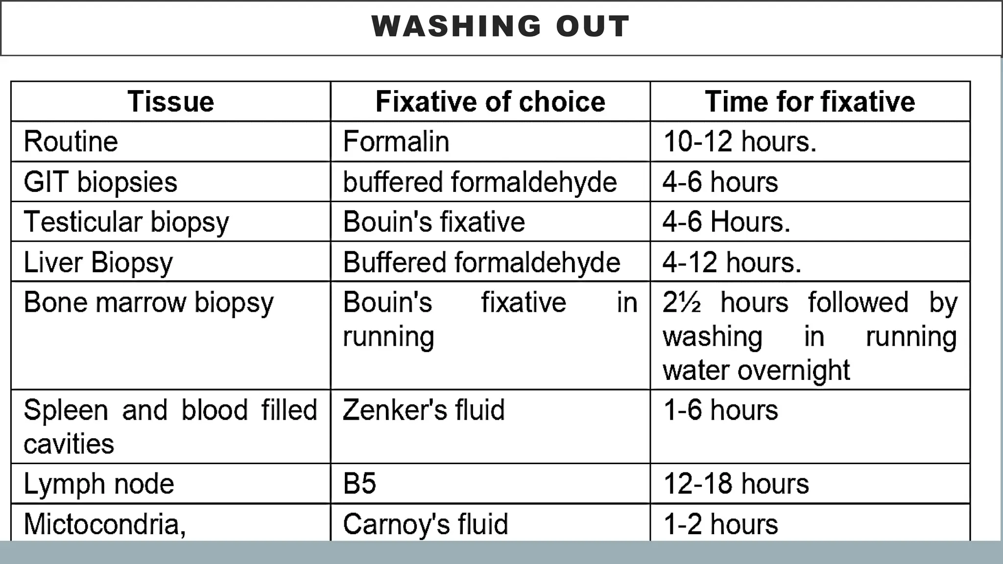
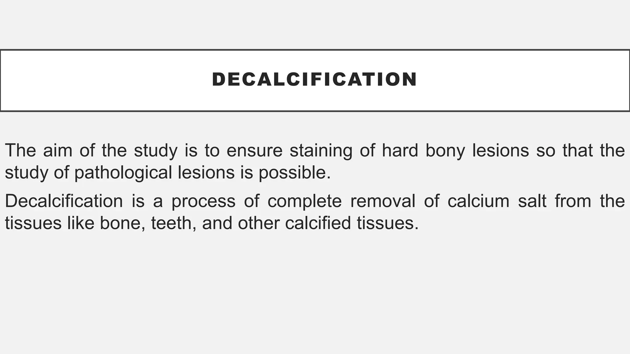
GENERAL INTRODUCTON AND BASIC TECHNIQUES
TO
HISTOPATHOLOGY
OBAJE Godwin Sunday
Department of Anatomy, Faculty of Basic Medical
Sciences
Alex Ekwueme Federal University Ndufu Alike, Nigeria
godwin.sunday@funai.edu.ng, +2348068638121
ANATOMY 361, AEFUNAI, NIGERIA, 2024
DEFINITION
• Histopathology, it’s a branch of
pathology which deals with the
study of disease in a tissue
section
Anatomical pathology
 The term histochemistry means study of chemical nature of the
tissue components by histological methods.
 The basic details of tissue handling, processing and staining
form the objective of histopathology.
Class work 01. Histopathology is a branch of
pathology which deals with the study of
disease in a tissue section but involves some
histological techniques. Discuss!
STRUCTURAL UNITS
IN
HISTOPATHOLOGY
• The cell is the single structural
unit of all tissues
• The study of cell is called cell
biology
• A tissue is a group of cells
specialized and differentiated to
perform a specialized function
• Collection of different type of cells
forms an organ
TYPES OF HISTOLOGICAL PREPARATION
Whole mount
Section
Smears.
WHOLE MOUNTS
• These are preparation of
entire animal eg. fungus,
parasite.
• These preparations should
be no more than 0.2-0.5 mm
in thickness.
Representative of H and E
stained whole mount of
interlobular duct
WHOLE MOUNT
• Refers to a technique where an entire specimen is stained and
examined without sectioning.
• Whole mount staining allows researchers to study the entire
structure in its natural three-dimensional form.
WHOLE MOUNT
Procedure is similar to
immunocytochemistry (ICC) or staining
of cryosections
Sample size is larger and thicker than
typical slide sections
Using fixation, commonly done with 4%
paraformaldehyde (PFA), but other
fixatives like methanol may be used
In permeability, longer incubations are
needed to allow permeabilization
throughout the sample.
SECTIONS
• The majority of the preparations in
histology are sections.
• The tissue is cut in about 3-5 mm
thick pieces processed and 5
microns thick sections are cut on a
microtome.
• These are then stained and
permanently mounted.
Normal histology of seminal vesicle
SECTIONS
• Microtomes are special
instruments which have
automatic mechanism for cutting
very thin sections
• To cut the sections on the
microtome; the tissue must be
made hard enough to not get
crushed
SECTIONS
• 2 methods of hardening the
tissues
• One is by freezing them and the
other is by embedding them in a
hard material such at paraffin
wax or gelatin.
SMEARS
They allow us to assess cell morphology, size, and other
characteristics
Smears are made from blood, bone marrow or any fluid such as
pleural or ascitic fluid
These are immediately fixed in alcohol for cellular and structural
staining
Blood smear label
SMEARS
• Smears are also made by crushing soft tissue between two
slides or an impression made by pressing a clean slide in
contact with the moist surface of a tissue
• By doing this, the cells are imprinted on the slide and these
may be stained for cytological examination
PRINCIPLE OF FIXATION
FIXATION
It is a complex series of chemical events which brings about
changes in the various chemical constituents of cell. The cell
morphology and structural detail is preserved
Unless a tissue is fixed soon after the removal from the body it
will undergo degenerative changes due to autolysis and
putrefaction
AIMS AND EFFECTS OF FIXATION
• To preserve the tissue
• To prevent postmortem changes
• Preservation of chemical compounds of tissue
• The hardening effect of fixatives allows easy manipulation of soft
tissue like brain and intestines
PROPERTIES OF FIXATIVES
• Coagulation and precipitation
• Penetration
• Solubility of fixatives
• Concentration (isotonic or hypotonic)
• Reaction (, like most preservatives are acidic)
AMOUNT OF FIXATIVE
• Depend on the thickness of tissue (1.5mm thick), the fixative
should be atleast 15-20 times the bulk of tissue. However, for
museum specimens the volume of fixative is > 50 times.
REAGENTS EMPLOYED AS FIXATIVES (SIMPLE
FIXATIVES)
• Formaldehyde is a gas but is soluble in water to the extent of 37-
40% w/v. This solution of formaldehyde in water is called formalin
or full strength formalin.
• Formalin is one of the commonly used fixative in all laboratories
since it is cheap penetrates rapidly and does not over harden the
tissues.
• It preserves the proteins by forming crosslinkage with them and
the tissue component. • It denatures the proteins.
Formaldehyde
REAGENTS EMPLOYED AS FIXATIVES (SIMPLE
FIXATIVES)
• Glycogen is partially preserved hence formalin is not a fixative
choice for carbohydrates
• It neither preserves nor destroys fat
• Artifacts are formed by by-product of formalin (formic acid, which
is brown pigment) after oxidation. To remove artifacts, sodium
hydroxide is used.
• Formalin on prolonged exposure can cause either dermatitis. Its
vapour may damage the nasal mucosa and cause sinusitis
ALCOHOL (ETHYL ALCOHOL)
• It acts as a reducing agents
• Become oxidized to acetaldehyde and then to acetic acid
• It is slow to penetrate, hardens and shrinks the tissue
• Ethanol preserves some proteins (like in immunoflourence)
• It is a fat solvent hence it dissolve fats and lipids
• Methyl alcohol is used for fixing blood and bone marrow smears
ACETONE
• Cold acetone is sometimes used as a fixative for the histochemical
demonstration of some tissue enzymes like phosphatases and lipases
• Its mode of action as fixative is similar to that of alcohol
MERCURIC CHLORIDE
(HGCL2)
• Good salt employed in fixing but is rarely used alone because it causes
shrinkage of the tissue.
• It brings about precipitation of the proteins. However, it can be removed by
potassium iodide
• It penetrates rapidly without destroying lipids
• It neither fixes nor destroys carbohydrates.
PICRIC ACID
• It produces marked cells shrinkage. Hence it is not used alone
• Stored in a damp place because of its explosive nature
• Preferably stored under a layer of water
• It penetrates well and fixes rapidly
POTASSIUM DICHROMATE
• It fixes the cytoplasm without precipitation
• Used for fixing phosphatides and mitochondria
• It is a strong oxidizing agent and brings about fixation by forming
cross links with proteins
• It gives excellent preservation of details of a cell, therefore
exclusively used for electron microscopy
• It fixes fat e.g. myelin
• It also demonstrates fat when 0.5-2% aqueous solution gives a
black colour to fat
OSIMIUM TETRAOXIDE
ACETIC ACID
• It causes the cells to swell hence can never be used alone but
should be used with fixatives causing cell shrinkage
GLUTARADEHYDE
• It is used alone or in combination with osimium tetroxide for
electron microscopy.
PREPARATION OF THE SPECIMEN
FOR FIXATION
A. For achieving good fixation it is important that the fixative
penetrates the tissue well.
• Hence the tissue section should be > 4mm thick, so that fixation
fluid penetrates from the periphery to the centre of the tissue.
• For hollow viscera fixative is injected into the cavity e.g. urinary
bladder, eyeball etc.
PREPARATION OF THE SPECIMEN
FOR FIXATION
B. Ratio of volume of fixative to the specimen should be 1:20
C. Time necessary for fixation is important. Routinely, 10%
aqueous formalin at room temperature takes 12 hours to fix the
tissue
FIXATIVES ARE DIVIDED INTO THREE MAIN GROUPS
Microanatomical fixatives - such fixatives preserves the anatomy
of the tissue
Cytological fixatives - such fixation are used to preserve
intracellular structures or inclusion
Histochemical fixatives : Fixative used to preserve the chemical
nature of the tissue
MICROANATOMICAL FIXATIVES
• 10% (v/v) formalin in 0.9% sodium chloride (normal saline)
• Buffered formation (formalin 10ml, acid sodium phosphate - 0.4
gm (monohydrate), anhydrous disodium – phosphate, and water
to 100 ml)
MICROANATOMICAL FIXATIVES
• Formal calcium: Formalin : 10 ml Calcium acetate 2.0 gm, water
to 100 ml
• Buffered formal sucrose (formalin, sucrose : : 10ml 7.5 gm, and
M/15 phosphate to 100 ml buffer (pH 7.4)
MICROANATOMICAL FIXATIVES
• Bouin's fluid (Saturated aqueous picric acid and formalin glacial
acetic acid). Specific features are:
a) Penetrates rapidly and evenly and causes little shrinkage
b) Excellent fixative for testicular and intestinal biopsies because it
gives very good nuclear details. In testes, is used for
oligospermia and infertility studies
c) Good fixative for glycogen
CYTOLOGICAL FIXATIVES
• Subdivided into 2: Nuclear fixatives and cytoplasmic fixatives
NUCLEAR FIXATIVES
As the name suggests it gives good nuclear fixation. This group
includes
a)Carnoy's fluid (Absolute alcohol, chloroform 60ml 30ml, glacial
acetic acid 10 ml)
b)It penetrates very rapidly and gives excellent nuclear fixation
c)Good fixative for carbohydrates
d)Nissil substance and glycogen are preserved
e)It causes considerable shrinkage.
CYTOPLASMIC FIXATIVES
• Champy's fluid (3g/dl Potassium dichromate, 1% (V/V) chromic
acid, and 2gm/dl osmium tetraoxide)
CYTOPLASMIC FIXATIVES
a) This fixative cannot be kept hence prepared fresh
b) It preserves the mitochondria
c) Penetration is poor and uneven
WASHING OUT
DECALCIFICATION
The aim of the study is to ensure staining of hard bony lesions so that the
study of pathological lesions is possible.
Decalcification is a process of complete removal of calcium salt from the
tissues like bone, teeth, and other calcified tissues.
PREPARED SOLUTION FOR DECALCIFICATION
• Nitric acid- 5-10% aqueous solution used
• Hydrochloric acid - 5-10% aqueous solution
• Weak acid e.g. formic, acetic and picric acid of these formic acids
are extensively used as acid decalcifier.
TISSUE PROCESSING
• The term tissue processing refers to treatment of the tissue
necessary to impregnate it into a solid medium so that the tissue
is rendered sufficiently firm yet elastic for the tissue sections of
desirable thickness to be cut on microtome.
TISSUE PROCESSING
• Tissue must be sectioned
• Processed tissue must be properly stored
• Must be properly labelled
PRINCIPLE OF TISSUE PROCESSING
• Removal of water from the tissue (dehydration) and replaced with
paraffin wax (embedding)
• The embedding medium must be permeable for easy
solidification of tissue without damage to tissue
STEPS
• Fixation
• Dehydration
• Clearing - with a substance which is totally miscible with both the
dehydrating agent
• Embedding
FACTORS INFLUENCING THE RATE OF IMPREGNATION
Agitation - Tissue placed in liquid is agitated so that the fluid
immediately in contact with the surface of tissue is replaced by the
fresh immersing liquid
• Efficient agitation reduces the processing time by 25-30% with
improved impregnation of the tissue
FACTORS INFLUENCING THE RATE OF IMPREGNATION
• Heat - Heat increases the rate of penetration
• Viscosity - Larger the molecule the higher is the viscosity slower
is the rate of penetration
FACTORS INFLUENCING THE RATE OF IMPREGNATION
• Ultrasonic : Use of ultrasonics increases the penetration rate
• Vacuum : Use of reduced pressure in well known in the
impregnation of tissue by molten paraffin wax
CLEARING
• Clearing agents: Xylene, Toluene Blue, benzene, chloroform,
carbon tetrachloride, and Cedar wood oil
PARAPLAST
• This is mixture of highly purified paraffin and several plastic
polymers
• It has greater elasticity than normal paraffin wax, therefore, the
results are superior
• It should not be used for thin walled structures as it prevents
complete expansion of the specimen
AUTOMATIC TISSUE PROCESSOR
It has 2 advantages
a)Transferring the tissue mechanically from one reagent to another
can be done both by day and night
b)Reduces processing time by the action of continuous agitation.
c)This eliminates the possibility of human errors of leaving the
tissue for long time in one solution due to forgetfulness
THANKS!!!!!!!!!!!
General views of Histopathology and step

General views of Histopathology and step

  • 1.
    GENERAL INTRODUCTON ANDBASIC TECHNIQUES TO HISTOPATHOLOGY OBAJE Godwin Sunday Department of Anatomy, Faculty of Basic Medical Sciences Alex Ekwueme Federal University Ndufu Alike, Nigeria godwin.sunday@funai.edu.ng, +2348068638121 ANATOMY 361, AEFUNAI, NIGERIA, 2024
  • 2.
    DEFINITION • Histopathology, it’sa branch of pathology which deals with the study of disease in a tissue section Anatomical pathology
  • 3.
     The termhistochemistry means study of chemical nature of the tissue components by histological methods.  The basic details of tissue handling, processing and staining form the objective of histopathology. Class work 01. Histopathology is a branch of pathology which deals with the study of disease in a tissue section but involves some histological techniques. Discuss!
  • 4.
    STRUCTURAL UNITS IN HISTOPATHOLOGY • Thecell is the single structural unit of all tissues • The study of cell is called cell biology • A tissue is a group of cells specialized and differentiated to perform a specialized function • Collection of different type of cells forms an organ
  • 5.
    TYPES OF HISTOLOGICALPREPARATION Whole mount Section Smears.
  • 6.
    WHOLE MOUNTS • Theseare preparation of entire animal eg. fungus, parasite. • These preparations should be no more than 0.2-0.5 mm in thickness. Representative of H and E stained whole mount of interlobular duct
  • 7.
    WHOLE MOUNT • Refersto a technique where an entire specimen is stained and examined without sectioning. • Whole mount staining allows researchers to study the entire structure in its natural three-dimensional form.
  • 8.
    WHOLE MOUNT Procedure issimilar to immunocytochemistry (ICC) or staining of cryosections Sample size is larger and thicker than typical slide sections Using fixation, commonly done with 4% paraformaldehyde (PFA), but other fixatives like methanol may be used In permeability, longer incubations are needed to allow permeabilization throughout the sample.
  • 9.
    SECTIONS • The majorityof the preparations in histology are sections. • The tissue is cut in about 3-5 mm thick pieces processed and 5 microns thick sections are cut on a microtome. • These are then stained and permanently mounted. Normal histology of seminal vesicle
  • 10.
    SECTIONS • Microtomes arespecial instruments which have automatic mechanism for cutting very thin sections • To cut the sections on the microtome; the tissue must be made hard enough to not get crushed
  • 11.
    SECTIONS • 2 methodsof hardening the tissues • One is by freezing them and the other is by embedding them in a hard material such at paraffin wax or gelatin.
  • 12.
    SMEARS They allow usto assess cell morphology, size, and other characteristics Smears are made from blood, bone marrow or any fluid such as pleural or ascitic fluid These are immediately fixed in alcohol for cellular and structural staining Blood smear label
  • 13.
    SMEARS • Smears arealso made by crushing soft tissue between two slides or an impression made by pressing a clean slide in contact with the moist surface of a tissue • By doing this, the cells are imprinted on the slide and these may be stained for cytological examination
  • 14.
  • 15.
    FIXATION It is acomplex series of chemical events which brings about changes in the various chemical constituents of cell. The cell morphology and structural detail is preserved Unless a tissue is fixed soon after the removal from the body it will undergo degenerative changes due to autolysis and putrefaction
  • 16.
    AIMS AND EFFECTSOF FIXATION • To preserve the tissue • To prevent postmortem changes • Preservation of chemical compounds of tissue • The hardening effect of fixatives allows easy manipulation of soft tissue like brain and intestines
  • 17.
    PROPERTIES OF FIXATIVES •Coagulation and precipitation • Penetration • Solubility of fixatives • Concentration (isotonic or hypotonic) • Reaction (, like most preservatives are acidic)
  • 18.
    AMOUNT OF FIXATIVE •Depend on the thickness of tissue (1.5mm thick), the fixative should be atleast 15-20 times the bulk of tissue. However, for museum specimens the volume of fixative is > 50 times.
  • 19.
    REAGENTS EMPLOYED ASFIXATIVES (SIMPLE FIXATIVES) • Formaldehyde is a gas but is soluble in water to the extent of 37- 40% w/v. This solution of formaldehyde in water is called formalin or full strength formalin. • Formalin is one of the commonly used fixative in all laboratories since it is cheap penetrates rapidly and does not over harden the tissues. • It preserves the proteins by forming crosslinkage with them and the tissue component. • It denatures the proteins. Formaldehyde
  • 20.
    REAGENTS EMPLOYED ASFIXATIVES (SIMPLE FIXATIVES) • Glycogen is partially preserved hence formalin is not a fixative choice for carbohydrates • It neither preserves nor destroys fat • Artifacts are formed by by-product of formalin (formic acid, which is brown pigment) after oxidation. To remove artifacts, sodium hydroxide is used. • Formalin on prolonged exposure can cause either dermatitis. Its vapour may damage the nasal mucosa and cause sinusitis
  • 21.
    ALCOHOL (ETHYL ALCOHOL) •It acts as a reducing agents • Become oxidized to acetaldehyde and then to acetic acid • It is slow to penetrate, hardens and shrinks the tissue • Ethanol preserves some proteins (like in immunoflourence) • It is a fat solvent hence it dissolve fats and lipids • Methyl alcohol is used for fixing blood and bone marrow smears
  • 22.
    ACETONE • Cold acetoneis sometimes used as a fixative for the histochemical demonstration of some tissue enzymes like phosphatases and lipases • Its mode of action as fixative is similar to that of alcohol
  • 23.
    MERCURIC CHLORIDE (HGCL2) • Goodsalt employed in fixing but is rarely used alone because it causes shrinkage of the tissue. • It brings about precipitation of the proteins. However, it can be removed by potassium iodide • It penetrates rapidly without destroying lipids • It neither fixes nor destroys carbohydrates.
  • 24.
    PICRIC ACID • Itproduces marked cells shrinkage. Hence it is not used alone • Stored in a damp place because of its explosive nature • Preferably stored under a layer of water • It penetrates well and fixes rapidly
  • 25.
    POTASSIUM DICHROMATE • Itfixes the cytoplasm without precipitation • Used for fixing phosphatides and mitochondria
  • 26.
    • It isa strong oxidizing agent and brings about fixation by forming cross links with proteins • It gives excellent preservation of details of a cell, therefore exclusively used for electron microscopy • It fixes fat e.g. myelin • It also demonstrates fat when 0.5-2% aqueous solution gives a black colour to fat OSIMIUM TETRAOXIDE
  • 27.
    ACETIC ACID • Itcauses the cells to swell hence can never be used alone but should be used with fixatives causing cell shrinkage
  • 28.
    GLUTARADEHYDE • It isused alone or in combination with osimium tetroxide for electron microscopy.
  • 29.
    PREPARATION OF THESPECIMEN FOR FIXATION A. For achieving good fixation it is important that the fixative penetrates the tissue well. • Hence the tissue section should be > 4mm thick, so that fixation fluid penetrates from the periphery to the centre of the tissue. • For hollow viscera fixative is injected into the cavity e.g. urinary bladder, eyeball etc.
  • 30.
    PREPARATION OF THESPECIMEN FOR FIXATION B. Ratio of volume of fixative to the specimen should be 1:20 C. Time necessary for fixation is important. Routinely, 10% aqueous formalin at room temperature takes 12 hours to fix the tissue
  • 31.
    FIXATIVES ARE DIVIDEDINTO THREE MAIN GROUPS Microanatomical fixatives - such fixatives preserves the anatomy of the tissue Cytological fixatives - such fixation are used to preserve intracellular structures or inclusion Histochemical fixatives : Fixative used to preserve the chemical nature of the tissue
  • 32.
    MICROANATOMICAL FIXATIVES • 10%(v/v) formalin in 0.9% sodium chloride (normal saline) • Buffered formation (formalin 10ml, acid sodium phosphate - 0.4 gm (monohydrate), anhydrous disodium – phosphate, and water to 100 ml)
  • 33.
    MICROANATOMICAL FIXATIVES • Formalcalcium: Formalin : 10 ml Calcium acetate 2.0 gm, water to 100 ml • Buffered formal sucrose (formalin, sucrose : : 10ml 7.5 gm, and M/15 phosphate to 100 ml buffer (pH 7.4)
  • 34.
    MICROANATOMICAL FIXATIVES • Bouin'sfluid (Saturated aqueous picric acid and formalin glacial acetic acid). Specific features are: a) Penetrates rapidly and evenly and causes little shrinkage b) Excellent fixative for testicular and intestinal biopsies because it gives very good nuclear details. In testes, is used for oligospermia and infertility studies c) Good fixative for glycogen
  • 35.
    CYTOLOGICAL FIXATIVES • Subdividedinto 2: Nuclear fixatives and cytoplasmic fixatives
  • 36.
    NUCLEAR FIXATIVES As thename suggests it gives good nuclear fixation. This group includes a)Carnoy's fluid (Absolute alcohol, chloroform 60ml 30ml, glacial acetic acid 10 ml) b)It penetrates very rapidly and gives excellent nuclear fixation c)Good fixative for carbohydrates d)Nissil substance and glycogen are preserved e)It causes considerable shrinkage.
  • 37.
    CYTOPLASMIC FIXATIVES • Champy'sfluid (3g/dl Potassium dichromate, 1% (V/V) chromic acid, and 2gm/dl osmium tetraoxide)
  • 38.
    CYTOPLASMIC FIXATIVES a) Thisfixative cannot be kept hence prepared fresh b) It preserves the mitochondria c) Penetration is poor and uneven
  • 39.
  • 40.
    DECALCIFICATION The aim ofthe study is to ensure staining of hard bony lesions so that the study of pathological lesions is possible. Decalcification is a process of complete removal of calcium salt from the tissues like bone, teeth, and other calcified tissues.
  • 41.
    PREPARED SOLUTION FORDECALCIFICATION • Nitric acid- 5-10% aqueous solution used • Hydrochloric acid - 5-10% aqueous solution • Weak acid e.g. formic, acetic and picric acid of these formic acids are extensively used as acid decalcifier.
  • 42.
    TISSUE PROCESSING • Theterm tissue processing refers to treatment of the tissue necessary to impregnate it into a solid medium so that the tissue is rendered sufficiently firm yet elastic for the tissue sections of desirable thickness to be cut on microtome.
  • 43.
    TISSUE PROCESSING • Tissuemust be sectioned • Processed tissue must be properly stored • Must be properly labelled
  • 44.
    PRINCIPLE OF TISSUEPROCESSING • Removal of water from the tissue (dehydration) and replaced with paraffin wax (embedding) • The embedding medium must be permeable for easy solidification of tissue without damage to tissue
  • 45.
    STEPS • Fixation • Dehydration •Clearing - with a substance which is totally miscible with both the dehydrating agent • Embedding
  • 46.
    FACTORS INFLUENCING THERATE OF IMPREGNATION Agitation - Tissue placed in liquid is agitated so that the fluid immediately in contact with the surface of tissue is replaced by the fresh immersing liquid • Efficient agitation reduces the processing time by 25-30% with improved impregnation of the tissue
  • 47.
    FACTORS INFLUENCING THERATE OF IMPREGNATION • Heat - Heat increases the rate of penetration • Viscosity - Larger the molecule the higher is the viscosity slower is the rate of penetration
  • 48.
    FACTORS INFLUENCING THERATE OF IMPREGNATION • Ultrasonic : Use of ultrasonics increases the penetration rate • Vacuum : Use of reduced pressure in well known in the impregnation of tissue by molten paraffin wax
  • 49.
    CLEARING • Clearing agents:Xylene, Toluene Blue, benzene, chloroform, carbon tetrachloride, and Cedar wood oil
  • 50.
    PARAPLAST • This ismixture of highly purified paraffin and several plastic polymers • It has greater elasticity than normal paraffin wax, therefore, the results are superior • It should not be used for thin walled structures as it prevents complete expansion of the specimen
  • 51.
    AUTOMATIC TISSUE PROCESSOR Ithas 2 advantages a)Transferring the tissue mechanically from one reagent to another can be done both by day and night b)Reduces processing time by the action of continuous agitation. c)This eliminates the possibility of human errors of leaving the tissue for long time in one solution due to forgetfulness
  • 54.