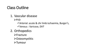
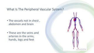
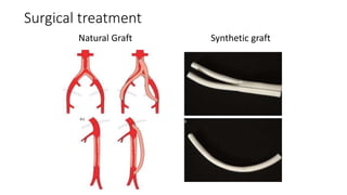
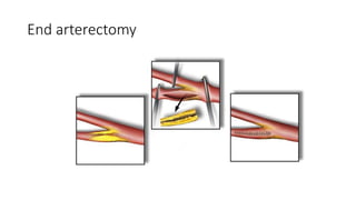
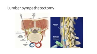
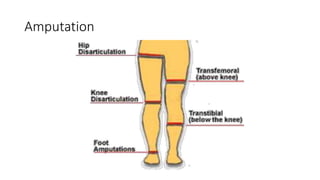
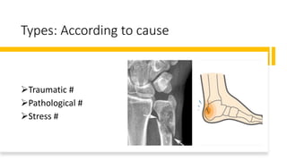
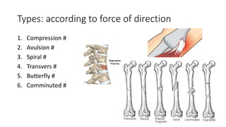
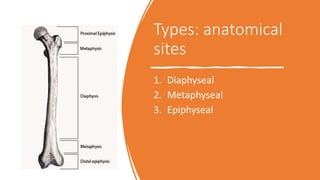
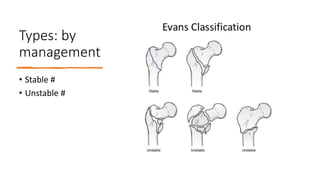
Special class for final prof
examinees
Dr. Nabarun Biswas
Registrar Surgery
MMCH
Class Outline
1. Vascular disease
PVD
Arterial: acute & chr limb ischaemia, Burger’s,
Venous : Varicose, DVT
2. Orthopedics
Fracture
Osteomyelitis
Tumour
Vascular
What Is The Peripheral Vascular System?
• The vessels not in chest ,
abdomen and brain
• These are the veins and
arteries in the arms,
hands, legs and feet
Diseases commonly found in peripheral vasculature
PVD
Arterial
Occlusive
Acute
ischemia
Emboli
Chronic
ischemia
Atherosclerosi
s / buerger’s
Functional
Vasospastis
Raynaud’s
disease
Venous
Thrombosis
Superficial Deep
Varicose
Mixed
Arterio-venous
fistula
Arterial
stenosis and
occlusion
•Cause:
•Atheroma
•Emboli
•Trauma
•Type:
•Acute
•Chronic
RISK FACTORS
• Positive family history of premature heart attacks or
strokes
• Older than 50 years
• Male sex
• Overweight or obesity
• Inactive (sedentary) lifestyle
• Smoking
• Diabetes
• High blood pressure
• High cholesterol or LDL (the "bad cholesterol"), plus
high triglycerides and low HDL
Chronic Limb ischaemia
Chronic ischaemia
Features:
1. Intermittent claudication
2. Rest pain
3. Ulcer and gangrene
4. Colour change
5. Temperature
6. Sensation & movement
7. Capillary refill time
8. Peripheral pulses
Intermittent claudication
• Claudication means cramp like pain in muscle
during walking due to muscle ischaemia
• Not in the 1st step (in osteoarthritis)
• Relieved by stand still/rest (in PLID posture
change/rest)
• Commonly felt in calf
• Leriche’s syndrome: Buttock claudication +
Impotency due to aorto-ileac obstruction
Grading of Intermittent claudication
•Grade 1:
• Pain goes away & patient continue walking (pain producing
substances washed away by collaterals)
•Grade 2
• Pain starts after some distance & can walk with pain
•Grade 3
• When pain starts patient can't walk without taking rest
Rest pain
Continuous pain occurs at rest throughout day and
night due to severe ischaemia & felt in foot
 Exacerbate by lying down or foot elevation
Worse @ night due to decreased heart rate.
Comfort by hanging the foot
This pain is due to ischaemia of nerve (crying of
dying nerve)
Ulcer & gangrene
• Ulcer due to severe ischaemia
• Painful ulcer
• Site: between toes, around maleoli
• Black, mummified
• may be wet if infection occurs
Colour change of foot (a) elevation pallor and
(b) dependent rubor
Relationship between occlusion site & Clinical
features
Aorto-iliac
⁻ Claudication both buttock
& thigh
⁻ Leriche’s syndrome
⁻ Absent femoral pulses
Iliac
⁻ Unilateral claudication in
thigh/ calf
⁻ Unilateral absent pulse
Femoro-popliteal
Distal obstructions
Stage of limb ischaemia: Fontaine's staging
• Stage I: Asymptomatic
• Stage II: Intermittent claudication
• Stage III: Rest pain
• IIIa: Rest pain + ankle pressure > 50 mm of Hg
• IIIb: Rest pain + ankle pressure < 50 mm of Hg
• Stage IV: Ulcer / Gangrene
Investigations
For diagnosis
Doppler USG
Duplex scanning
Angiography
CT angiography
MR
angiography
DSA
For assessment
CBC
S. Lipid profile
S. Creatinine
Blood sugar
S. Electrolite
ECG
CXR
Doppler USG
Measured by Doppler USG
Blood flow
Site of stenosis
Systolic pressure of small arteries
ABPI
ABPI
• Ratio of systolic pressure @ ankle to arm
• Measured by Doppler USG
• Measured artery: dorsalis pedis, ant. tibial, & post. Tibial
• Value:
• Normal= 1
• Claudication = < 0.9
• Rest pain = < 0.5
• Imminent necrosis = <0.3
• Calcification = > 1
Duplex scanning
• Combination of Doppler
study & B mood USG
• Gives idea about-
• Stenosis
• Visual impression of small
vessel
• Turbulence
• Velocity of flow
• Changes in direction of flow
Angiography
• Invasive & indicated when
intervention is think about
• Using Sheldinger technique
• Usually through femoral artery
• Advantages
• Visualization of artery, site of
occlusion, collaterals seen
• Disadvantages
• Bleeding, haematoma, thrombosis,
embolism
• Arterial dissection, false aneurism
Angiography
• CT Angiography • MR Angiography
Treatment
Nonsurgical
I. General
II. Drugs
Interventional
Surgical
Nonsurgical treatment
•General
1. Stop smoking
2. Walk within limit of disability
3. Dietary advice to loss weight
4. Care of ischaemic foot, avoid trauma
Nonsurgical treatment
• Drugs
1. Pentoxiphylin ↓blood viscosity , improve circulation; 400 mg bd.
2. Cilostazole  inhibit platelet aggregation; 100mg bd
3. Analgesic
4. Low dose aspirin
5. Clopidogrel
6. Anti HTN
7. Anti DM
8. Statin  lipid lowering
9. Antibiotics
Anti platelet; 75 mg daily
Interventional treatment
PTA: Percuteneoans transluminal
angioplasty
• By balloon Catheter
• Via femoral artery
• Fluoroscopic guidance
• Suitable for short segment, < 5 cm
Transluminal stenting
• Self expanding metallic stent
• Suitable for long segment; > 5 cm
• PTA failure
Surgical treatment
Indication:
• Severe symptom
• Angioplasty failed/not
possible
Options:
1. Bypass graft
 Natural
 Saphanous (long/short) graft
 Arm vein graft
 Synthetic
 Dacron
 PTFE
2. End arterectomy
3. Lumber sympathetectomy
4. Amputation
Surgical treatment
Natural Graft Synthetic graft
End arterectomy
Lumber sympathetectomy
Amputation
Critical Limb Ischaemia
Def:
Critical limb ischaemia is a late sign of progressive limb ischaemia,
characterized by-
—Rest pain; requiring regular analgesic >2 weeks
—Ulcer due to arterial insufficiency
—Gangrene
—Systolic pressure of limb < 50 mm of Hg
• Management: same as chronic limb ischaemia
Acute occlusive condition
• Causes
• Emboli
• Thrombi
• Trauma
Embolus
An embolus is a body that is foreign to the blood stream & which may lodge into
blood vessel & cause obstruction.
Type
• Thrombo-embolic
• Lt. atrium  fibrillation/MI/ endocarditis
• Aneurism
• Atherosclerotic plaque
• Fat
• Air
• Infective
• Parasitic
• Malignant
Site of acute arterial occlusion
Limbs
Brain - middle cerebral artery
Retina – central retinal artery
Amourosis fugux
Gut
Kidney
Symptoms
• Sudden, severe onset of constant pain/numbness
• May have H/O cardiac disease
• May have H/O trauma/ arterial catheterization
• May have H/O arterial graft
Signs
5P
• Pain, pallor, paresthesia, paralysis & pulselessness
• Limb is cold, tender, swollen
• Toe cannot be moved
• If occlusion is > 6 hours  irreversible damage occurs & line of
demarcation may present
Immediate management
• It is a surgical emergency, so if a patient have a H/O cardiac disease with
acute limb ischaemic features immediate management should be done
• So the Mx is:
1. 5000 U of heparin I/V
• To reduce extension of thrombi
• To maintain patency of vessel
2. Analgesic
Investigation
• In treating acute limb ischaemia (ALI) TIME is everything
• in worst case ALI may progress to critical limb ischaemia
So, the choice of INV. Is
Colour Doppler study
Evaluates lesion into 3 categories
1. Viable
2. Threatened
3. Irreversible
Surgical management
1. Embolectomy
• Rx of choice
• Under LA
• By Fogarty balloon catheter
2. Intra arterial thrombolysis
• With tissue plasminogen
activator
• Or streptokinase/ urokinase
Intra arterial thrombolysis….cont.
• Injected into clot via catheter
• Success achieved within 24 hours
• Regular angiogram should be done
• Contraindication
• Recent stroke
• Bleeding diathesis
• Pregnancy
• Age> 80
After managing ALI total evaluation of the cause should be
attempted & managed accordingly
Varicose vein
Cause:
• Prolong standing
• Valve defect
• DVT
• Obesity
• Pregnancy
• Familial
• Idiopathic
CEAP classification
Varicose vein
Clinical features:
• Dull aching pain
• Pigmentation
• Spider formation
• Saphena varix
• Itching
• Eczema
• Ulceration over gaiter area
Varicose vein
Investigation:
• Duplex ultrasound
imaging
• Venography
Varicose vein
Treatment
1. Avoid long standing
2. Compression
stocking
3. USG guided foam
sclerotherapy
4. EVLA
5. Radio-frequency
ablation
6. Surgery:
• Striping
• Phlebectomy
Deep venous thrombosis
Factors responsible: (Virchow’s
triad)
1. Endothelial injury
2. Alteration of normal blood
flow
3. Hypercoagulability of blood
I. Primary
• Antithrombin III deficiency
• Protein S or C deficiency
• Fibrinogen defect
II. Secondary
• DIC
• OCP
• Malignancy
• Smoking
• Pregnancy
• Massive tissue injury
• immobilization
Clinical features:
• Asymptomatic/ occult DVT (80%)
• Dull aching pain
• Redness
• Swelling
• Increased temperature
• Dilated superficial veins
• Calf stiffness
Investigations:
• Duplex scanning
• Ascending venography
• MR venography
• Baseline investigations
Treatment:
• General measure
i. Bed rest
ii. Elevation
iii. Analgesics & antibiotics
• Stablished DVT
1. LM wt Heparin  S/C for 5-7 days
+
Warfarin : 10 mg on day 1
10 mg on day 2
5 mg on day 3  continue 2-3 days (NB: PT & INR measured on day 2 & 3)
2. Intervention:
I. Venous thrombectomy
II. Thrombolysis
III. Stent grafting
Prophylaxis:
indication
Prophylaxis against DVT
1.Pre-operative
• Weight reduction
• Stop OCP 1 month before surgery
• Correction of high-risk conditions
2.Per-operative
Mechanical
Graduated compressive stocking
Electrical stimulator of calf muscle
Electrical external pneumatic
compression of calf
Chemical
LMW Heparin  30 mg daily  S/C
 5 days (started 12 hours before
surgery)
3.Post-operative
Graduated compressive stocking
Early mobilization & massage
Adequate hydration
Orthopedics
Fracture:
Fracture is the break in the structural continuity of
bone
Types: According to
skin involvement
•Open
•Closed
Types: According to completeness of involved
bone fracture
1. Complete
Transvers
Oblique
Comminuted
2. Incomplete
Greenstick
Compression
Types: According to cause
Traumatic #
Pathological #
Stress #
Types: according to force of direction
1. Compression #
2. Avulsion #
3. Spiral #
4. Transvers #
5. Butterfly #
6. Comminuted #
Types: anatomical
sites
1. Diaphyseal
2. Metaphyseal
3. Epiphyseal
Types: by
management
• Stable #
• Unstable #
Diagnosis of fracture:
History Examination Investigation
Management of fracture:
Principal of management of fracture :
Early treatment: ATLS +
Assessment and management of
neurovascular status
Reduction
Immobilization
Rehabilitation
Pathological #
General Local
Benign Malignant
Primary
Secondary
Causes of pathological fracture
Osteomyelitis
Osteomyelitis : Infection of all parts of bone
Acute : Chronic
1 bone infection but not bone
death
bone infection associated
with bone death
2 Common in children Common in adult
3 Develops within 2 weeks Develops after few months
4 Usually, hematogenous Usually, traumatic
Pathogenesis: bacteria lodgment in metaphysis
a. Stage I: Inflammation
b. Stage II: Suppuration
c. Stage III: Necrosis
d. Stage IV: New bone formation
e. Stage V: Resolution
(ইনসাননর ননউ ররভ্য ুলুশন)
Diagnosis:
Clinical features:
• Symptoms
• Local pain
• Fever
• Swelling
• Immobility
• History of trauma
• Sign
• Local sign of inflammation
Investigations:
• Imaging
• Plane X-ray
• USG
• MRI
• CT
• Biomarker
• Tissue sampling
Treatment:
Principle of treatment
1. Rest
2. Elevation
3. Surgical drainage
4. Appropriate antibiotics in right length of time
5. Nutrition & Anaemia correction
Bone tumour
Bone
tumour
EG
MO
DE
Bone tumour
Primary
Meyeloma
MM
Solitary
plasmacytoma
Osteogenic
Osteoid
osteoma
Osteoblastoma
osteosarcoma
Chondrogenic
Osteochondroma
Enchondroma
Chondrosarcoma
Other
Simple bone
cyst
ABC
GCT
Fibrous Dys
Ewing’s
sarcoma
Secondary
(BBKPT)
Osteosarcoma
• Common site: Metaphysis of knee,
proximal humerus
• Age: child & adolescent
• Osteoblastic
• C/F: pain, swelling, fracture
• Metastasis: lung
• X-ray: sunburst, codma’s triangle,
pathological #
• Rx: NACT, Surgery
Ewing’s sarcoma
• Site: diaphysis of long bone
• Age: 10-20 years
• Round cell sarcoma
• C/F: pain, swelling, fever
• X-ray: onion peel, moth eaten appearance
• Rx: CT,RT, surgery
Giant cell tumour
• Site: Epiphysis of long bone
• Age: young adult (20-45yr)
• Osteoclast like giant cell
• C/F: Pain, swelling, pathological #
• X-ray: soap bubble appearance
• Rx: surgery followed by RT
vascular & ortho.pptx

vascular & ortho.pptx

  • 1.
    Special class forfinal prof examinees Dr. Nabarun Biswas Registrar Surgery MMCH
  • 2.
    Class Outline 1. Vasculardisease PVD Arterial: acute & chr limb ischaemia, Burger’s, Venous : Varicose, DVT 2. Orthopedics Fracture Osteomyelitis Tumour
  • 3.
  • 4.
    What Is ThePeripheral Vascular System? • The vessels not in chest , abdomen and brain • These are the veins and arteries in the arms, hands, legs and feet
  • 5.
    Diseases commonly foundin peripheral vasculature PVD Arterial Occlusive Acute ischemia Emboli Chronic ischemia Atherosclerosi s / buerger’s Functional Vasospastis Raynaud’s disease Venous Thrombosis Superficial Deep Varicose Mixed Arterio-venous fistula
  • 6.
  • 7.
    RISK FACTORS • Positivefamily history of premature heart attacks or strokes • Older than 50 years • Male sex • Overweight or obesity • Inactive (sedentary) lifestyle • Smoking • Diabetes • High blood pressure • High cholesterol or LDL (the "bad cholesterol"), plus high triglycerides and low HDL
  • 8.
  • 11.
    Chronic ischaemia Features: 1. Intermittentclaudication 2. Rest pain 3. Ulcer and gangrene 4. Colour change 5. Temperature 6. Sensation & movement 7. Capillary refill time 8. Peripheral pulses
  • 12.
    Intermittent claudication • Claudicationmeans cramp like pain in muscle during walking due to muscle ischaemia • Not in the 1st step (in osteoarthritis) • Relieved by stand still/rest (in PLID posture change/rest) • Commonly felt in calf • Leriche’s syndrome: Buttock claudication + Impotency due to aorto-ileac obstruction
  • 13.
    Grading of Intermittentclaudication •Grade 1: • Pain goes away & patient continue walking (pain producing substances washed away by collaterals) •Grade 2 • Pain starts after some distance & can walk with pain •Grade 3 • When pain starts patient can't walk without taking rest
  • 14.
    Rest pain Continuous painoccurs at rest throughout day and night due to severe ischaemia & felt in foot  Exacerbate by lying down or foot elevation Worse @ night due to decreased heart rate. Comfort by hanging the foot This pain is due to ischaemia of nerve (crying of dying nerve)
  • 15.
    Ulcer & gangrene •Ulcer due to severe ischaemia • Painful ulcer • Site: between toes, around maleoli • Black, mummified • may be wet if infection occurs
  • 16.
    Colour change offoot (a) elevation pallor and (b) dependent rubor
  • 17.
    Relationship between occlusionsite & Clinical features Aorto-iliac ⁻ Claudication both buttock & thigh ⁻ Leriche’s syndrome ⁻ Absent femoral pulses Iliac ⁻ Unilateral claudication in thigh/ calf ⁻ Unilateral absent pulse Femoro-popliteal Distal obstructions
  • 18.
    Stage of limbischaemia: Fontaine's staging • Stage I: Asymptomatic • Stage II: Intermittent claudication • Stage III: Rest pain • IIIa: Rest pain + ankle pressure > 50 mm of Hg • IIIb: Rest pain + ankle pressure < 50 mm of Hg • Stage IV: Ulcer / Gangrene
  • 19.
    Investigations For diagnosis Doppler USG Duplexscanning Angiography CT angiography MR angiography DSA For assessment CBC S. Lipid profile S. Creatinine Blood sugar S. Electrolite ECG CXR
  • 20.
    Doppler USG Measured byDoppler USG Blood flow Site of stenosis Systolic pressure of small arteries ABPI
  • 21.
    ABPI • Ratio ofsystolic pressure @ ankle to arm • Measured by Doppler USG • Measured artery: dorsalis pedis, ant. tibial, & post. Tibial • Value: • Normal= 1 • Claudication = < 0.9 • Rest pain = < 0.5 • Imminent necrosis = <0.3 • Calcification = > 1
  • 22.
    Duplex scanning • Combinationof Doppler study & B mood USG • Gives idea about- • Stenosis • Visual impression of small vessel • Turbulence • Velocity of flow • Changes in direction of flow
  • 23.
    Angiography • Invasive &indicated when intervention is think about • Using Sheldinger technique • Usually through femoral artery • Advantages • Visualization of artery, site of occlusion, collaterals seen • Disadvantages • Bleeding, haematoma, thrombosis, embolism • Arterial dissection, false aneurism
  • 24.
  • 25.
  • 26.
    Nonsurgical treatment •General 1. Stopsmoking 2. Walk within limit of disability 3. Dietary advice to loss weight 4. Care of ischaemic foot, avoid trauma
  • 27.
    Nonsurgical treatment • Drugs 1.Pentoxiphylin ↓blood viscosity , improve circulation; 400 mg bd. 2. Cilostazole  inhibit platelet aggregation; 100mg bd 3. Analgesic 4. Low dose aspirin 5. Clopidogrel 6. Anti HTN 7. Anti DM 8. Statin  lipid lowering 9. Antibiotics Anti platelet; 75 mg daily
  • 28.
    Interventional treatment PTA: Percuteneoanstransluminal angioplasty • By balloon Catheter • Via femoral artery • Fluoroscopic guidance • Suitable for short segment, < 5 cm
  • 29.
    Transluminal stenting • Selfexpanding metallic stent • Suitable for long segment; > 5 cm • PTA failure
  • 30.
    Surgical treatment Indication: • Severesymptom • Angioplasty failed/not possible Options: 1. Bypass graft  Natural  Saphanous (long/short) graft  Arm vein graft  Synthetic  Dacron  PTFE 2. End arterectomy 3. Lumber sympathetectomy 4. Amputation
  • 31.
  • 32.
  • 33.
  • 34.
  • 35.
    Critical Limb Ischaemia Def: Criticallimb ischaemia is a late sign of progressive limb ischaemia, characterized by- —Rest pain; requiring regular analgesic >2 weeks —Ulcer due to arterial insufficiency —Gangrene —Systolic pressure of limb < 50 mm of Hg • Management: same as chronic limb ischaemia
  • 36.
    Acute occlusive condition •Causes • Emboli • Thrombi • Trauma
  • 37.
    Embolus An embolus isa body that is foreign to the blood stream & which may lodge into blood vessel & cause obstruction. Type • Thrombo-embolic • Lt. atrium  fibrillation/MI/ endocarditis • Aneurism • Atherosclerotic plaque • Fat • Air • Infective • Parasitic • Malignant
  • 38.
    Site of acutearterial occlusion Limbs Brain - middle cerebral artery Retina – central retinal artery Amourosis fugux Gut Kidney
  • 39.
    Symptoms • Sudden, severeonset of constant pain/numbness • May have H/O cardiac disease • May have H/O trauma/ arterial catheterization • May have H/O arterial graft
  • 40.
    Signs 5P • Pain, pallor,paresthesia, paralysis & pulselessness • Limb is cold, tender, swollen • Toe cannot be moved • If occlusion is > 6 hours  irreversible damage occurs & line of demarcation may present
  • 41.
    Immediate management • Itis a surgical emergency, so if a patient have a H/O cardiac disease with acute limb ischaemic features immediate management should be done • So the Mx is: 1. 5000 U of heparin I/V • To reduce extension of thrombi • To maintain patency of vessel 2. Analgesic
  • 42.
    Investigation • In treatingacute limb ischaemia (ALI) TIME is everything • in worst case ALI may progress to critical limb ischaemia So, the choice of INV. Is Colour Doppler study Evaluates lesion into 3 categories 1. Viable 2. Threatened 3. Irreversible
  • 43.
    Surgical management 1. Embolectomy •Rx of choice • Under LA • By Fogarty balloon catheter 2. Intra arterial thrombolysis • With tissue plasminogen activator • Or streptokinase/ urokinase
  • 44.
    Intra arterial thrombolysis….cont. •Injected into clot via catheter • Success achieved within 24 hours • Regular angiogram should be done • Contraindication • Recent stroke • Bleeding diathesis • Pregnancy • Age> 80 After managing ALI total evaluation of the cause should be attempted & managed accordingly
  • 45.
    Varicose vein Cause: • Prolongstanding • Valve defect • DVT • Obesity • Pregnancy • Familial • Idiopathic
  • 46.
  • 47.
    Varicose vein Clinical features: •Dull aching pain • Pigmentation • Spider formation • Saphena varix • Itching • Eczema • Ulceration over gaiter area
  • 48.
    Varicose vein Investigation: • Duplexultrasound imaging • Venography
  • 49.
    Varicose vein Treatment 1. Avoidlong standing 2. Compression stocking 3. USG guided foam sclerotherapy 4. EVLA 5. Radio-frequency ablation 6. Surgery: • Striping • Phlebectomy
  • 50.
    Deep venous thrombosis Factorsresponsible: (Virchow’s triad) 1. Endothelial injury 2. Alteration of normal blood flow 3. Hypercoagulability of blood I. Primary • Antithrombin III deficiency • Protein S or C deficiency • Fibrinogen defect II. Secondary • DIC • OCP • Malignancy • Smoking • Pregnancy • Massive tissue injury • immobilization
  • 51.
    Clinical features: • Asymptomatic/occult DVT (80%) • Dull aching pain • Redness • Swelling • Increased temperature • Dilated superficial veins • Calf stiffness
  • 52.
    Investigations: • Duplex scanning •Ascending venography • MR venography • Baseline investigations
  • 53.
    Treatment: • General measure i.Bed rest ii. Elevation iii. Analgesics & antibiotics • Stablished DVT 1. LM wt Heparin  S/C for 5-7 days + Warfarin : 10 mg on day 1 10 mg on day 2 5 mg on day 3  continue 2-3 days (NB: PT & INR measured on day 2 & 3) 2. Intervention: I. Venous thrombectomy II. Thrombolysis III. Stent grafting
  • 54.
  • 55.
    Prophylaxis against DVT 1.Pre-operative •Weight reduction • Stop OCP 1 month before surgery • Correction of high-risk conditions 2.Per-operative Mechanical Graduated compressive stocking Electrical stimulator of calf muscle Electrical external pneumatic compression of calf Chemical LMW Heparin  30 mg daily  S/C  5 days (started 12 hours before surgery) 3.Post-operative Graduated compressive stocking Early mobilization & massage Adequate hydration
  • 56.
  • 57.
    Fracture: Fracture is thebreak in the structural continuity of bone
  • 58.
    Types: According to skininvolvement •Open •Closed
  • 59.
    Types: According tocompleteness of involved bone fracture 1. Complete Transvers Oblique Comminuted 2. Incomplete Greenstick Compression
  • 60.
    Types: According tocause Traumatic # Pathological # Stress #
  • 61.
    Types: according toforce of direction 1. Compression # 2. Avulsion # 3. Spiral # 4. Transvers # 5. Butterfly # 6. Comminuted #
  • 62.
  • 63.
  • 64.
    Diagnosis of fracture: HistoryExamination Investigation
  • 65.
    Management of fracture: Principalof management of fracture : Early treatment: ATLS + Assessment and management of neurovascular status Reduction Immobilization Rehabilitation
  • 66.
    Pathological # General Local BenignMalignant Primary Secondary Causes of pathological fracture
  • 67.
  • 68.
    Osteomyelitis : Infectionof all parts of bone Acute : Chronic 1 bone infection but not bone death bone infection associated with bone death 2 Common in children Common in adult 3 Develops within 2 weeks Develops after few months 4 Usually, hematogenous Usually, traumatic
  • 69.
    Pathogenesis: bacteria lodgmentin metaphysis a. Stage I: Inflammation b. Stage II: Suppuration c. Stage III: Necrosis d. Stage IV: New bone formation e. Stage V: Resolution (ইনসাননর ননউ ররভ্য ুলুশন)
  • 70.
    Diagnosis: Clinical features: • Symptoms •Local pain • Fever • Swelling • Immobility • History of trauma • Sign • Local sign of inflammation Investigations: • Imaging • Plane X-ray • USG • MRI • CT • Biomarker • Tissue sampling
  • 71.
    Treatment: Principle of treatment 1.Rest 2. Elevation 3. Surgical drainage 4. Appropriate antibiotics in right length of time 5. Nutrition & Anaemia correction
  • 72.
  • 73.
  • 74.
  • 76.
    Osteosarcoma • Common site:Metaphysis of knee, proximal humerus • Age: child & adolescent • Osteoblastic • C/F: pain, swelling, fracture • Metastasis: lung • X-ray: sunburst, codma’s triangle, pathological # • Rx: NACT, Surgery
  • 77.
    Ewing’s sarcoma • Site:diaphysis of long bone • Age: 10-20 years • Round cell sarcoma • C/F: pain, swelling, fever • X-ray: onion peel, moth eaten appearance • Rx: CT,RT, surgery
  • 78.
    Giant cell tumour •Site: Epiphysis of long bone • Age: young adult (20-45yr) • Osteoclast like giant cell • C/F: Pain, swelling, pathological # • X-ray: soap bubble appearance • Rx: surgery followed by RT