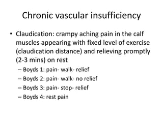
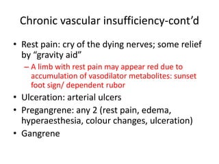
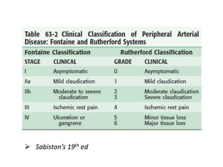
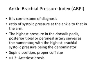
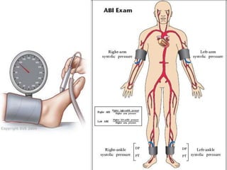
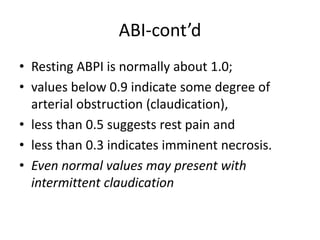
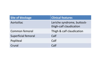
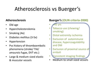
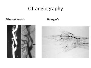
Ischaemia of Lower Limbs
By
Dr. Shampile
16/11/2017
Terminology
• Acute: <14 days
• Acute on chronic: worsening in <14 days
• Chronic: stable for >14 days
• Incomplete: no limb threat
• Complete: limb threatened
• Irreversible: limb non viable
Definition
• peripheral vascular disease refers to the
obstruction or deterioration of arteries other
than those supplying the heart and within the
brain.
Aetiology-ALI
Embolism- 90% from heart, 9% from great
vessels, 1% other.
• Left atrium in patients in atrial fibrillation
• Mural thrombus after myocardial infarct
• Prosthetic and diseased heart valves
• Aneurysm or atheromatous stenosis
• Tumour, foreign body
Aetiology-cont’d
Thrombosis; Atherosclerosis, aneurysm, graft stenosis,
low flow, e.g. hypovolaemia, thrombotic states.
Other
• Trauma ( general or operative)
• Arteritis,
• Congenital anomalies
• Dissecting aneurysm
• Raynaud's Syndrome
• Metabolic (DM)
• Drugs e.g. Acute ischaemia due to intra-arterial
temazepam injection
Pathophysiology-ALI
• Sudden interruption of blood supply.
• Emboli tend to lodge at sites of vessel
bifurcation.
• There are two phases of cell injury: ischaemic
injury as tissues are deprived of blood supply
and reperfusion injury if blood flow is
restored
Clinical features of limb ischaemia
• Symptoms and signs depend on the site of
occlusion, duration of ischaemia and degree
of collateral circulation.
• Embolus more likely if severe, sudden onset
and potential source identifiable, e.g. atrial
fibrillation.
• Thrombosis usually if less severe (collaterals
present), history of claudication
Acute arterial occlusion
• Pain (absent in incomplete ischemia)
• Pallor (present in chronic ischemia)
• Pulselessness (present in chronic ischemia)
• Perishing cold (unreliable)
• Paresthesia (leading to anaesthesia)
• Paralysis (contrasts with venous
occlusion when muscle function is not
affected)
• Ischaemia beyond 6 hours is usually
irreversible and results in limb loss.
• No history of claudication
EXAMINATION
• Limb is pale with absent pulses; capillary
return is slow.
• After several hours, there is venous
stagnation.
• Altered sensation, and if ischaemia is severe,
paraesthesia with muscle paralysis
INVESTIGATIONS
• Bloods: FBC, U&Es, coagulation profile,
thrombophilia screen.
• Imaging: CXR, Doppler or duplex scanning of
blood flow, arteriography to demonstrate the
site of occlusion and plan intervention if limb
viable.
• ECG: Looking for atrial fibrillation.
Management
Diagnois
• Is this ischaemia ?
• Is this an embolism or thrombosis ? clinical
features include:
 Rapidity of onset of symptoms
 Features of pre-existing chronic arterial disease
 Potential source of embolus
 State of pedal pulses in contralateral leg
• Is this limb viable ?
MANAGEMENT
• Immediate: ABCs, analgesia, heparin
anticoagulation to prevent thrombus
propagation.
• Treat associated cardiac disease
• Surgical: Revascularisation within 6 hours in
order to salvage limb. Operative risk is often
high due to underlying heart disease.
Postoperative anticoagulation is essential
MANAGEMENT
Treatment options are:
• Embolic disease - embolectomy or intra-
arterial thrombolysis
• Thrombotic disease - intra-arterial
thrombolysis / angioplasty or bypass surgery
• Amputation ; if non viable limb
MANAGEMENT
Emergency embolectomy
• Can be performed under either general or local
anaesthesia
• Display and control arteries with slings
• Transverse artereotomy performed over common
femoral artery
• Fogarty balloon embolectomy catheters used to
retrieve thrombus
• If embolectomy fails - ontable angiogram and consider
Bypass graft or intraoperative thrombolysis
MANAGEMENT
• If acute or chronic thrombosis: The limb may
remain viable for a longer time due to
collateral formation, and percutaneous
intervention is an option, e.g. aspiration,
intraarterial thrombolysis with local infusion
of, e.g., t-PA, and angioplasty of underlying
stenoses.
MANAGEMENT
Intra-arterial thrombolysis
• Arteriogram and catheter advanced into
thrombus
• Streptokinase 5000u/hr + heparin 250u/hr
• Alternative thrombolytic agents include
Urokinase or tissue plasminogen activator (tPA)
• Repeat arteriogram at 6 -12 hours
• Advance catheter and continue thrombolysis for
48 hours or until clot lysis
MANAGEMENT
• Angioplasty of chronic arterial stenosis may be
necessary
• Success 60-70% but needs careful case selection
• Not suitable if severe neuro-sensory deficit
• Thrombolysis can be accelerated by
 Pulse spray through multiple side hole
catheter
Aspiration thrombectomy - debulking thrombus
aspiration
High dose over shorter time
MANAGEMENT
• If thrombosis but the limb is not likely to
remain viable for 12–24 hours necessary for
this procedure: Urgent reconstructive surgery
is required,
• if technically possible with autogenous
(saphenous vein) or synthetic (e.g. PTFE or
Dacron) bypass grafting.
• If risk of compartment syndrome, fasciotomy
is required.
Vascular Trauma of the Extremity
• Often the extremities are placed at the
bottom of the ‘‘to-do’’ list in patients with life-
threatening multisystem trauma, only to find
an ischemic limb hours later.
• Therefore, a high index of suspicion is
warranted in patients with extremity
fracture/dislocations
Vascular Trauma of the Extremity
• Blunt trauma results in a stretch injury and
subsequent occlusion of the artery at the level
of the associated fracture or joint dislocation.
• Penetrating wounds can produce either vessel
transection or a pseudoaneurysm
• Angiography, to discern between vessel spasm
and overt injury, can be performed in the
angiography suite or in the OR.
COMPLICATIONS
• From disease: Gangrene, limb loss, death.
• From intra-arterial thrombolysis: Mortality
(1–2%), CVA, major haemorrhage
• Post-treatment: Reperfusion syndrome,
compartment syndrome, rhabdomyolysis,
rethrombosis.
PROGNOSIS
• Risk of limb loss is up to 30%; mortality 10%,
with major mortality factor underlying cardiac
disease.
Chronic Arterial Insufficiency
• Asymptomatic to gangrenous tissue loss
• Intermittent claudication
• Rest pain
• Dependent rubor
• Ulceration
• Gangrene
• Arterial pulsation diminished or absent
• Arterial bruit
• Slow capillary refilling
Chronic vascular insufficiency
• Claudication: crampy aching pain in the calf
muscles appearing with fixed level of exercise
(claudication distance) and relieving promptly
(2-3 mins) on rest
– Boyds 1: pain- walk- relief
– Boyds 2: pain- walk- no relief
– Boyds 3: pain- stop- relief
– Boyds 4: rest pain
Chronic vascular insufficiency-cont’d
• Rest pain: cry of the dying nerves; some relief
by “gravity aid”
– A limb with rest pain may appear red due to
accumulation of vasodilator metabolites: sunset
foot sign/ dependent rubor
• Ulceration: arterial ulcers
• Pregangrene: any 2 (rest pain, edema,
hyperaesthesia, colour changes, ulceration)
• Gangrene
 Sabiston’s 19th ed
Ankle Brachial Pressure Index (ABPI)
• It is cornerstone of diagnosis
• ratio of systolic pressure at the ankle to that in
the arm.
• The highest pressure in the dorsalis pedis,
posterior tibial or peroneal artery serves as
the numerator, with the highest brachial
systolic pressure being the denominator
• Supine position, proper cuff size
• >1.3: Arteriosclerosis
ABI-cont’d
• Resting ABPI is normally about 1.0;
• values below 0.9 indicate some degree of
arterial obstruction (claudication),
• less than 0.5 suggests rest pain and
• less than 0.3 indicates imminent necrosis.
• Even normal values may present with
intermittent claudication
Site of blockage Clinical features
Aortoiliac Leriche syndrome, buttock-
thigh-calf claudication
Common femoral Thigh & calf claudication
Superficial femoral Calf
Popliteal Calf
Crural Calf
Atherosclerosis vs Buerger’s
Atherosclerosis Buerger’s (OLIN criteria-2000)
• Old age
• Hypercholesterolemia
• Smoking (4x)
• Diabetes mellitus (3-5x)
• Hypertension
• Pvs history of thromboembolic
phenomena (stroke/ TIA/
amaurois fugax, DVT etc.)
• Large & medium sized elastic
& muscular vessels
• <45 yrs
• Tobacco use (chewing/
smoking)
• Distal extremity ischemia
• Exclusion of autoimmune
disease, hypercoagulability
and DM
• Exclusion of proximal source of
emboli
• Consistent imaging findings
• Medium to small sized vessels
CT angiography
Atherosclerosis Buerger’s
Other Diseases
• Takayasu arteritis: pulseless disease of asian
young females
• Raynaud phenomenon: pallor followed by
cyanosis & rubor
• Raynaud’s syndrome: peripheral arterial
manifestation of a collagen disease, such as
systemic lupus erythematosus or rheumatoid
arthritis or vibration white finger.
• Raynaud disease: Raynaud phenomenon in a
young female without any other cause
Pathogenesis of Atherosclerosis
Pathogenesis of Buerger’s Disease
• Cellular, segmental, occlusive inflammatory
thrombi of the vessel wall
• Small to medium sized vessels
• Involves all layers of the vessels
• Integrity of the vessel wall structure is
maintained including internal elastic lamina
• Later on spreads to the adjoining nerve and
vein
Pathogenesis of Buerger’s Disease
• Induced by an unknown agent from smoking
& tobacco chewing, nicotine patch
• Rickettsia infection
• Increased antiendothelial antibodies
• Impaired relaxation of the peripheral vessels
• HLA-A9, HLA-54, HLA-B5
• Increased sensitivity to type 1 & type III
collagen
Assessment
• Disappearing pulse: decreased pulse pressure
on exercise: pulse disappears on exercise
• Allen’s test: positive in young patient with foot
ulceration: Buerger’s Disease
• doppler: harmless, defines level
Investigations
• ECG: rule out embolic phenomenon
• Angio(Arterio)graphy: (convntional/ MR/ CT)
– Buerger’s (TAO): corkscrew collaterals, tree root/ spider leg appearance
– Atherosclerosis (ASO): level of obstruction
Other investigations
General
• FBC
• U/E
• Lipid profile
• ECG
• ESR
• CRP
• Homocystine
• PT/ aPTT/ INR, Protein C/S/
Factor V leiden, Antithrombin
III/ Lupus anticoag
• Carotid duplex
Buerger’s Disease specific treatments
• Prostaglandins (limaprost/iloprost) : antiplatelets
& vasodilators
– Increase cutaneous & muscular blood supply
• Bosentan: endothelin antagonist
• Epidural anaesthesia
• Hyperbaric Oxygen therapy
• Genetic therapy by VEGF gene intramuscular
transfer
• Stem cell & VEGF injections intramuscularly
• Debridement
Buerger’s: Surgeries
• Lumbar Sympathectomy-
open or endoscopic,
remove 2nd -4th lumbar
sympathetic ganglia
• Amputation
• Chemical Sympathectomy
Atherosclerosis: specific T/T
Endovascular Surgical
• Self expandable metallic
stents
• Balloon expandable stents
• Endarterectomy
• Bypass:
– Aortobifemoral
– Axillobifemoral
– Femorofemoral
– Obturator bypass
– Axillopopliteal
– Thoracofemoral
– Infrainguinal
REFERENCES
• Beard J D Gaines P A. Management of the acutely
ischaemic leg. Curr Pract Surg 1995; 7: 123-130.
• Beattie D K Davies A H. Management of the
acutely ischaemic limb. Br J Hosp Med 1996; 55:
204-208.
• Earnshaw J J. Thrombolysis in acute limb
ischaemia. Ann R Coll Surg Eng 1994; 76: 216-
222.
• Golledge J Galland R B. Lower limb intra-arterial
thrombolysis. Postgrad Med J 1995; 71: 146-150.
REFERENCES
• Carroll PR, McAninch JW, Klosterman P, Greenblatt M
(1984) Renovascular trauma: risk assessment, surgical
management, and outcome. J Trauma 30:547
• Courcy PA, Brotman S, Oster-Grantie ML, et al. (1984)
Superior mesenteric artery and vein injuries from blunt
abdominal trauma. J Trauma 24:843
• Dajee H, Richardson IW, Iype MO (1979) Seatbelt aorta:
acute dissection and thrombosis of the abdominal aorta.
Surgery 85:263
• Feliciano DV (1998) Approach to major abdominal vascular
injury. J Vasc Surg 7:730

Tutorial ischaemia of lower limbs

  • 1.
    Ischaemia of LowerLimbs By Dr. Shampile 16/11/2017
  • 2.
    Terminology • Acute: <14days • Acute on chronic: worsening in <14 days • Chronic: stable for >14 days • Incomplete: no limb threat • Complete: limb threatened • Irreversible: limb non viable
  • 3.
    Definition • peripheral vasculardisease refers to the obstruction or deterioration of arteries other than those supplying the heart and within the brain.
  • 4.
    Aetiology-ALI Embolism- 90% fromheart, 9% from great vessels, 1% other. • Left atrium in patients in atrial fibrillation • Mural thrombus after myocardial infarct • Prosthetic and diseased heart valves • Aneurysm or atheromatous stenosis • Tumour, foreign body
  • 5.
    Aetiology-cont’d Thrombosis; Atherosclerosis, aneurysm,graft stenosis, low flow, e.g. hypovolaemia, thrombotic states. Other • Trauma ( general or operative) • Arteritis, • Congenital anomalies • Dissecting aneurysm • Raynaud's Syndrome • Metabolic (DM) • Drugs e.g. Acute ischaemia due to intra-arterial temazepam injection
  • 6.
    Pathophysiology-ALI • Sudden interruptionof blood supply. • Emboli tend to lodge at sites of vessel bifurcation. • There are two phases of cell injury: ischaemic injury as tissues are deprived of blood supply and reperfusion injury if blood flow is restored
  • 7.
    Clinical features oflimb ischaemia • Symptoms and signs depend on the site of occlusion, duration of ischaemia and degree of collateral circulation. • Embolus more likely if severe, sudden onset and potential source identifiable, e.g. atrial fibrillation. • Thrombosis usually if less severe (collaterals present), history of claudication
  • 8.
    Acute arterial occlusion •Pain (absent in incomplete ischemia) • Pallor (present in chronic ischemia) • Pulselessness (present in chronic ischemia) • Perishing cold (unreliable) • Paresthesia (leading to anaesthesia) • Paralysis (contrasts with venous occlusion when muscle function is not affected)
  • 9.
    • Ischaemia beyond6 hours is usually irreversible and results in limb loss. • No history of claudication
  • 10.
    EXAMINATION • Limb ispale with absent pulses; capillary return is slow. • After several hours, there is venous stagnation. • Altered sensation, and if ischaemia is severe, paraesthesia with muscle paralysis
  • 11.
    INVESTIGATIONS • Bloods: FBC,U&Es, coagulation profile, thrombophilia screen. • Imaging: CXR, Doppler or duplex scanning of blood flow, arteriography to demonstrate the site of occlusion and plan intervention if limb viable. • ECG: Looking for atrial fibrillation.
  • 12.
    Management Diagnois • Is thisischaemia ? • Is this an embolism or thrombosis ? clinical features include:  Rapidity of onset of symptoms  Features of pre-existing chronic arterial disease  Potential source of embolus  State of pedal pulses in contralateral leg • Is this limb viable ?
  • 13.
    MANAGEMENT • Immediate: ABCs,analgesia, heparin anticoagulation to prevent thrombus propagation. • Treat associated cardiac disease • Surgical: Revascularisation within 6 hours in order to salvage limb. Operative risk is often high due to underlying heart disease. Postoperative anticoagulation is essential
  • 14.
    MANAGEMENT Treatment options are: •Embolic disease - embolectomy or intra- arterial thrombolysis • Thrombotic disease - intra-arterial thrombolysis / angioplasty or bypass surgery • Amputation ; if non viable limb
  • 15.
    MANAGEMENT Emergency embolectomy • Canbe performed under either general or local anaesthesia • Display and control arteries with slings • Transverse artereotomy performed over common femoral artery • Fogarty balloon embolectomy catheters used to retrieve thrombus • If embolectomy fails - ontable angiogram and consider Bypass graft or intraoperative thrombolysis
  • 16.
    MANAGEMENT • If acuteor chronic thrombosis: The limb may remain viable for a longer time due to collateral formation, and percutaneous intervention is an option, e.g. aspiration, intraarterial thrombolysis with local infusion of, e.g., t-PA, and angioplasty of underlying stenoses.
  • 17.
    MANAGEMENT Intra-arterial thrombolysis • Arteriogramand catheter advanced into thrombus • Streptokinase 5000u/hr + heparin 250u/hr • Alternative thrombolytic agents include Urokinase or tissue plasminogen activator (tPA) • Repeat arteriogram at 6 -12 hours • Advance catheter and continue thrombolysis for 48 hours or until clot lysis
  • 18.
    MANAGEMENT • Angioplasty ofchronic arterial stenosis may be necessary • Success 60-70% but needs careful case selection • Not suitable if severe neuro-sensory deficit • Thrombolysis can be accelerated by  Pulse spray through multiple side hole catheter Aspiration thrombectomy - debulking thrombus aspiration High dose over shorter time
  • 19.
    MANAGEMENT • If thrombosisbut the limb is not likely to remain viable for 12–24 hours necessary for this procedure: Urgent reconstructive surgery is required, • if technically possible with autogenous (saphenous vein) or synthetic (e.g. PTFE or Dacron) bypass grafting. • If risk of compartment syndrome, fasciotomy is required.
  • 20.
    Vascular Trauma ofthe Extremity • Often the extremities are placed at the bottom of the ‘‘to-do’’ list in patients with life- threatening multisystem trauma, only to find an ischemic limb hours later. • Therefore, a high index of suspicion is warranted in patients with extremity fracture/dislocations
  • 21.
    Vascular Trauma ofthe Extremity • Blunt trauma results in a stretch injury and subsequent occlusion of the artery at the level of the associated fracture or joint dislocation. • Penetrating wounds can produce either vessel transection or a pseudoaneurysm • Angiography, to discern between vessel spasm and overt injury, can be performed in the angiography suite or in the OR.
  • 22.
    COMPLICATIONS • From disease:Gangrene, limb loss, death. • From intra-arterial thrombolysis: Mortality (1–2%), CVA, major haemorrhage • Post-treatment: Reperfusion syndrome, compartment syndrome, rhabdomyolysis, rethrombosis.
  • 23.
    PROGNOSIS • Risk oflimb loss is up to 30%; mortality 10%, with major mortality factor underlying cardiac disease.
  • 24.
    Chronic Arterial Insufficiency •Asymptomatic to gangrenous tissue loss • Intermittent claudication • Rest pain • Dependent rubor • Ulceration • Gangrene • Arterial pulsation diminished or absent • Arterial bruit • Slow capillary refilling
  • 25.
    Chronic vascular insufficiency •Claudication: crampy aching pain in the calf muscles appearing with fixed level of exercise (claudication distance) and relieving promptly (2-3 mins) on rest – Boyds 1: pain- walk- relief – Boyds 2: pain- walk- no relief – Boyds 3: pain- stop- relief – Boyds 4: rest pain
  • 26.
    Chronic vascular insufficiency-cont’d •Rest pain: cry of the dying nerves; some relief by “gravity aid” – A limb with rest pain may appear red due to accumulation of vasodilator metabolites: sunset foot sign/ dependent rubor • Ulceration: arterial ulcers • Pregangrene: any 2 (rest pain, edema, hyperaesthesia, colour changes, ulceration) • Gangrene
  • 27.
  • 28.
    Ankle Brachial PressureIndex (ABPI) • It is cornerstone of diagnosis • ratio of systolic pressure at the ankle to that in the arm. • The highest pressure in the dorsalis pedis, posterior tibial or peroneal artery serves as the numerator, with the highest brachial systolic pressure being the denominator • Supine position, proper cuff size • >1.3: Arteriosclerosis
  • 30.
    ABI-cont’d • Resting ABPIis normally about 1.0; • values below 0.9 indicate some degree of arterial obstruction (claudication), • less than 0.5 suggests rest pain and • less than 0.3 indicates imminent necrosis. • Even normal values may present with intermittent claudication
  • 31.
    Site of blockageClinical features Aortoiliac Leriche syndrome, buttock- thigh-calf claudication Common femoral Thigh & calf claudication Superficial femoral Calf Popliteal Calf Crural Calf
  • 32.
    Atherosclerosis vs Buerger’s AtherosclerosisBuerger’s (OLIN criteria-2000) • Old age • Hypercholesterolemia • Smoking (4x) • Diabetes mellitus (3-5x) • Hypertension • Pvs history of thromboembolic phenomena (stroke/ TIA/ amaurois fugax, DVT etc.) • Large & medium sized elastic & muscular vessels • <45 yrs • Tobacco use (chewing/ smoking) • Distal extremity ischemia • Exclusion of autoimmune disease, hypercoagulability and DM • Exclusion of proximal source of emboli • Consistent imaging findings • Medium to small sized vessels
  • 33.
  • 34.
    Other Diseases • Takayasuarteritis: pulseless disease of asian young females • Raynaud phenomenon: pallor followed by cyanosis & rubor • Raynaud’s syndrome: peripheral arterial manifestation of a collagen disease, such as systemic lupus erythematosus or rheumatoid arthritis or vibration white finger. • Raynaud disease: Raynaud phenomenon in a young female without any other cause
  • 35.
  • 36.
    Pathogenesis of Buerger’sDisease • Cellular, segmental, occlusive inflammatory thrombi of the vessel wall • Small to medium sized vessels • Involves all layers of the vessels • Integrity of the vessel wall structure is maintained including internal elastic lamina • Later on spreads to the adjoining nerve and vein
  • 37.
    Pathogenesis of Buerger’sDisease • Induced by an unknown agent from smoking & tobacco chewing, nicotine patch • Rickettsia infection • Increased antiendothelial antibodies • Impaired relaxation of the peripheral vessels • HLA-A9, HLA-54, HLA-B5 • Increased sensitivity to type 1 & type III collagen
  • 38.
    Assessment • Disappearing pulse:decreased pulse pressure on exercise: pulse disappears on exercise • Allen’s test: positive in young patient with foot ulceration: Buerger’s Disease • doppler: harmless, defines level
  • 39.
    Investigations • ECG: ruleout embolic phenomenon • Angio(Arterio)graphy: (convntional/ MR/ CT) – Buerger’s (TAO): corkscrew collaterals, tree root/ spider leg appearance – Atherosclerosis (ASO): level of obstruction
  • 40.
    Other investigations General • FBC •U/E • Lipid profile • ECG • ESR • CRP • Homocystine • PT/ aPTT/ INR, Protein C/S/ Factor V leiden, Antithrombin III/ Lupus anticoag • Carotid duplex
  • 41.
    Buerger’s Disease specifictreatments • Prostaglandins (limaprost/iloprost) : antiplatelets & vasodilators – Increase cutaneous & muscular blood supply • Bosentan: endothelin antagonist • Epidural anaesthesia • Hyperbaric Oxygen therapy • Genetic therapy by VEGF gene intramuscular transfer • Stem cell & VEGF injections intramuscularly • Debridement
  • 42.
    Buerger’s: Surgeries • LumbarSympathectomy- open or endoscopic, remove 2nd -4th lumbar sympathetic ganglia • Amputation • Chemical Sympathectomy
  • 44.
    Atherosclerosis: specific T/T EndovascularSurgical • Self expandable metallic stents • Balloon expandable stents • Endarterectomy • Bypass: – Aortobifemoral – Axillobifemoral – Femorofemoral – Obturator bypass – Axillopopliteal – Thoracofemoral – Infrainguinal
  • 45.
    REFERENCES • Beard JD Gaines P A. Management of the acutely ischaemic leg. Curr Pract Surg 1995; 7: 123-130. • Beattie D K Davies A H. Management of the acutely ischaemic limb. Br J Hosp Med 1996; 55: 204-208. • Earnshaw J J. Thrombolysis in acute limb ischaemia. Ann R Coll Surg Eng 1994; 76: 216- 222. • Golledge J Galland R B. Lower limb intra-arterial thrombolysis. Postgrad Med J 1995; 71: 146-150.
  • 46.
    REFERENCES • Carroll PR,McAninch JW, Klosterman P, Greenblatt M (1984) Renovascular trauma: risk assessment, surgical management, and outcome. J Trauma 30:547 • Courcy PA, Brotman S, Oster-Grantie ML, et al. (1984) Superior mesenteric artery and vein injuries from blunt abdominal trauma. J Trauma 24:843 • Dajee H, Richardson IW, Iype MO (1979) Seatbelt aorta: acute dissection and thrombosis of the abdominal aorta. Surgery 85:263 • Feliciano DV (1998) Approach to major abdominal vascular injury. J Vasc Surg 7:730

Editor's Notes

  • #11 signifying the limb may be nonviable.
  • #16 Fogarty balloon-tipped catheter that is passed beyond the embolus, the balloon inflated and withdrawn
  • #21 careful peripheral vascular examination, including Doppler pressure measurements comparing extremities, and associated neurologic examination
  • #22 Motor/sensory defects may be the first sign of arterial injury due to ischemia, rather than an absent pulse