
Recommended
More Related Content
What's hot
What's hot (20)
Viewers also liked
Viewers also liked (13)
Similar to CBCT in Implants- Summary
Similar to CBCT in Implants- Summary (20)
Recently uploaded
Recently uploaded (20)
CBCT in Implants- Summary
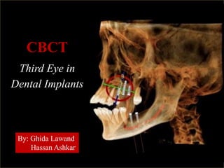
- 1. CBCT Third Eye in Dental Implants By: Ghida Lawand Hassan Ashkar
- 3. 1. Examine patient’s current and status medical history to assess risk profile
- 5. 2. Periodontal/tissue biotype Thin gingival tissue Thick gingival tissue - Highly scalloped, thin, nonkeratinzied and flabby mucosa more susceptible to gingival recession and mechanical trauma soft tissue grafting indicated - Thick, keratinized and non-mobile mucosa Less susceptible to gingival recession following implant treatment - Esthetics: thick mucosa is able to mask the implants’ color and their submucosal metallic components, reducing the risk of a nonesthetic result.
- 6. 3) Lip line The lip line is associated with the amount of tooth and supporting tissues visible when patient chews, speaks, or smiles. High lip line: patient’s entire maxillary teeth and part of his/her alveolar process is revealed presents highest esthetic risk in implant treatment provide a diagnostic set up or a temporary prosthesis for the patient to try on before implant treatment to assess appearance regarding the lip line provisional prosthesis can also serve as a model for a surgical guide or stent to aid placing implants LowMediumHigh
- 7. 4) Occlusal status and functional exam 1. Parafunctional habits such as bruxism and clenching must first be diagnosed and treated before implant treatment to ensure occlusal stability 2. Parafunctional habits cause excessive loading on implants causing technical complications such as screw or framework fractures
- 8. 5) Evaluate edentulous ridge 1. Visually assess the contour, height and width of the edentuglous ridge 2. Fibrous overlying tissue can be deceiving 3. Palpate to detect any concavities or depressions 4. This step is essential to detemine if patient needs bone augmentation 5. Nevertheless, edentulous ridge must be radiographically examined 6. Observe crown to bone relationship (distance between the ideal position of the clinical crowns and the underlying bone) 7. measure the distance between the dentulous ridge and the opposing dentition to ensure there’s enough space for the restoration 8. length of endentulous area can be measured to determine the approximate number of implants needed only verified through radiographs Labially, this ridge reveals a slight loss in the center and the presence of healthy keratinized gingiva Occlusally, ridge appears wide enough to accommodate implants verified through radiography
- 9. a. Characteristics of Ideal Imaging Techniques in Implants: 1. Ability to visualize the site of implant site in the mesiodistal, faciolingual, and superoinferior dimensions 2. ability to determine axial orientation of implants 3. ability to allow reliable, accurate measurements 4. capacity to evaluate trabecular bone density and cortical thickness 5. capacity to correlate the imaged site with the clinical site 6. reasonable access and cost to the patient 7. minimal radiation risk Preoperative Radiographic Assessment
- 10. b. Preoperative radiographs should reveal: (15) 1. Position and size of relevant normal anatomic structures, including the: a. Inferior alveolar canals b. Mental foramina c. Incisive or nasopalatine foramen and canal d. Nasal floor 2. Shape and size of the antra, including the position of the antral floor and its relation to adjacent teeth 3. Presence of any underlying disease that could compromise the outcome of treatment e.g. osteoscleorosis 4. Presence of any buried teeth or retained roots 5. Quantity of alveolar crest/basal bone, allowing direct measuremtns of the height, width and shape 6. Quality (density) of bone, noting: a. Amount of cortical bone present b. Density of the cancellous bone c. the trabecular spaces size
- 11. CBCT Scans The Standard of Care in implants?
- 12. Advantages of CBCT in implant dentistry: 1. Evaluates all possible sites and anotmical structures: 2. No superimposition 3. Uniform magnification 4. Measurements accurate withing about 1 mm 5. Simulates implant pacement with implant planning software 6. CBCT dose of radiation exposure is less than that produced by CT scanning 7. Can limit radiation exposure according to Field of View chosen (FOV): small, medium, or large 8. provides precise information about pathology 9. Less expensive than CT scanning 10. allows for the 3-D evaluation of an arch Limitations of CBCT in implants: 1. Moderate cost and radiation risk as compared to other imaging techniques 2. Some metallic image artifacts 3. Special training for interpretation 4. Relative bone density measurements (HU) not calibrated
- 13. Pre-operative Diagnostic Planning To decide the precise healing scope of a prosthetic implant therapy to re-establish chewing function, it’s imperative to assess Bone availability and the existing anatomical landmarks. This is easier when using 3D diagnostics than using illustration with traditional images
- 14. A) Bone Availability The most frequently reported indication for the use of CBCT in implant planning area are to measure the alveolar ridge and map the bone morphology of possible implant sites for osseointegration of the implant. 1. Quantitative Evaluation of Bone Availability Bone compactness is an significant factor for implant placement to expect the ability to stabilize the implant when minimal bone height is otherwise available. Precise estimation of the alveolar bone height and width are mandatory: 1. For selecting the suitable implant size 2. Establish the degree of angulation of the edentulous alveolar ridge.
- 15. Bone Height: Bone Width: Measured from the crest of the alveolar ridge to the opposing border of the bone or an anatomical structure. According to the bone height, implant length is determined It is the bucco-lingual width of the alveolar bone. Implant diameter is selected based on this parameter.
- 17. Superior border of the mandible
- 18. Height of the maxillary alveolar ridge measured at a specific point at a specific point prior to implant placement Width of the maxillary alveolar ridge measured at a specific point prior to implant placement
- 19. After adequate height is available, the next most significant criterion affecting long-term survival of implants is the width of the available bone. Axial cross-section on the root level showing the space around the implant.
- 20. Note the little space between the mesial and distal surfaces of the implant and the adjacent teeth
- 21. 2. Qualitative Evaluation of Bone Availability
- 22. Hounsfield scale Referred to as HU, it is a scale used to measure the bone density in the CBCT scans. It can measure accurately the density of the selected point, whether it’s related to bone or other tissues. HU gives a quantitative assessment of the bone density where it’s measured by the ability of the tissue to attenuate x-ray beam. HU scale range from -1000 (air) to +3000 (enamel). While bone quality refers to both thickness and density of the cortical plates as well as trabecular portion of the bone. This parameter affects to high limit the success of the treatment.
- 23. Example of Trabecular bone density in the anterior region of maxilla. Example of Trabecular bone density in the anterior region of mandible. Example of Cortical bone density in the anterior region of mandible.
- 25. Length of the Edentulous Area Missing upper left lateral incisor to be restored with implant. In 3D and Axial Slice.
- 27. 3- Ridge Orientation and morphology: it is the inclination of the alveolar edentulous ridge. Virtual implant can be positioned in a cross sectional alveolar bone using special planning software. CBCT Shows the type of alveolar bone present - Cross-sectional CBCT revealing three types of mandibular ridge morphology: - line A represents a the line of reference 2 millimeter coronal to the inferior alveolar nerve canal
- 28. Choosing the suitable length and diameter of the implant. Note the angular inclination of the implant
- 29. According to Tolstunov, a Diplomate of the American Board of Oral Implantology/Implant Dentistry the alveolar jaw can be divided into four regions or “functional implant zones” with each region posessing "unique characteristics of anatomy, blood supply, pattern of bone resorption, bone quality and quantity, need for bone grafting and other supplemental surgical procedures, and a location related implant success rate.” Anatomical factors assessed by CBCT imaging
- 30. Also known as the “traumatic zone” consists of alveolar ridge of pre-maxilla and the teeth from right to left premolar exhibits the greatest loss of bone after tooth loss/extraction occurs in the facio-palatal or horizontal direction and mainly on the facial side of the alveolar ridge Any bone loss in this area is crucial due to its esthetic risk with dental implants if severe bone loss occurs, implants are harder to place in an esthetically favorable position. Functional Implant Zone 1 1) Nasopalatine canal Nasopalatine canal contains: a. nasopalatine nerve b. descending branch of the nasopalatine artery c. fibrous connective tissue if an implant contacts neural tissue in this canal, it could lead to failure of osseointegration or sensory dysfunction. Thus, when one or both central incisors have been lost for a while, limited CBCT imaging is indicated to determine the dimensions and morphology of the nasopalatine canal before dental implant surgery. Nasopalatine canal on a CBCT Coronal section
- 31. Cross-sectional views showing location of naso-palatine canal
- 32. • the sinus zone • located at the base of the maxillary sinuses CBCT imaging is indicated to evaluate: a. bone height between the floor of the maxillary sinus and alveolar bone must be evaluated through CBCT before placing dental implants in this zone b. When tooth is lost or extracted pneumatization of Sinus into the area initially occupied by the tooth vertical augmentation via a sinus lift procedure c. presence of sinus septa which could complicate the sinus lift procedure if encountered interoperatively Functional Implant Zone 2 CBCT sagittal section showing septa in left maxillary sinus
- 33. d) Detecting superior alveolar artery A. CBCT shows the posterior alveolar artery before creating a lateral windown into the maxillary sinus. B. The artery is seen during the sinus lift procedure located at 15 mm from the alveolar crest diagnosed with CBCT
- 34. “Inter-foraminal zone” This zone is comprised of the area of the mandibular alveolar ridge between mental foramen and right and left premolar associated with a thin alveolar ridge a) Sublingual undercuts If the alveolar ridge is not properly evaluatedd preoperatively perforation of lingual cortex severe bleeding with the formation of expanding sublingual hematomas Perforated lingual cortical plate Severe hematoma on the anterior floor of the mouth after implant placement in the anterior mandible Echymosis on the chin after implant placement in the anterior mandible Functional Implant Zone 3
- 35. b) Position of mental foramen Dental implants in the mandibular pre-molar region are dictated by the size and the location of the mental foramen. Its location can vary from the mandibular canine to the first molar. Once a zone of safety is detected through CBCT, implants can be placed anterior to, posterior to, or above the mental foramen.
- 36. d) Detection of Accessory Mental Foramens (AMF) Detecting AMFs may decrase the risk of haemorrhage, postoperative pain and paralysis in implant surgeries.
- 37. Functional Implant Zone 4 This zone is located behind the mental foramen on each side and extends from the 2nd premolar to retromolar pad. a. Mandibular canal through which the Inferior Alveolar Nerve (IAN) passes • Any damage to this nerve can result in persistent dysesthesia as it supplies the sensory innervation of the lower lip. • A minimum distance of 2 mm from is to be maintained when placing implants in this zone.
- 39. Sinus Floor Elevation Procedure: The procedure of sinus floor lifting is an inner augmentation of the maxillary sinus sheath, with /without grafts Purpose: Proliferation of the upright bony measurement of the sinus cavity in the side maxilla. This shaped space customarily allows the possibility of a dental implant to be inserted from the alveolar ridge to this chamber, to thereafter wait for osseo-integration from the renewing implanted bone. Indication: When an implant must be located in the posterior area of the maxilla Pre-operative valuation of the maxillary sinus is vital for the achievement of this surgery. It is significant for the surgeon to be conscious of the sizes of the facial maxillary sinus wall earlier in the begining . Existing 2 D radiological methods do not deliver any treasured data in this field. Pre-operative cone-beam computed tomography scan (CBCT) prior open sinus lift surgery has been recommended: If the quantity of bone, among the crest of the ridge and the maxillary sinus bottom is insufficient, (<5 mm) then open sinus lift process is specified. The CBCT technology permits us to quantify the quantity of bone in this zone: Post-operative assessment of this procedure using CBCT is also necessary: The height is more than 5 so a Sinus lift is not recommended
- 40. Existing 2 D radiological methods do not deliver any treasured data in this field. Pre-operative cone-beam computed tomography scan (CBCT) prior open sinus lift surgery has been recommended: If the quantity of bone, among the crest of the ridge and the maxillary sinus bottom is insufficient, (<5 mm) then open sinus lift process is specified. The CBCT technology permits us to quantify the quantity of bone in this zone: Post-operative assessment of this procedure using CBCT is also necessary: The height is more than 5 so a Sinus lift is not recommended
- 41. Features (other than the height and the width of the residual alveolar ridge) that should be assessed in pre-operative CBCT scan for sinus floor lifting include: Medial wall of the sinus Lateral wall of the sinus Sinus membrane: Schneiderian membrane a) Wideness of the lateral maxillary sinus wall. - If the lateral maxillary wall is dense open sinus lift procedure turn into a more difficult one and more time consuming - Plummeting the width of bone is essential to diminish problems. - Extreme convexity of this wall forces the doctor to select exact methods for this process
- 42. b) Occurrence of alveolar antral artery and its distance. - A large diameter artery in the sinus lifting procedure can provoke profuse bleeding concealing the vision in the surgical field. This blood vessel is accountable for intraoperative bleeding which is the additional recurrent problem of sinus lift procedure after membrane puncture. Alveolar antral blood vessel with a diameter more than 0.5 mm can be observed on CBCT images and profuse bleeding should be predictable if the artery has a diameter > 3 mm. - If the entire blood vessel is surrounded by the bone, organization by thermal cautery is all that is wanted but when this blood vessel is in near contact with sinus membrane, use of thermal membrane perforation. Alveolar antral artery in in close proximity to the Schneiderian membrane
- 43. c) Maxillary sinus floor width. - It’s the length between the lateral maxillary sinus partition and the medial maxillary sinus partition (lateral nasal cavity border) and the formed viewpoint. - In actual thin and very extensive sinuses and sharp angulation between these two constructions, the open sinus lift surgery becomes hard. Cone-beam computed tomography (CBCT) scans representing three types of maxillary sinuses with different widths: Narrow sinus Average sinus Wide Sinus a) Indiscretion of sinus floor. Wrongdoings of the maxillary sinus bottom make the surgery more problematic in contrast with the flat-surface bone .These irregularities can be effortlessly noticed using CBCT. a) Close relative of Schneiderian membrane with roots of nearby teeth If Schneiderian membrane contacts the adjacent the roots of the teeth head- to-head to the non-dentate area, the probability of casing puncture during sinus lift process upsurges.
- 44. Postoperative assessment The patient must be assessed every 6 months, starting from the first 4-6 months after initial healing of the implants. Postoperative Clinical Assessment Clinical Signs of Implant Failure: 1) Implants lack sensory feedback of occlusal forces. Thus, occlusion must be monitored, to detect a. Screw loosening b. Fracture of the prosthesis c. Redness, swelling, discomfort d. Denture ulcers
- 45. 2) Examine for signs of peri-implant mucositis: inflammation of the soft tissues surrounding the implant reversible 3) Examine for signs of peri-implantitis: an inflammatory process affecting the tissues around an osseointegrated implant in function: 1. Diagnostic feature: crestal bone loss 2. Mobility is its terminal stage 3. Infectious disease causes of inflammation must be excluded such as retained cement or cementitis 4) Presence of signs of pain, infection, neuropathies, paresthesia 5) Signs of inflammation such as sinusitis indicating involvement of maxillary sinus
- 46. Postoperative Radiographic Assessment Implants should be radiographically assessed on an annual or biannual basis. In the implant’s first year of service, annual vertical bone loss be less than 0.2 mm is normal Choice of radiographic technique: 1. For asymptomatic implants: a. intraoral periapical b. panoramic imaging in extensive cases c. CBCT is not indicated for periodic review of asymptomatic implants 2. CBCT imaging a. Implant mobility b. altered sensation c. implant retrievel Radiographic signs of implant failure: Peri-implant radiolouceny indicating bone loss: (2mm of loss is acceptable in the first year, and 0.2mm each year after) widening of the PDL space of adjacent natural teeth which indicates poor stress distribution Evidence of saucerization indicating loss of crestal bone apical migration of the alveolar bone or indistinct osseous margins.
- 47. Examination of Implant Failures and Complications Using CBCT a. Altered sensation: Neurovascular disturbances following most often damage to several canals is often easily detected by CBCT images allowing the implantologist to indicate the reason of the patient’s constant pain or numbness the implant should be removed from the nerve canal, so that any additional compression of the nerve is eliminated 1. Disturbance to the mandibular canal : Sagittal CBCT image Coronal CBCT image
- 48. 2. Damage to the nasopalatine canal b) Infection or postoperative integration failure Maxillary sinus infection following sinus floor elevation procedures with simultaneous dental implant placement may be one of the problems that lead to postoperative complications. This infection cannot be examined on normal panoramic x-rays.
- 49. c) Implant displacement (malpostion) Displacement of the dental implant into the maxillary sinus. CBCT allows us to examine the position of the displaced implant in different aspects allowing the implantologist to locate it for removal. An intraoral approach consisting is needed where an elevation of a mucoperiosteal flap and creation of a bony window pedicled to the Schneiderian membrane must be adopted in reference to the CBCT images because perforation of the membrane is highly relevant in such a procedure.
- 50. d) Perforations the implant tip may be shortened until soft tissue coverage is attained. 1) Perforation of the lingual bone plate in the anterior mandible and resulting severe hemorrhages and life-threatening airway obstructions. 2)Perforation of the floor of the nose during implant bed preparation and/or placement difficulty in breathing and infection