Downloaded 164 times


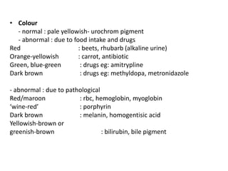


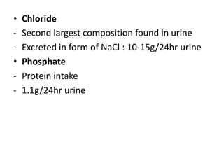


Urine analysis involves collecting a urine sample and using reagent strips or microscopic examination to test for various substances. Reagent strips can detect glucose, bilirubin, ketones, protein, blood, and other parameters. Microscopic examination looks for cells, casts, crystals, and other features. Normal urine composition includes urea, uric acid, creatinine, chloride, and phosphate. Abnormal results may indicate diseases like infections, kidney problems, or other disorders. Care must be taken to collect samples properly for accurate analysis and diagnosis.