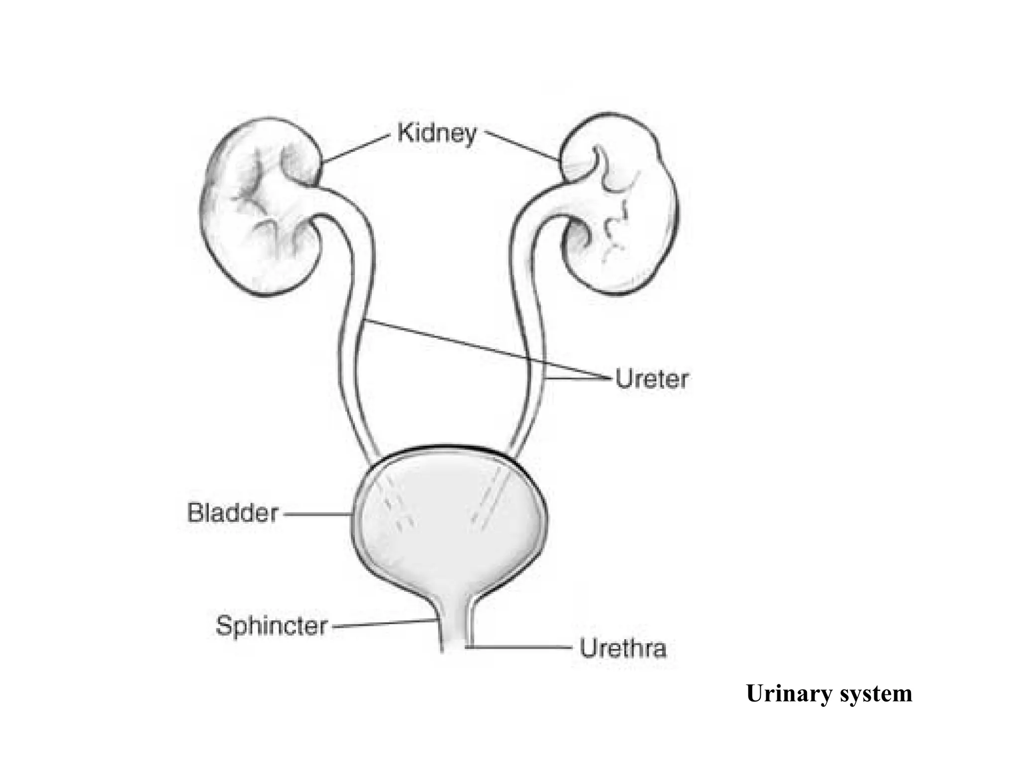
Urinary system
• Urinarysystem consists of pair of kidneys and urinary
tracts which includes (two ureters, urinary bladder and
urethra). Each kidney contains 1.3 million urinary units
called
nephrons.
• Each nephron consist of glomerulus and urinary ducts
(Bowman's capsule, proximal convoluted tubule, Henley
loop, distal convoluted tubule and collecting duct).
The Composition ofUrine
Normal Urine Constituents
Abnormal Urine Constituents
Water (about 95% of urine)
Glucose
Urea
Protein
Creatinine
Bile pigments
Uric acid
Blood cells
Electrolytes
Cast parasites and
Bacterial microbes
6.
The Factors AffectingThe
composition of Urine
• Diet and nutritional status
• Condition of body metabolism
• Ability of kidney function
• Level of contamination with pathogenic
microorganisms ( bacteria) or even non-pathogenic
microflora
7.
Collection And PreservationOf Urine
Specimen
• the urine must be properly collected
– Urine Containers
There are many types of containers used for collecting urine
– Disposable containers of plastic or coated paper are available in
many sizes and are provided with lids to reduce bacterial and
other types of contamination
8.
Types of Specimen
1.First Morning Specimen - a specimen obtained during the first urination
of the day.
– Most concentrated
– Bladder incubated
• good for:
• Nitrite
• Protein
• Microscopic examination
1. Random Specimen - a specimen obtained at any time during
examination.
– Most convenient
– Most common
– Good for:
• ƒChemical Screen
• Microscopic examination
9.
3. Second-voided Specimen- In this case first morning specimen is
discarded and the second specimen is collected and tested. Such
type of specimen is good for:
– Reflection of blood glucose.
– Keeping of formed elements intact
4. Postprandial : a specimen obtained 2 hours after meal.
– Good for glucose.
5. 24- Hour specimen - a specimen obtained within 24 hours.
3. Necessary for quantitative tests, especially for quantitative
determination of protein.
• Mid- stream Specimen - a specimen obtained from the middle
part of the first urine.
It is commonly used for routine urinalysis.
It is also important for bacteriological urine culture.
10.
7. Clean CatchUrine Specimen
Used for microbial culture and routine urinalysis. When
specimens are collected for bacteriological examination
they should be collected by the clean catch’ method or by
catheterization into sterilized container
– Catheterization is the process of passing a tube through the urethra to
the bladder for the withdrawal of urine (it may introduce urinary tract
infection)
11.
Procedure for Collectionof 24 hour
Urine Specimen
1. Inform or Direct the patient to completely empty his bladder and
discard his urine exactly at the beginning of the 24 hour time
collection (let say at 6:00 a.m.).
2. Collect all urine voided during the following 24 hours, including that
voided exactly at the end of the 24 hour period in a container (at
6:00 a.m.) of the following (second) day.
3. All the urine collected must be preserved.
4. The container should be labeled with :
5. The test order
6. The patient’s name
7. Time of collection
8. The preservative added
12.
Preservation of UrineSpecimen
• Urine should be examined immediately as much as possible after
it is passed, because some urinary components are unstable
• Long standing of urine at room temperature can cause :
– Growth of bacteria
– Break down of urea to ammonia by bacteria leading to an increase in the pH of the
urine and this may cause the precipitation of calcium and phosphates.
– Oxidation of urobilingen to urobilin.
– Destruction of glucose by bacteria
– Lysis of RBCs, WBCs and casts
13.
Method of Preservationof Urine Specimen
a. Physical Method
- Refrigeration
- Freezing
b. Chemical Method
Use of chemical preservatives such as :
- Thymol
- Toluene
- Formaldehyde
- Hydrochloric acid ( HCl)
- Chloroform
- Boric acid
- Chlorhexidine
- Sodium carbonate
15.
Type of Examinationin Routine Urinalysis
1. Physical Examination
of Urine
Volume
Color
Odor
Appearance
pH
Specific gravity
2) Chemical Examination of
Urine
Glucose
Protein
Ketones
Bilirubin
Urobilinogen
Blood
Nitrite
Leukocyte Esterase
Melanin
Categories of UrineTests
According to their degree of accuracy urine tests are grouped into
three broad categories:
1.Screening tests: Screening tests tell only whether a substance is present or
absent, and the results are reported as positive or negative
2.Qualitative tests: determine accurately the amount of the substances to be tested
3.Quantitative test :are usually reported in milligrams per deciliter, gram per
deciliter, and per liter. For quantitative test, a complete 24-hour urine specimen is
needed.
18.
Physical Examination OfUrine
Urine volume: This is dependent normally up on fluid intake,
environmental condition, diet and activity of the human .
•Value above or below the normal value (1.5 L/Day) can be considered
as pathological disorder but it should be combined with clinical and
laboratory examination
•above normal (polyuria) urine volume (< 2.5-3L/Day) due to large
quantities intake of liquids, diuretics, alcohol, in sufficient of urinary
ductsinreabsorption of water and urine concentrated as in diabetes
mellitus or diabetes insipidus.
•- under normal (Oligourea) urine volume (< 400 ml/Day)
•- Anuria, urine volume ( < 50 ml/Day), due to: hot weather,
sweating, low water intake, or due to disease in kidney or urinary
ducts.
19.
Color: Can beobserved in a test tube or in a urinometer tube
•Yellow to amber (Normal); the color comes primarily from the
presence of urobilin.
Urobilin is a final waste product resulting from the breakdown
of heme from hemoglobin during the destruction of aging blood
cells.
•Colorless to pale yellow; dilute urine with low specific gravity
and polyuria.
• Dark yellow or yellow brown; concentrated urine with a high
specific gravity and small quantity
•Yellow brown or greenish yellow; yellow green foam when
urine is shakenUrobilinoids – chromagon derived from heme
green biliverdin yellow-brownbillirubin-and urobilin.
20.
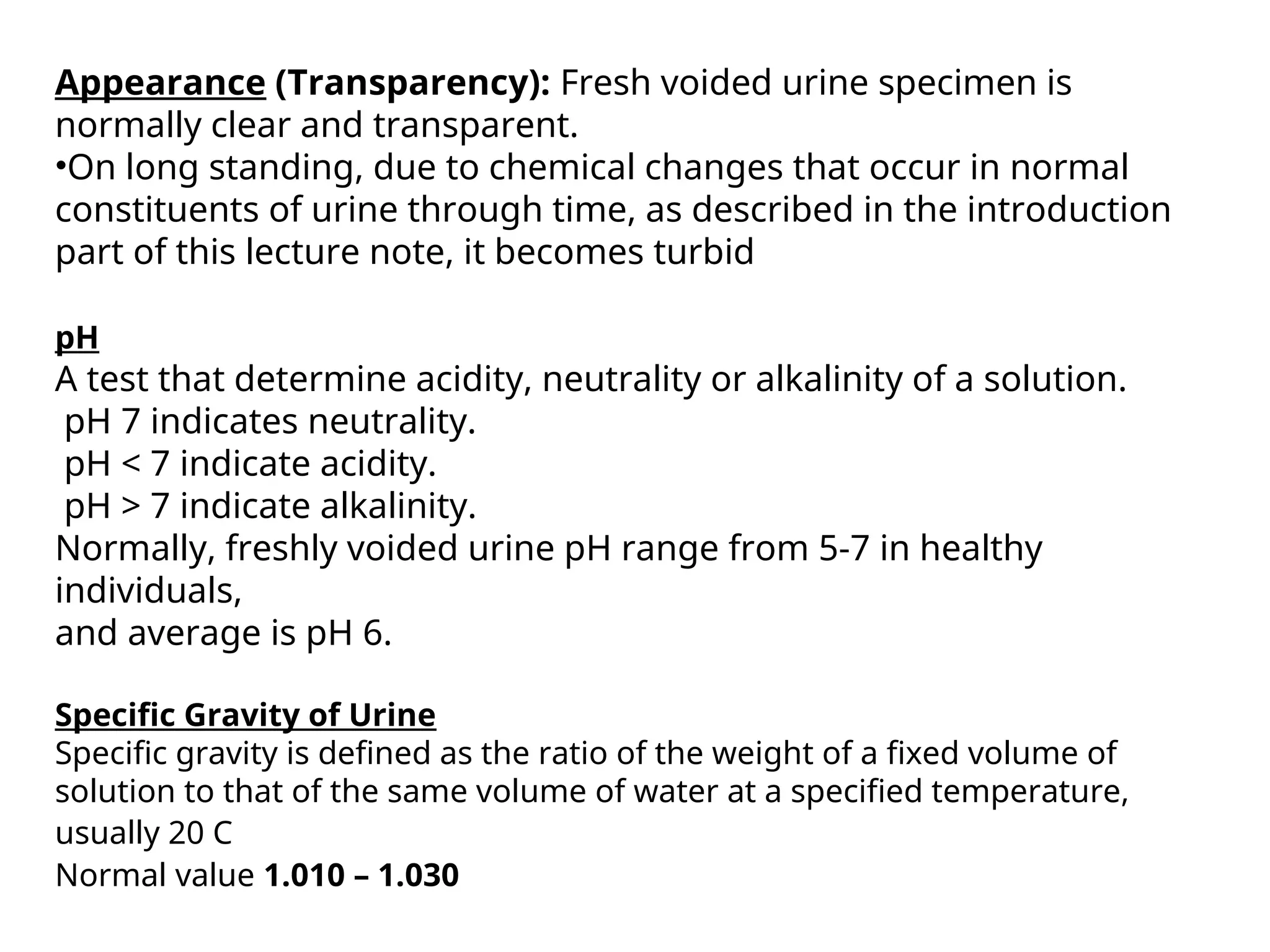
Appearance (Transparency): Freshvoided urine specimen is
normally clear and transparent.
•On long standing, due to chemical changes that occur in normal
constituents of urine through time, as described in the introduction
part of this lecture note, it becomes turbid
pH
A test that determine acidity, neutrality or alkalinity of a solution.
pH 7 indicates neutrality.
pH < 7 indicate acidity.
pH > 7 indicate alkalinity.
Normally, freshly voided urine pH range from 5-7 in healthy
individuals,
and average is pH 6.
Specific Gravity of Urine
Specific gravity is defined as the ratio of the weight of a fixed volume of
solution to that of the same volume of water at a specified temperature,
usually 20 C
Normal value 1.010 – 1.030
21.
Chemical Analysis OfUrine
Determination of Urinary Sugar (Glucose):
Glucose is the sugar most commonly found in the urine, although
other sugars , such as lactose, fructose , galactose, and pentose, may
be found under certain condition. Normally, urine doesnot contain a
sufficient amount of sugar to react with any of the popular enzyme
or reducing tests.
Causes of Glycosuria
• Physiological
• Pathological
Physiological
Sometimes under physiological situations, glycosuria can occur
a. After large ingestion of carbohydrates
b. Anything that stimulates sympathetic nervous system such as
excitement, stress etc.
c. 15 to 20% cases of pregnancy may be associated with
physiological glycosuria.
d. Renal Glycosuria: In some persons, glycosuria is found when
blood glucose is in normal range. This is known as renal
glycosuria. This is again due to lowered renal threshold. Usually
this is a benign condition .
22.
Pathological Glycosuria
A. Diabetesmellitus
The most common condition for glycosuria is diabetes mellitus, a
metabolic disorder due to deficiencies of insulin. Glucose is not properly
metabolized and blood glucose concentration rises, and when it is in
range of 170 - 180 mg /dl , glucose starts appearing in urine.
B. Glycosuria due to other endocrine disorders
Deranged function of a number of endocrine disorders can cause
hyperglycemia and this may result in glycosuria,
e.g. - Hyperthyroidism
- Hyperadrenalism
- Hyperpitutarism
- Some diseases of pancreas
Dipstick chemical analysis
•Urine dipstick is a narrow plastic strip
which has several squares of different
colors attached to it.
• Each small square represents a component
of the test used to interpret urinalysis.
Colors generated by each pad are visually
compared against a range of colors on
brand-specific color charts
• The entire strip is dipped in the urine
sample and color changes in each square
are noted.
26.
The squares onthe dipstick represent
the following components in the urine
Nitrite (suggestive of bacteria in urine)
Bilirubin ( possible liver disease or red blood cell break down)
Urobilinogen ( possible liver disease)
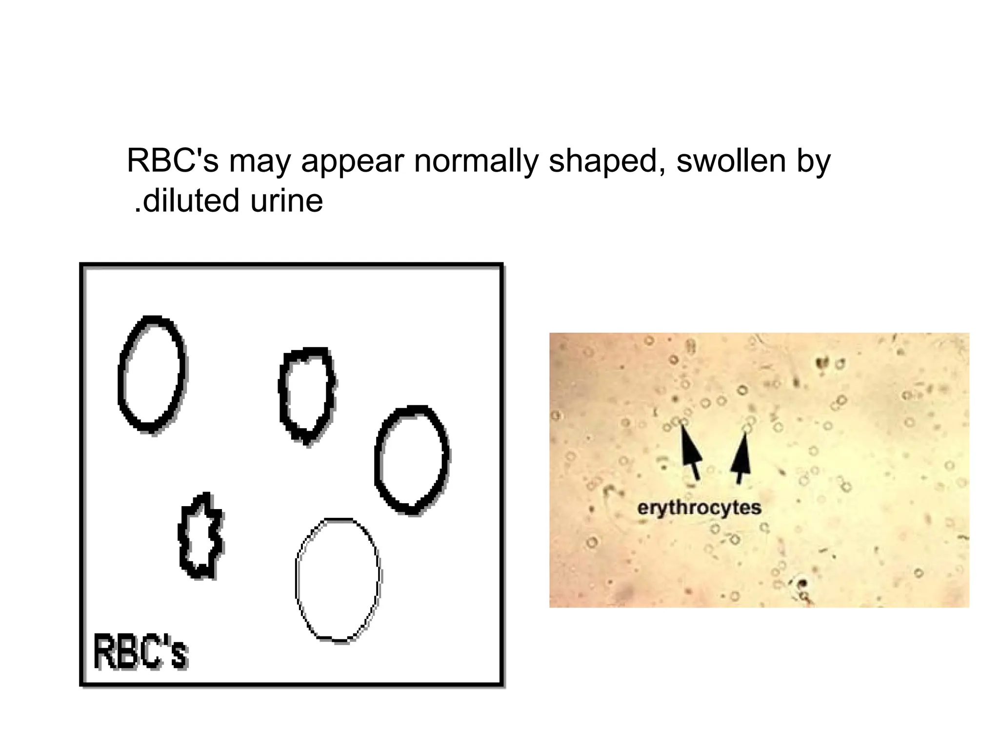
MICROSCOPIC URINALYSIS
Microscopic examinationused to view elements that are
not visible without microscope. e.g cells
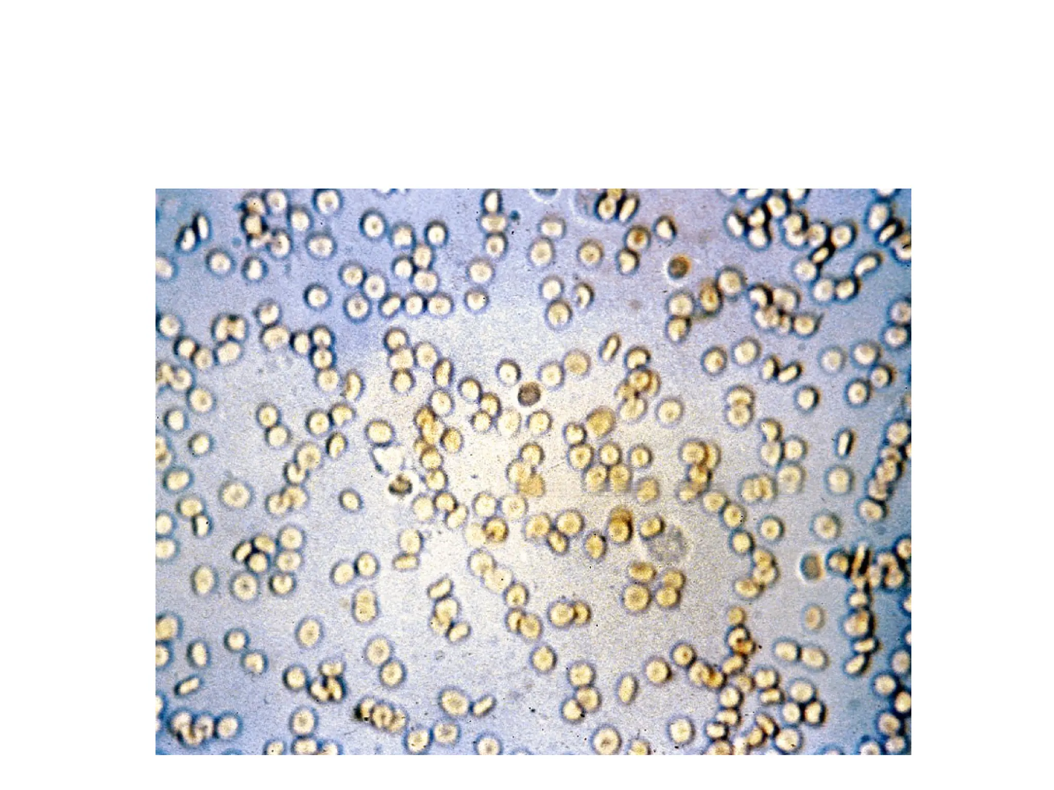
1. Red Blood Cells:
Hematuria is the presence of abnormal numbers of red cells in
urine due to:
a. Glomerular damage
b. Tumors
c. Urinary tract stones
d. Upper and lower urinary tract infections
29.
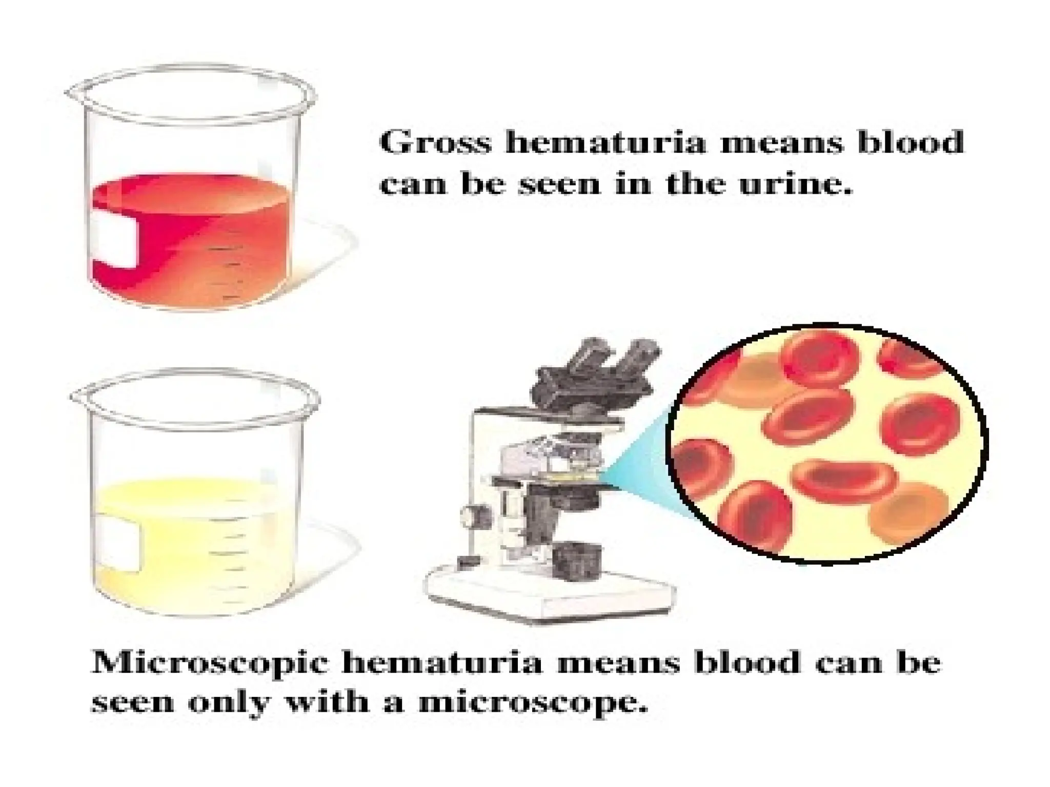
Haematuria
Two Types ofHematuria
• Gross hematuria means that the blood can
be seen by the naked eye. The urine may
look pinkish, brownish, or bright red.
• Microscopic hematuria means that the urine
is clear, but blood cells can be seen under a
microscope.
2
.
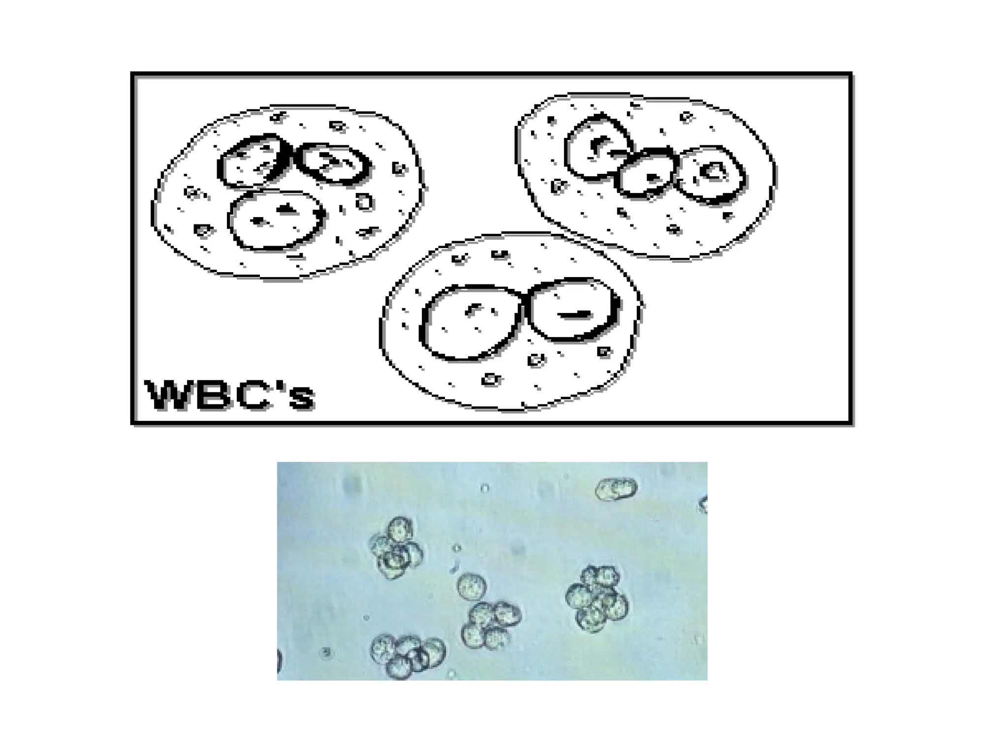
White Blood Cells
Pyuriarefers to the presence of abnormal
numbers of leukocytes that may appear
with infection in either the upper or lower
urinary tract or with acute
glomerulonephritis.
Usually, the WBC's are granulocytes
WBCs - ≤2-5 WBCs/hpf
34.
3
.
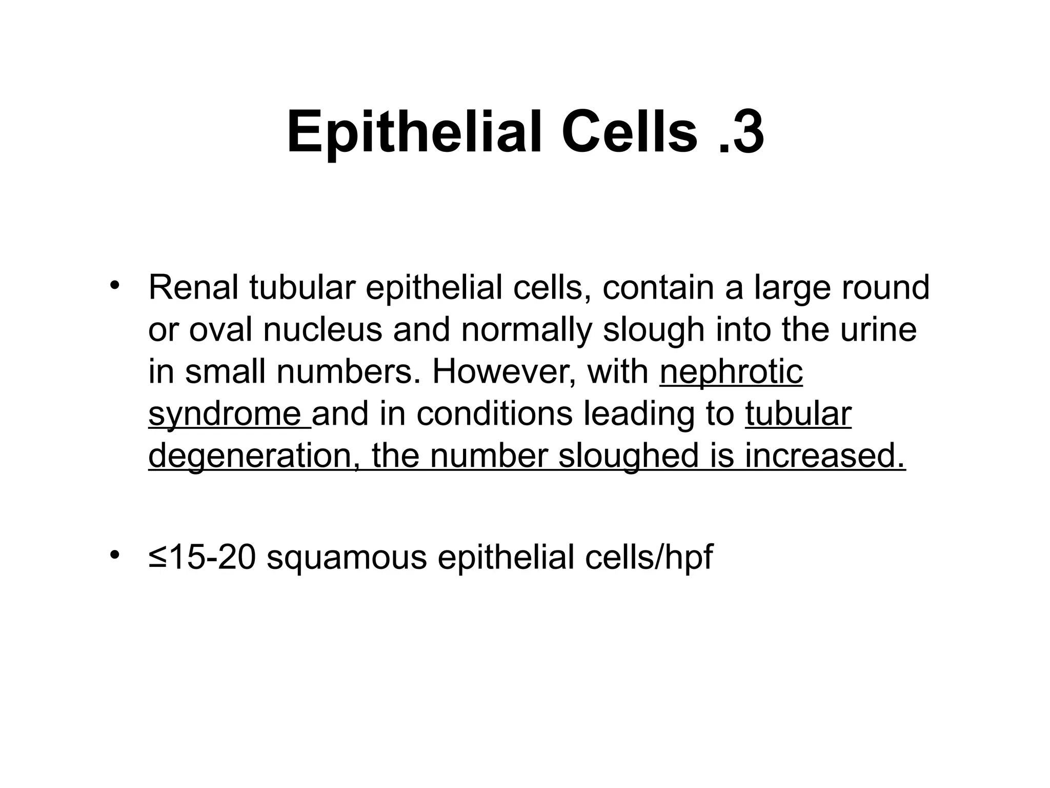
Epithelial Cells
• Renaltubular epithelial cells, contain a large round
or oval nucleus and normally slough into the urine
in small numbers. However, with nephrotic
syndrome and in conditions leading to tubular
degeneration, the number sloughed is increased.
• ≤15-20 squamous epithelial cells/hpf
36.
4
.
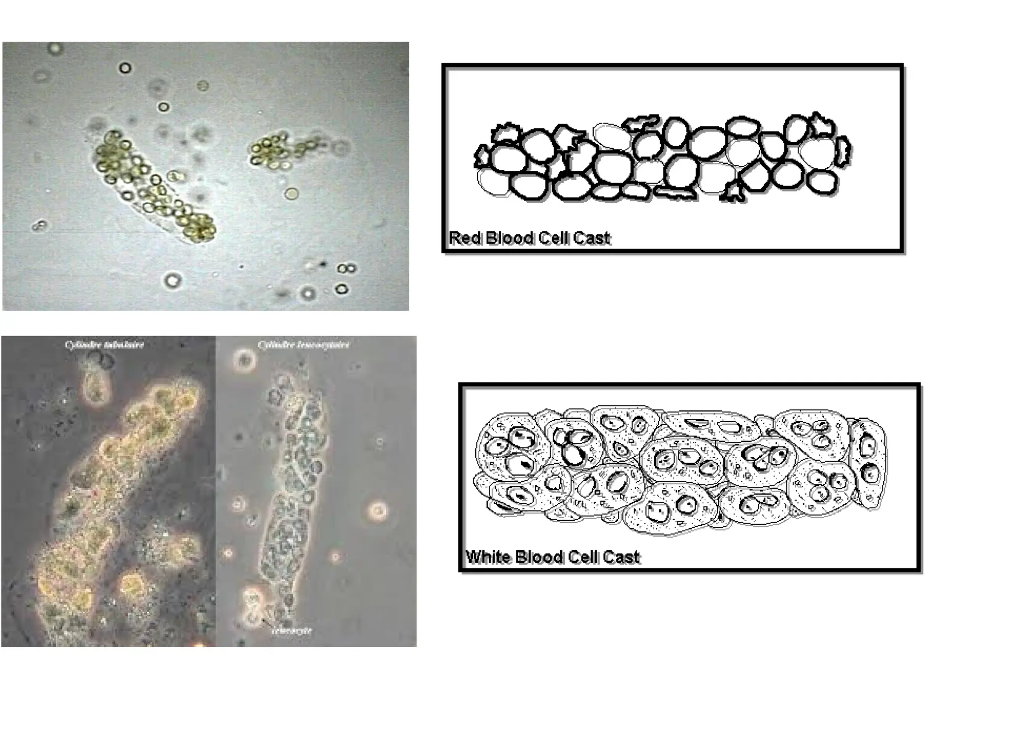
Casts
• Urinary castsare cylindrical structures produced by the
kidney and present in the urine in certain disease states.
• They are formed in the distal convoluted tubule (DCT)
and collecting ducts of nephrons, then dislodge and pass
into the urine, where they can detected by microscopy.
-Urinary casts may be made up of cells (such as white
blood cells, red blood cells, kidney cells) or substances
such as protein.
38.
The factors whichfavor protein cast formation
1
.
low flow rate of the filtrate
2
.
high salt concentration
3
.
low pH
all of which favor protein denaturation and
precipitation, particularly that of the Tamm-Horsfall
protein
.
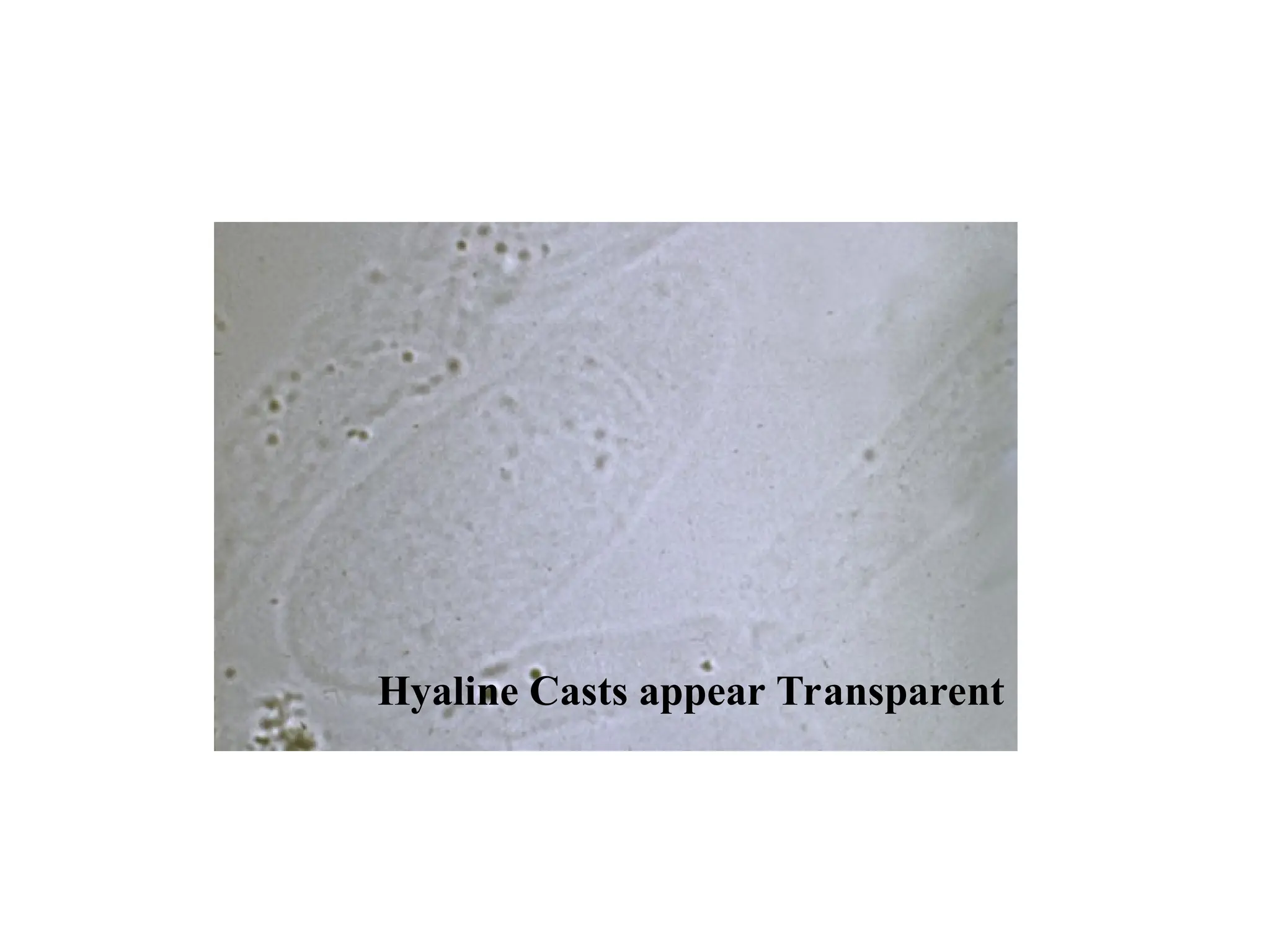
Protein casts with long, thin tails formed at the junction
of Henle's loop and the distal convoluted tubule are
called cylindroids. Hyaline casts (Tamm-Horsfall
proteins) can be seen even in healthy people
.
39.
Hyaline casts arecomposed primarily of a
mucoprotein (Tamm-Horsfall proteins)
secreted by tubule cells. The Tamm-Horsfall
protein secretion (green dots) is illustrated in the
diagram below, forming a hyaline cast in the
collecting duct
40.
Red blood cellsmay stick together and form
red blood cell casts. Such casts are indicative
of glomerulonephritis, with leakage of RBC's
from glomeruli
White blood cell casts may also be present
with glomerulonephritis. Their presence
indicates inflammation of the kidney,
because such casts will not form except in
the kidney
.
Bence Jones proteinsare small proteins found in the
urine. Testing for these proteins is done to diagnose and
monitor multiple myeloma and other similar diseases
.
Bence Jones proteins are considered the first tumor
marker.
A tumor marker is a substance, made by the body, that
is linked to a certain cancer, or malignancy. Bence
Jones proteins are made by plasma cells, a type of white
blood cell. The presence of these proteins in a person's
urine is associated with a malignancy of plasma cells.
Bence Jones proteins
47.
Bence Jones proteincast (myeloma cast) from the urinary sediment
of a patient with lambda-Bence Jones type multiple myeloma.
Sternheimer stein, X200