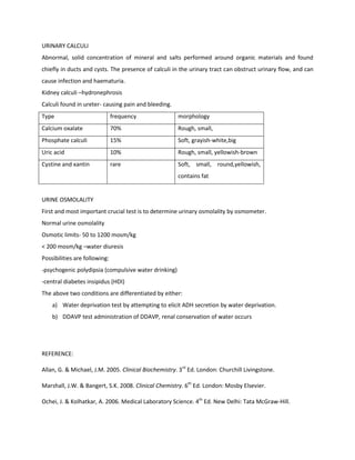
RENAL FUNCTION

1. MAINTENANCE OF WATER AND ELECTROLYTE BALANCE OF THE BODY
The proximal renal tubules reabsorb about 85% of water filtered by the glomerulus, while the rest
passes through the loop of Henle where some of the water is reabsorbed.
The cell lining the collecting ducts which are normally impermeable to water are rendered permeable by
the action of an antidiuretic hormone (ADH, vasopressin). Thus, water is allowed to re-enter circulation.
When the body needs to excrete more water, resulting in more water being excreted in the urine (dilute
urine).

2. MAINTENANCE OF BLOOD PH
Substances that cause the pH of blood to rise or fall are removed by the kidneys. Substances of acid
reaction are waste products of protein metabolism-urea, uric acid and creatinine.

3. EXCRETION
Waste products of drugs which are left in the body are later on excreted via kidneys. Toxic substances
which are rendered harmless in the liver are excreted by the kidneys in the urine.

4. FILTRATION
The glomerular filtration rate (GFR) measures the efficiency of this filtration and it is determined by urea
and creatinine clearance tests. The filtrate passes from the Bowman’s capsule into the renal tubule.
During this passage, the tubular cells reabsorb those substances that are needed by the body into the
bloodstream.

5. PRODUCTION OF ERYTHROPOEITIN
The substance that is necessary for the normal production of rbc in the bone marrow is the
erythropoietin. It is produced by the kidneys.

6. REABSORPTION
Desirable elements in the filtrate such as glucose and amino acids are reabsorbed. This reabsorption
occurs mainly in the proximal convulated tubules.

AETIOLOGY

      -      Acute renal failure (ARF)-the kidneys fail over a period of hours or days. May be reversed and
             normal renal function regained.

      -      Chronic Renal Failure (CRF)-develops over months or years and leads eventually to end stage of
             renal failure (ESRF). Irreversible.

ARF

      •      Sudden deterioration of renal function indicated by rapid rising of serum urea and creatinine
             concentration.

Kidney failure can be classified as:

          -Pre-renal : the kidney fails to receive a proper blood supply

          -Post-renal : the urinary drainage of the kidney is impaired because of an obstruction.
-Renal : intrinsic damage to the kidney tissue. This may be due to a variety of diseases, or the renal

        damage may be a consequence of prolonged pre-renal or post-renal problems.

DIAGNOSIS

         Decreased plasma volume because of blood loss, burns, prolonged vomiting or diarrhea
         Diminished cardiac output
         Local factors, such as occlusion of renal artery


BIOCHEMICAL FINDINGS

         Serum urea and creatinine are increased
         Metabolic acidosis
         Hyperkalaemia
         A high urine osmolality

          Post-renal factor: filtration at the glomeruli is reduced caused by blockage
          e.g; renal stones, carcinoma of cervix, prostate or occasionally bladder


ACUTE TUBULAR NECROSIS

         Acute blood loss in severe trauma
         Septic shock
         Specific renal disease- glomerulonephritis

MANAGEMENT

         Correction of pre-renal factors, if present, by replacement of any ECF volume deficit.
         Treatment of underlying disease
         Biochemical monitoring
         Dialysis

Consequences of CRF

Sodium and water metabolism

         Retain ability to reabsorb sodium
         Or renal tubules may loose ability to reabsorb water and there for urine is concentrated.

Potassium metabolism

         Sudden deterioration of renal function may precipitate a rapid rise in serum potassium
          concentration.

Acid-base balance

         Retention of hydrogen ions-metabolic acidosis
Calcium and phosphate metabolism
     The ability of renal cells to make vitamin D decrease
     Calcium absorption is reduced-hypercalcemia
     Parathyroid hormone is stimulated in a attempt to restore plasma calcium to normal, but
       adverse effects of renal osteodystrophy

Erythropoietin synthesis
      Normochromic normocytic anaemia is due to failure of erythropoietin production.
      Biosynthesized human erythropoietin may be used to treat the anaemia of CRF.

Management

       Water and sodium intake should be carefully matched to the losses.
       Hyperkalaemia may be controlled by oral ion-exchange resins (resonium A).
       Hyperphosphataemia may be controlled by oral aluminium or magnesium salts
       The administration of hydroxylated vitamin D metabolites may prevent the development of
        secondary hyperparathyroidism.
       Dietary restriction of protein, to reduce the formation of nitrogenous waste product.
       Dialysis & renal transplant


RENAL FUNCTION TESTS
UREA
Urea is carried by the plasma to the kidney where it is filtered from the plasma by the glomerulus. About
40% of the urea in the glomerular filtrate is reabsorbed by the renal tubules.
ESTIMATION OF UREA
Normal range of blood urea
Adults: 2.5-7.0 mmol/L (15-40 mg/dl)
Excess of urea in blood- uraemia
Result of kidney impairment
Urea clearance test
Sample: 24 hour urine specimen & blood sample
Normal range: 40-65 ml/min
< 10 ml/min during renal failure
CREATININE
A nitrogenous product which is produce from the metabolism of creatinine from the metabolism of
creatine in skeletal muscles. It is filtered ion kidneys and excreted in the urine.
CREATININE CLEARANCE TEST
Sample: 24 hour urine specimen & blood sample
Normal range for creatinine       serum/plasma
Adult men 88-135 umol/L
Adult female 62-115 umol/L
Creatinine clearance
Adult men 97-137 ml/min
Adult female 88-128 ml/min
High serum or plasma creatinine levels- kidney impairment


        Creatinine clearance is calculated from the formula:


        U
                 urinary creatinine concentration (μmol/L)
        V
                 urine flow rate (mL/min i.e., 24hr urine volume(ml)divided by 1440(min))
        P
                 plasma creatinine concentration (μmol/L)

URIC ACID
Uric acid is the final breakdown product of purine metabolism.
Converted in liver into uric acid and filtered in kidney.
Nearly all of the uric acid in plasma is in the form of monosodium urate- insoluble
At high levels (> 6.4 mg/dl), the plasma is saturated; urate crystals may form and precipitate in tissues
Normal range:
Adult males: 3.5-7.2 mg/dl
Adult female: 2.6-6.0 mg/dl
Gout is an arthropathy caused by the precipitation of monosodium urate crystals in synovial joints. This
occurs when plasma urate concentrations are elevated. Secondary causes of hyperuricaemia include
renal disease, thiazide diuretics, increased cell turnover and a high intake of purine-rich foods. Gout and
hyperuricaemia show a strong familial incidence. Some very rare inherited defects in purine metabolism
that cause hyperuricaemia have been described, but in the majority of patients, there is no such defect,
and hyperuricaemia is thought to be due to decreased renal urate excretion. Gout usually presents as an
acute arthritis, but can lead to chronic joint disease, and crystals of monosodium urate can be deposited
in tissues and in the renal tubules, causing an obstructive uropathy.
URINARY CALCULI
Abnormal, solid concentration of mineral and salts performed around organic materials and found
chiefly in ducts and cysts. The presence of calculi in the urinary tract can obstruct urinary flow, and can
cause infection and haematuria.
Kidney calculi –hydronephrosis
Calculi found in ureter- causing pain and bleeding.
Type                           frequency              morphology
Calcium oxalate                70%                    Rough, small,
Phosphate calculi              15%                    Soft, grayish-white,big
Uric acid                      10%                    Rough, small, yellowish-brown
Cystine and xantin             rare                   Soft, small, round,yellowish,
                                                      contains fat


URINE OSMOLALITY
First and most important crucial test is to determine urinary osmolality by osmometer.
Normal urine osmolality
Osmotic limits- 50 to 1200 mosm/kg
< 200 mosm/kg –water diuresis
Possibilities are following:
-psychogenic polydipsia (compulsive water drinking)
-central diabetes insipidus (HDI)
The above two conditions are differentiated by either:
    a) Water deprivation test by attempting to elicit ADH secretion by water deprivation.
    b) DDAVP test administration of DDAVP, renal conservation of water occurs


REFERENCE:

Allan, G. & Michael, J.M. 2005. Clinical Biochemistry. 3rd Ed. London: Churchill Livingstone.

Marshall, J.W. & Bangert, S.K. 2008. Clinical Chemistry. 6th Ed. London: Mosby Elsevier.

Ochei, J. & Kolhatkar, A. 2006. Medical Laboratory Science. 4th Ed. New Delhi: Tata McGraw-Hill.

Renal function

  • 1.
    RENAL FUNCTION 1. MAINTENANCEOF WATER AND ELECTROLYTE BALANCE OF THE BODY The proximal renal tubules reabsorb about 85% of water filtered by the glomerulus, while the rest passes through the loop of Henle where some of the water is reabsorbed. The cell lining the collecting ducts which are normally impermeable to water are rendered permeable by the action of an antidiuretic hormone (ADH, vasopressin). Thus, water is allowed to re-enter circulation. When the body needs to excrete more water, resulting in more water being excreted in the urine (dilute urine). 2. MAINTENANCE OF BLOOD PH Substances that cause the pH of blood to rise or fall are removed by the kidneys. Substances of acid reaction are waste products of protein metabolism-urea, uric acid and creatinine. 3. EXCRETION Waste products of drugs which are left in the body are later on excreted via kidneys. Toxic substances which are rendered harmless in the liver are excreted by the kidneys in the urine. 4. FILTRATION The glomerular filtration rate (GFR) measures the efficiency of this filtration and it is determined by urea and creatinine clearance tests. The filtrate passes from the Bowman’s capsule into the renal tubule. During this passage, the tubular cells reabsorb those substances that are needed by the body into the bloodstream. 5. PRODUCTION OF ERYTHROPOEITIN The substance that is necessary for the normal production of rbc in the bone marrow is the erythropoietin. It is produced by the kidneys. 6. REABSORPTION Desirable elements in the filtrate such as glucose and amino acids are reabsorbed. This reabsorption occurs mainly in the proximal convulated tubules. AETIOLOGY - Acute renal failure (ARF)-the kidneys fail over a period of hours or days. May be reversed and normal renal function regained. - Chronic Renal Failure (CRF)-develops over months or years and leads eventually to end stage of renal failure (ESRF). Irreversible. ARF • Sudden deterioration of renal function indicated by rapid rising of serum urea and creatinine concentration. Kidney failure can be classified as: -Pre-renal : the kidney fails to receive a proper blood supply -Post-renal : the urinary drainage of the kidney is impaired because of an obstruction.
  • 2.
    -Renal : intrinsicdamage to the kidney tissue. This may be due to a variety of diseases, or the renal damage may be a consequence of prolonged pre-renal or post-renal problems. DIAGNOSIS  Decreased plasma volume because of blood loss, burns, prolonged vomiting or diarrhea  Diminished cardiac output  Local factors, such as occlusion of renal artery BIOCHEMICAL FINDINGS  Serum urea and creatinine are increased  Metabolic acidosis  Hyperkalaemia  A high urine osmolality Post-renal factor: filtration at the glomeruli is reduced caused by blockage e.g; renal stones, carcinoma of cervix, prostate or occasionally bladder ACUTE TUBULAR NECROSIS  Acute blood loss in severe trauma  Septic shock  Specific renal disease- glomerulonephritis MANAGEMENT  Correction of pre-renal factors, if present, by replacement of any ECF volume deficit.  Treatment of underlying disease  Biochemical monitoring  Dialysis Consequences of CRF Sodium and water metabolism  Retain ability to reabsorb sodium  Or renal tubules may loose ability to reabsorb water and there for urine is concentrated. Potassium metabolism  Sudden deterioration of renal function may precipitate a rapid rise in serum potassium concentration. Acid-base balance  Retention of hydrogen ions-metabolic acidosis
  • 3.
    Calcium and phosphatemetabolism  The ability of renal cells to make vitamin D decrease  Calcium absorption is reduced-hypercalcemia  Parathyroid hormone is stimulated in a attempt to restore plasma calcium to normal, but adverse effects of renal osteodystrophy Erythropoietin synthesis  Normochromic normocytic anaemia is due to failure of erythropoietin production.  Biosynthesized human erythropoietin may be used to treat the anaemia of CRF. Management  Water and sodium intake should be carefully matched to the losses.  Hyperkalaemia may be controlled by oral ion-exchange resins (resonium A).  Hyperphosphataemia may be controlled by oral aluminium or magnesium salts  The administration of hydroxylated vitamin D metabolites may prevent the development of secondary hyperparathyroidism.  Dietary restriction of protein, to reduce the formation of nitrogenous waste product.  Dialysis & renal transplant RENAL FUNCTION TESTS UREA Urea is carried by the plasma to the kidney where it is filtered from the plasma by the glomerulus. About 40% of the urea in the glomerular filtrate is reabsorbed by the renal tubules. ESTIMATION OF UREA Normal range of blood urea Adults: 2.5-7.0 mmol/L (15-40 mg/dl) Excess of urea in blood- uraemia Result of kidney impairment Urea clearance test Sample: 24 hour urine specimen & blood sample Normal range: 40-65 ml/min < 10 ml/min during renal failure CREATININE A nitrogenous product which is produce from the metabolism of creatinine from the metabolism of creatine in skeletal muscles. It is filtered ion kidneys and excreted in the urine.
  • 4.
    CREATININE CLEARANCE TEST Sample:24 hour urine specimen & blood sample Normal range for creatinine serum/plasma Adult men 88-135 umol/L Adult female 62-115 umol/L Creatinine clearance Adult men 97-137 ml/min Adult female 88-128 ml/min High serum or plasma creatinine levels- kidney impairment Creatinine clearance is calculated from the formula: U urinary creatinine concentration (μmol/L) V urine flow rate (mL/min i.e., 24hr urine volume(ml)divided by 1440(min)) P plasma creatinine concentration (μmol/L) URIC ACID Uric acid is the final breakdown product of purine metabolism. Converted in liver into uric acid and filtered in kidney. Nearly all of the uric acid in plasma is in the form of monosodium urate- insoluble At high levels (> 6.4 mg/dl), the plasma is saturated; urate crystals may form and precipitate in tissues Normal range: Adult males: 3.5-7.2 mg/dl Adult female: 2.6-6.0 mg/dl Gout is an arthropathy caused by the precipitation of monosodium urate crystals in synovial joints. This occurs when plasma urate concentrations are elevated. Secondary causes of hyperuricaemia include renal disease, thiazide diuretics, increased cell turnover and a high intake of purine-rich foods. Gout and hyperuricaemia show a strong familial incidence. Some very rare inherited defects in purine metabolism that cause hyperuricaemia have been described, but in the majority of patients, there is no such defect, and hyperuricaemia is thought to be due to decreased renal urate excretion. Gout usually presents as an acute arthritis, but can lead to chronic joint disease, and crystals of monosodium urate can be deposited in tissues and in the renal tubules, causing an obstructive uropathy.
  • 5.
    URINARY CALCULI Abnormal, solidconcentration of mineral and salts performed around organic materials and found chiefly in ducts and cysts. The presence of calculi in the urinary tract can obstruct urinary flow, and can cause infection and haematuria. Kidney calculi –hydronephrosis Calculi found in ureter- causing pain and bleeding. Type frequency morphology Calcium oxalate 70% Rough, small, Phosphate calculi 15% Soft, grayish-white,big Uric acid 10% Rough, small, yellowish-brown Cystine and xantin rare Soft, small, round,yellowish, contains fat URINE OSMOLALITY First and most important crucial test is to determine urinary osmolality by osmometer. Normal urine osmolality Osmotic limits- 50 to 1200 mosm/kg < 200 mosm/kg –water diuresis Possibilities are following: -psychogenic polydipsia (compulsive water drinking) -central diabetes insipidus (HDI) The above two conditions are differentiated by either: a) Water deprivation test by attempting to elicit ADH secretion by water deprivation. b) DDAVP test administration of DDAVP, renal conservation of water occurs REFERENCE: Allan, G. & Michael, J.M. 2005. Clinical Biochemistry. 3rd Ed. London: Churchill Livingstone. Marshall, J.W. & Bangert, S.K. 2008. Clinical Chemistry. 6th Ed. London: Mosby Elsevier. Ochei, J. & Kolhatkar, A. 2006. Medical Laboratory Science. 4th Ed. New Delhi: Tata McGraw-Hill.