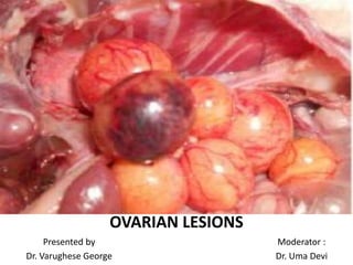
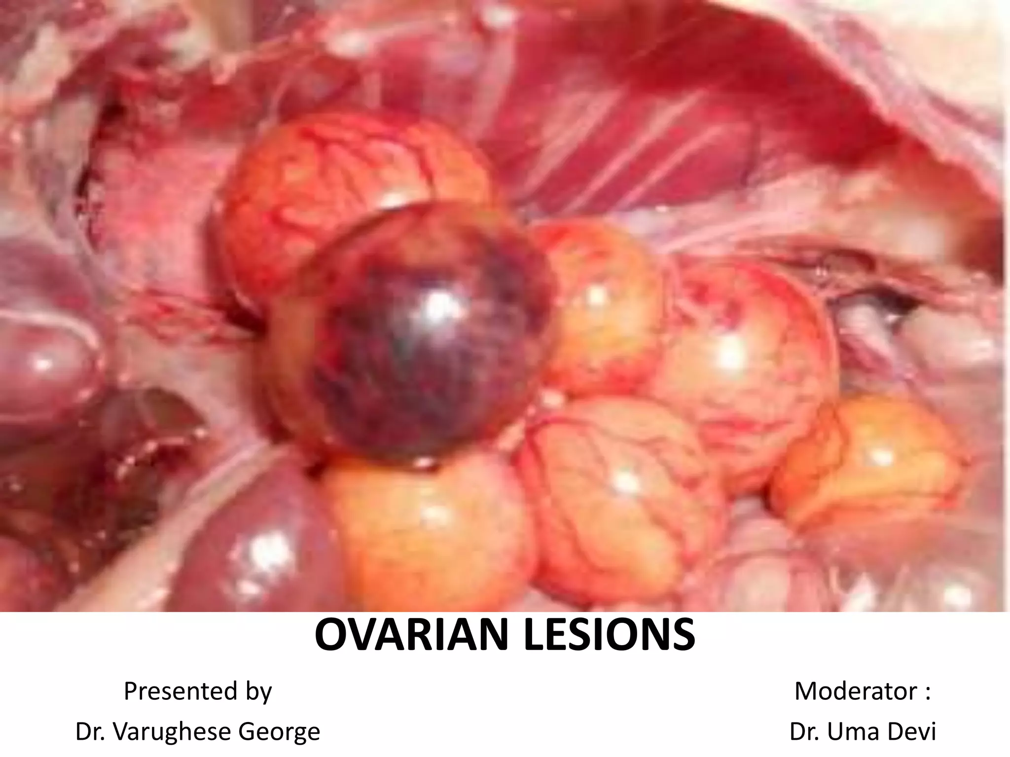
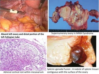
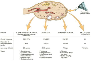
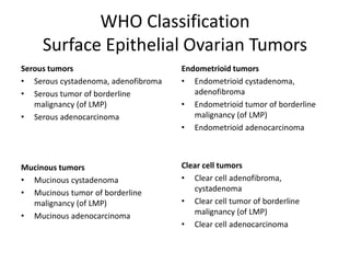
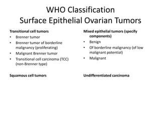
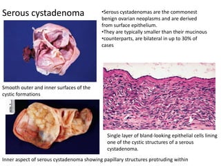
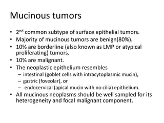
SlideShare a Scribd company logo
OVARIAN LESIONS
Presented by
Dr. Varughese George
Moderator :
Dr. Uma Devi
NON-NEOPLASTIC OVARIAN LESIONS
Congenital Lesions and Ectopic Tissues
• Absent Ovary
• Lobulated, Accessory, and Supernumerary Ovary
• Adrenal Cortical Rests
• Uterus-Like Ovarian Mass
• Splenic–Gonadal Fusion
• Ectopic prostatic tissue
Adrenal cortical rest within mesovarium
Splenic–gonadal fusion - A nodule of splenic tissue i
contiguous with the surface of the ovary
Supernumerary ovary in MRKH SyndromeAbsent left ovary and distal portion of the
left Fallopian tube
Infections
Bacterial
• Pelvic inflammatory disease (PID)
of bacterial origin accounts for
most ovarian infections.
• Ovarian involvement by PID is
almost always secondary to
salpingitis and typically takes the
form of a tuboovarian abscess.
• A mixed flora with a
preponderance of anaerobic
• organisms is typically recovered
from the contents of the
• abscess
Tuboovarian abscess.
Lining consists of inflammatory debris
with subjacent granulation tissue
Infections
Bacterial
Xanthogranuloma of ovary.
Inflammatory reaction consists
predominantly of foamy histiocytes
Ovarian xanthogranuloma,
sectioned surface
Rarely, a chronic ovarian abscess may result in a tumor-like mass, variably designated
ovarian xanthogranuloma
Infections
Bacterial
Malacoplakia of ovary - Mixed inflammatory
infiltrate with epithelioid histiocytes, some
containing Michaelis–Guttman bodies.
Associated with E coli and Enterococcus faecalis.
Actinomycosis of ovary - A colony of
actinomyces (sulfur granule) is
surrounded by a purulent exudate.
Infections
Bacterial
Tuberculosis
• Tuberculous oophoritis is uncommon and usually secondary to
tuberculous salpingitis.
Leprosy
• rarely involves the female genital tract.
• the ovary is the most commonly involved gynecologic site.
• In chronic forms of leprous oophoritis, a chronic inflammatory
cell infiltrate and fibrosis are seen, and bacilli usually are
demonstrable.
Syphilis
• Syphilitic involvement of the ovary is very rare.
• Luetic oophoritis has been described in congenital, secondary,
and tertiary forms of the disease.
Infections
Parasitic
Ovarian involvement by
Enterobius vermicularis.
Microscopic diagnosis showed cystic lesions with a cellular
membrane and the scolices in the daughter cyst
Macroscopic appearance of the resected
hydatid cyst
Inflammation
Granulomatous reaction on ovarian surface
potentially mimicking a malignant tumor.
Yellowish-brown granular deposits on ovarian
surface, representing implants of keratin derived
from an endometrial adenocarcinoma with
squamous differentation
Foreign body reaction to keratin implants on
ovarian serosal surface.
There was a coexistent endometrial
adenocarcinoma with squamous
differentiation, confined to the uterus
Inflammation
Gallstone implanted on ovarian surfacePostcautery carbon pigment granuloma in
ovary. Note black carbon pigment.
Palisading histiocytes are seen at the bottom
and right.
Inflammation
Isolated palisading granuloma of ovary Cortical granuloma.
Circumscribed collection of lymphocytes,
spindle cells, and an occasional multinucleated
giant cell lie within the ovarian stroma
Non-neoplastic Lesions of the
Follicular and Stromal Elements
Lining consists of an inner layer of
granulosa cells and an outer layer
of theca interna cells(reticulin).
Follicular cyst
Follicular cysts measure 2.5 to 10 cm in
diameter and contain serosanguineous
fluid.
Non-neoplastic Lesions of the
Follicular and Stromal Elements
A prominent inner layer of connective
tissue is present
.
Corpus luteum cyst
Corpus luteum cyst. Note the convoluted
yellow lining
Non-neoplastic Lesions of the
Follicular and Stromal Elements
Higher-power view of lining cells
showing marked nuclear
hyperchromasia and atypicality.
•Luteinized cells with abundant eosinophilic
•to clear cytoplasm line a cyst (upper) and lie
within its fibrous wall (lower).
•Note focal nuclear atypicality.
Large solitary luteinized follicle cyst of pregnancy and puerperium
Polycystic ovary disease.
Superficial cortical fibrosis and multiple cystic
follicles are present
Multiple cystic follicles lie beneath the
superficially fibrotic cortex.
A cystic follicle is lined by non-luteinized granulosa cells and an outer,
thicker layer of luteinized theca interna cells
Polycystic Ovary Syndrome
Stromal hyperplasia. (a) diffuse proliferation
of ovarian stromal cells within the cortex&
medulla is seen. (b)absence of follicular
derivatives
Stromal hyperthecosis. (a) A nest of luteinized
stromal cells is present within the ovarian
stroma. (b) Calretinin immunostain
highlighting luteinized stromal cells
Massive Ovarian Edema
•The involved ovary in massive edema is
enlarged, soft and fluctuant, ranging from 5.5
to 35 cm.
•The ovary has a shiny, white, smooth exterior
as a result of a white and fibrotic superficial
cortex and a sectioned surface that is
edematous or gelatinous and exudes watery
fluid
The edematous ovarian stroma
separates several corpora fibrosa
Luteinized stromal cells are present
Ovarian Fibromatosis
Bilateral ovarian fibromatosis.
Convoluted external surface with patchy areas
of white, fibrous tissue (right ovary)
Fibrotic spindle cell proliferation invests
follicular structures within the cortex
Fibrotic ovarian stroma surrounds a preantral follicle
Characterized by a fibromatoid proliferation of
collagen-producing spindle cells that typically
surrounds normal follicular structures and
thickens the superficial cortex
Ovarian Endometriosis
Multiple, hemorrhagic lesions involve the
ovarian surface
Microscopically, composed of endometrial
glands, endometrial stroma, and fresh and old
hemorrhagic foci with accumulation of
hemosiderin-laden macrophages.
Autoimmune oophoritis
• Poorly understood disorder
characterized microscopically
by lymphocytic and plasma
cell infiltration in relation to
developing follicles but not
primordial follicles.
• results in primary ovarian
failure with either primary or
secondary amenorrhea.
• Many of the reported cases
have been associated with
adrenal failure (Addison
disease), hypothyroidism, or
both conditions.
Lymphoplasmacytic infiltrate of ovary in
oophoritis.
NEOPLASTIC OVARIAN LESIONS
WHO Classification
Surface Epithelial Ovarian Tumors
Serous tumors
• Serous cystadenoma, adenofibroma
• Serous tumor of borderline
malignancy (of LMP)
• Serous adenocarcinoma
Mucinous tumors
• Mucinous cystadenoma
• Mucinous tumor of borderline
malignancy (of LMP)
• Mucinous adenocarcinoma
Endometrioid tumors
• Endometrioid cystadenoma,
adenofibroma
• Endometrioid tumor of borderline
malignancy (of LMP)
• Endometrioid adenocarcinoma
Clear cell tumors
• Clear cell adenofibroma,
cystadenoma
• Clear cell tumor of borderline
malignancy (of LMP)
• Clear cell adenocarcinoma
WHO Classification
Surface Epithelial Ovarian Tumors
Transitional cell tumors
• Brenner tumor
• Brenner tumor of borderline
malignancy (proliferating)
• Malignant Brenner tumor
• Transitional cell carcinoma (TCC)
(non-Brenner type)
Squamous cell tumors
Mixed epithelial tumors (specify
components)
• Benign
• Of borderline malignancy (of low
malignant potential)
• Malignant
Undifferentiated carcinoma
Serous tumors
• Most common subtype of surface epithelial tumors.
• Occur in women aged 30 to 40 years.
• Majority of serous tumors are benign(60%).
• 15% are borderline (also known as LMP or atypical
proliferating) tumors.
• 25% are malignant.
• The neoplastic epithelium, particularly in benign &
borderline tumors, resembles fallopian tube epithelium
and contains ciliated and secretory cells.
• The cystic spaces contain serous or serosanguineous
fluid.
Smooth outer and inner surfaces of the
cystic formations
Inner aspect of serous cystadenoma showing papillary structures protruding within
Serous cystadenoma
Single layer of bland-looking epithelial cells lining
one of the cystic structures of a serous
cystadenoma.
•Serous cystadenomas are the commonest
benign ovarian neoplasms and are derived
from surface epithelium.
•They are typically smaller than their mucinous
•counterparts, are bilateral in up to 30% of
cases
Serous cystadenofibroma
•The tumor slightly lobulated and made up of
cystic and solid structures.
• The tumor is firm and hard on cut section.
•It is yellowish gray.
•The cystic spaces show a smooth inner lining.
The stroma is composed of dense, fibrous
tissue and dilated cyst-like glands lined with
cuboidal epithelial cells.
solid neoplasms composed of tough, rubbery tissue with interspersed glandular spaces.
Serous tumor of borderline malignancy
•Inner aspect of an ovarian borderline serous
neoplasm.
•Numerous papillary projections facing the lumen
can be appreciated.
Borderline or low malignant potential
serous tumors are lined by a complex
branching papillary epithelium, without
stromal invasion.
Tumors show some or all of the features associated with carcinoma but lack definite
stromal invasion, i.e. irregular or destructive stromal infiltration by small glands or sheets
of cells
Serous tumor of borderline malignancy
Peritoneal implants associated with borderline tumors are classified as
non-invasive or invasive
Nests of neoplastic serous epithelium are
surrounded by a markedly desmoplastic
stroma; however, the entire implant is well
circumscribed without invasion of the
surrounding adipose tissue.
Nests of neoplastic serous epithelium
irregularly infiltrate the muscularis propria of
the bowel wall.
Serous tumor of borderline malignancy
Two additional architectural patterns may be seen:
micropapillary & cribriform
Long, thin, papillae emanate directly from
stromal cores giving a “medusa head”
appearance.
The tips of papillae fuse to form cribriform
structures.
Serous tumor of borderline malignancy with
microinvasion
Borderline serous neoplasm with foci of
microinvasion represented by clusters of cells
with abundant eosinophilic cytoplasm.
The center of the photo shows small
nests and individual cells with abundant
eosinophilic cytoplasm surrounded by a
clefted space and desmoplastic stroma.
This is accompanied in about half of the cases with lymphovascular invasion
Serous cystadenocarcinoma
The tumor is predominantly solid, with
multiple papillary excrescences, necrotic and
hemorrhagic areas.
Grossly, the tumors are large and friable
with multiloculated cysts and polypoid growths.
Serous cystadenocarcinoma
Low-grade serous carcinoma.
Nests of serous carcinoma with minimal
cytologic atypia and a low mitotic rate
(< 12 mitoses/1 0 high power fields)
High-grade serous carcinoma.
•The tumor forms papillary structures and slit-
like spaces.
•There is marked cytologic atypia and abnormal
mitotic figures.
•This tumor invades omental adipose tissue.
● Vimentin positive
● CA-125 positive
● > 60% of high-grade tumors and <
10% of low-grade tumors are positive
for p53
● WT-1 positive
Serous psammocarcinoma
A rare form of serous adenocarcinoma
•Ovarian surface is bosselated with breach in
capsule.
•Cut section shows solid with variegated areas
and >50% of the tumor showing gritty yellowish
areas of calcification
Tumor showing
•destructive invasion of stroma
•no more than moderate cytologic atypia
•abundant psammoma bodies in at least 75%
of the papillae
•no areas of solid growth > 15 cells.
Mucinous tumors
• 2nd common subtype of surface epithelial tumors.
• Majority of mucinous tumors are benign(80%).
• 10% are borderline (also known as LMP or atypical
proliferating) tumors.
• 10% are malignant.
• The neoplastic epithelium resembles
– intestinal (goblet cells with intracytoplasmic mucin),
– gastric (foveolar), or
– endocervical (apical mucin with no cilia) epithelium.
• All mucinous neoplasms should be well sampled for its
heterogeneity and focal malignant component.
Mucinous cystadenomas
•Tumors are usually unilateral, multiloculated, and large(up to 50 cm).
•They have a smooth external surface and are filled with thick viscous secretions.
•The cut surface reveals unilocular or multilocular mucin-filled cysts of varying size.
Outer and inner aspect
Mucinous cystadenomas
Cyst wall lined by a single layer of tall columnar
cells with clear cytoplasm and bland basal
nuclei, most often of intestinal type.
In this instance, the lining of mucinous
cystadenoma resembles endocervical
epithelium.
Mucinous tumor of borderline malignancy
•Gross appearance of a mucinous ovarian neoplasm that had borderline features at
the microscopic level.
•Grossly, these tumors have thick cyst walls sometimes with papillary excrescences.
These tumors do not demonstrate destructive stromal invasion.
Mucinous tumor of borderline malignancy
•Borderline mucinous tumor, intestinal type.
•Mucinous epithelium lines complicated glands
and papillae.
•Borderline mucinous tumor, endocervical
type with detached buds of epithelium and
hierarchical branching pattern.
•Usually associated with acute inflammatory
infiltrate and frequently arises in the setting
of endometriosis.
Note the absence of stromal invasion.
Mucinous tumor of borderline malignancy
with intraepithelial carcinoma with microinvasion
•The neoplastic cells have cytologic features of
malignancy with marked cytologic atypia and
large nucleoli.
•No stromal invasion is present.
Mucinous borderline tumor is present on the
right with small nests of more eosinophilic
invasive tumor in a desmoplastic stroma
toward the center.
These foci should not exceed 3 mm in greatest
linear extent or 10 mm² in area
Mucinous cystadenocarcinoma
The tumor is predominantly solid, but some
mucin-containing cystic spaces can still be
appreciated.
The ovary is replaced by a solid, haemorrhagic
mass composed of multiple papillae and
locules, containing viscid fluid.
Mucinous cystadenocarcinoma
These large areas of crowded neoplastic
mucinous glands shows an expansile pattern of
mucinous adenocarcinoma
Complex architecture and obvious nuclear
atypia in mucinous cystadenocarcinoma.
•The expansile pattern shows crowded glands, little stroma, and, sometimes a cribriform
architecture.
•The destructive pattern shows single glands or individual cells invading >3 mm in two
linear dimensions or >10 10 mm² in area.
● Vimentin - negative
● CK7 and 20 - positive
● CEA: positive cytoplasmic
staining
Approximately 5% of women with a mucinous ovarian tumor will
present with pseudomyxoma peritonei
Mucinous neoplasms often cause appendiceal distention (A) because of massive
Accumulation of extracellular mucin within the lumen(B). The neoplasms may spread to the
serosal surfaces of the viscera (C) or form gelatinous tumor deposits in the omentum (D).
Mucinous neoplasms typically display a circumferential growth pattern in the appendiceal
mucosa with a variable papillary architecture (A). The tumor cells contain abundant cytoplasmic
mucin and enlarged, hyperchromatic nuclei that are basally located, with minimal cytologic
atypia (B). Peritoneal mucin deposits contain scant strips and clusters of mucin-containing
epithelial cells (C) that are cytologically low grade, similar to the neoplastic cells present in the
appendix (D)
•Metastatic adenocarcinoma from the colorectum will show abundant luminal karyorrhectic
debris (dirty necrosis), a garland pattern of glands lining cystic spaces, an abrupt transition
between viable and necrotic epithelium
•Immunopositivity for CK20 and CEA
•Immunonegativity for CK7 and CA125
Metastatic adenocarcinoma
Endometrioid Tumors
• Majority of endometrioid tumors of the ovary are carcinomas.
• 10% to 20% are associated with endometriosis.
• 15% have a concomitant endometrioid tumor of the
endometrium.
• Squamous differentiation may be seen in all types of
endometrioid tumors.
Endometrioid (cyst)adenofibroma
•Solid fibrous tumors that average 8- 10 cm in
diameter.
• The cut surfaces are tan or white and contain
small cysts ranging from a few mm to several
cms in diameter.
Tubular or cystic glands lined by a single layer
of benign endometrial-type cells surrounded
and separated by dense fibrous stroma.
Endometrioid Carcinoma
•Only 10% to 20% are bilateral.
•Both tumors show a combination of solid and cystic appearances measuring 10-20cm.
•The solid areas can be firm or soft and are gray or tan.
•Areas of hemorrhage and necrosis are common.
Endometrioid Carcinoma
Well-differentiated endometrioid carcinoma of
ovary with focally villoglandular structures and
tubular glands composed of a stratified layer of
epithelial cells with smooth luminal borders.
Well-differentiated endometrioid ovarian
carcinoma with extensive squamous
metaplasia
Vimentin positive in malignant tumors
Borderline endometrioid tumor
•The glandular pattern is similar to that seen in complex hyperplasia of the
endometrium.
•The tumor cell nuclei show only mild atypia.
• Note the immature squamous metaplasia in the center of the field.
Clear cell tumors
Comprises of 5% of ovarian tumors.
Age > 50 years
Nulliparous women
Associated with endometriosis of ovary
Vast majority are frank adenocarcinomas.
Benign and borderline tumors are rare.
Clear cell carcinoma
The tumor is predominantly cystic, but it contains
several mural nodules.
Clear cell carcinoma
•Tumor cells have abundant clear cytoplasm, significant nuclear atypia,
and hyalinized stromal cores.
• The nuclei frequently jut into luminal spaces, a pattern referred to as
“hobnailing.”
● PAS highlights abundant
glycogen in clear cells
● Mucin negative
● α-Fetoprotein (AFP) rarely
stains positive
● Cytokeratin positive
Brenner tumor
• Constitute 1% and 2% of all ovarian tumors.
• Usually unilateral and incidental finding.
• About 5% are bilateral.
• Average age at presentation is approximately 50 years.
• Sometimes accompanied by signs of hyperestrinism uterine
bleeding from endometrial hyperplasia in postmenopausal
women.
• Slow rate of growth.
• Ascites is rare.
Benign Brenner tumor
•Well circumscribed, firm, pale yellow or gray-white, solid fibrous tumors.
•Occasional Brenner tumors are partly cystic.
•Firm white to tan whorled cut surface, may show cystic spaces and
calcification.
Benign Brenner tumor
•This benign tumor is composed of nests of cells of oval or polygonal epithelial
cells resembling urothelial cells of the bladder with bland, longitudinally grooved
nuclei in a fibromatous stroma.
• The epithelial nests may have central cystic spaces that may or may not be lined
by mucinous epithelium.
Borderline Brenner tumor
•Usually unilateral.
•They are circumscribed white or tan tumors that are considerably larger
than benign Brenner tumors with diameter ranges from 8-30 cm.
•Most are partially or largely cystic with polyps or friable papillae projecting
into the lumens of cysts or in some cases growing as nests of cells in a
fibromatous background.
Borderline Brenner tumor
•The tumor is composed of papillary structures lined by stratified urothelial
epithelium resembling low-grade urothelial carcinoma
Malignant Brenner tumor
•Large, partly cystic tumors with an average diameter of 15 cm.
•Cut sections show small cysts as well as are gray, white yellow, or tan solid regions which
frequently contain calcifications.
Malignant Brenner tumor
•The tumor is composed of nests of epithelial cells resembling high-grade
urothelial carcinoma invading the stroma.
Transitional Cell Carcinoma
• Solid and cystic mass that averages 10 cm in diameter with irregular
nodular surface.
•Cut section shows grayish white solid areas with areas of haemorrhage,
necrosis & few cystic spaces.
• Tumor might extends beyond the capsule.
Transitional Cell Carcinoma
Nests and islands of malignant transitional cells infiltrate a desmoplastic stroma.
● Cytokeratin 8/18 positive
● Cytokeratin 20 negative
Malignant mixed müllerian tumor
Resembles grossly and microscopically in every respect its more common uterine
counterpart
The tumors are large, variegated, solid and cystic, with hemorrhagic and necrotic areas.
Malignant mixed müllerian tumor
The mesenchymal component may be
A. homologous to the female genital tract
(e.g.,smooth muscle, endometrial stroma)
Or
B,C. heterologous elements not normally found
in the female genital tract {e.g., in the form of
bone and cartilage (B) or skeletal muscle (C) ,
fat}
A
B
C
Reticulin, CK, EMA and Vimentin +ve
Squamous Cell Carcinoma
Invasive squamous cell carcinoma in the ovary is most commonly due to malignant
transformation of a mature cystic teratoma
Microphotograph of right ovary showing
malignant squamous cells with Keratin Pearls
and areas of necrosis.
Undifferentiated Carcinomas
•Grossly, a large, predominantly solid tumor
with foci of hemorrhage and necrosis.
•Most are bilateral.
•Solid sheet of anaplastic malignant cells with
large nuclei, prominent nucleoli, and numerous
mitotic figures.
•Frequently show positive staining for p16, WT-1
and frequently for p53
WHO Classification
Germ Cell Ovarian Tumors
• Primitive germ cell tumors
• Dysgerminoma
• Yolk sac tumor
• Embryonal Carcinoma
• Polyembryoma
• Non-gestational choriocarcinoma
• Mixed germ cell tumor
• Biphasic/triphasic teratoma
– Mature teratoma
– Solid
– Cystic
– Fetiform teratoma (homunculus)
– Monodermal teratoma and
somatic -type tumors associated
with dermoid cysts.
• Thyroid tumor group
– Struma ovarii
– Benign
– Malignant.
Dysgerminoma
•Large and solid tumors with a smooth external surface and a lobulated gray-tan cut
surface.
•Special attention should be directed to hemorrhagic and cystic areas.
•Dysgerminoma constitutes < 1% of all ovarian tumors & approximately 5% of malignant ones
•Most patients are young, aged 20-30 years.
Tumor is composed of nests of large, round
tumor cells with abundant clear cytoplasm,
separated by a lymphohistocytic stroma.
Dysgerminoma with a large
syncytiotrophoblastic giant cell
Dysgerminoma
● Placental alkaline phosphatase (PLAP) and vimentin
positive
● Cytokeratin is usually negative and EMA is negative
● PAS highlights glycogen-rich cytoplasm of tumor
cells
Yolk Sac Tumor
•Grossly, tumors are unilateral and large, with a smooth external surface.
•Cut surface shows yellow to tan solid and cystic areas with areas of hemorrhage necrosis and
cystic degeneration.
•Also known as endodermal sinus tumors, occur in young women, usually with an
associated elevated serum alpha fetoprotein (AFP) level.
• Yolk sac tumors grow rapidly and have often spread outside the ovary at the time of
diagnosis.
Yolk Sac Tumor
Microcystic pattern
Tumor composed of small cystic spaces lined
by cuboidal to columnar cells with clear
cytoplasm and large hyperchromatic nuclei
Endodermal sinus pattern
This pattern features characteristic
Schiller-Duvall bodies (rounded fibrovascular
papillae containing a single central vessel and
lined by columnar tumor cells)
usually immunopositive for expression of keratins, alpha-1-antitrypsin, glypican 3, AFP, CK, PAS
Embryonal carcinoma
•Grossly, these tumors are large (approx.17cm)
Their external surface is smooth and glistening.
Cut surface is predominantly solid and
variegated, with extensive areas of necrosis
•and hemorrhage.
•Rare neoplasm in the ovary & is often a component of mixed germ cell tumors.
•Half of cases occur in prepubertal girls, and half have elevated beta-HCG levels.
•Tumor is composed of sheets of large
anaplastic cells with pale cytoplasm.
•Immunopositive for CD30, cytokeratin, PLAP,
SALL4, and OCT 3/4.
Polyembryoma
Bulky, unilateral solid mass with areas of
hemorrhage and necrosis.
The tumor is composed of embryoid bodies
showing amniotic cavity(right), embryonic disk
(center), and atypical yolk sac (left)
•A very rare, highly malignant tumor.
•Usually a component of a mixed germ cell tumor.
● AFP highlights yolk sac component
and hepatic elements
● α1-Antitrypsin may highlight yolk
sac component of embryoid bodies
and hepatic elements
● HCG highlight syncytiotrophoblastic
elements
Non-gestational Choriocarcinoma
A large unilateral solid purple-red tumor with a friable, hemorrhagic and necrotic cut surface.
Non-gestational Choriocarcinoma
composed of two cell populations :
• Cytotrophoblast
– Mononucleated, large, rounded
cells with pale cytoplasm present
centrally in tumor mass
– Positive for hCG
• • Syncytiotrophoblast
– Large basophilic, multinucleated,
vacuolated cells with irregular
outlines, varying shape.
– Nuclei are hyperchromatic,
pleomorphic
•Dilated vascular sinusoids, which are the source of the massive hemorrhage are also present.
• Vascular invasion is prominent in some cases
•Cytotrophoblast composed of medium-sized
cells are situated centrally
•Syncytiotrophoblast composed of very large
multinucleated cells are situated peripherally
Mixed germ cell tumors
•10% of germ cell tumors.
• Most common combination is dysgerminoma and yolk sac tumor
Mature Teratoma
• Most common ovarian germ cell tumor.
• Occur more often in adult women of reproductive age.
• Grossly, they are usually cystic with a single solid nodule that
may contain fat, teeth, bone, and many other tissue types.
• Cyst usually contain hair, soft yellow sebaceous debris.
• Microscopically, mature tissue from all three germ layers
• Ectoderm - skin or central nervous system elements
• Mesoderm - smooth muscle, teeth, bone
• Endoderm - respiratory epithelium, GI epithelium, thyroid
• Dermoid cyst refer to mature cystic teratomas lined by
squamous epithelium that containing skin appendages
may be present
Mature Teratomas
Mature solid teratoma has a predominantly
solid gross appearance, but multiple small
cystic areas also are present
Well-developed teeth in ovarian mature cystic teratoma.
Admixture of sebum and hair within the
cavity of an ovarian mature cystic teratoma.
Mature Teratomas
This mature teratoma, or dermoid cyst, has an
epithelial lining with adjacent skin
appendages.
This mature teratoma contains central nervous
system tissue (right), salivary gland tissue
(center), and cartilage (left).
Fetiform teratoma (homunculus)
• Extremely rare
teratomas in which an
ovarian cyst contains a
structure resembling a
malformed human fetus
(homunculus).
• Most occur in the 3rd or
4th decade.
• Absence of umbilical
cord & normal
extremities.
Immature Teratomas
• rapidly growing unilateral malignant tumors that occur
in children and young adults.
• Typically have solid and cystic components.
• Solid areas are generally more extensive than in a
mature teratoma.
• Microscopically, they contain immature or primitive
tissue (derived from any or all three germ cell layers)
usually mixed with areas of mature tissue.
• The most common immature element is
neuroectodermal and consists of rosettes, masses, or
tubules of primitive neural cells
Immature Teratomas
•Round, oval, or lobulated, soft or firm solid
mass, which frequently contains cystic
structures with solid areas present in the cyst
wall covered by an ill defined capsule.
• The cut surface is usually variegated,
trabeculated, and lobulated, varying in color
from gray to dark brown.
The most common immature element in
immature teratoma is neuroepithelium which
typically forms rosettes as shown in the figure
or tubules, and less commonly sheets of
primitive neuroepithelium.
Neural markers, including chromogranin
and synaptophysin; GAFP and S-100
protein highlight neuroectodermal
tissue
Monodermal teratomas
• Teratomas in which all the tissue is derived from one germ cell
layer.
• Struma ovarii is the most common monodermal teratoma and
consists of mature thyroid tissue, including follicles and colloid
• Secondary changes such as hyperplasia, adenoma, and even
carcinoma may be seen.
Struma ovarii
• Varies in size (< 10 cm)
•The external surface is
usually smooth.
•Cut surface may be
composed entirely of light
tan, glistening thyroid tissue.
•Hemorrhage, necrosis, and
foci of fibrosis may be
present.
•Solid tumors with small
amounts of colloid appear
less glistening and more
fleshy.
•Some tumors may be cystic
The thyroid tissue is represented by the solid
areas.
Struma ovarii
•The tumor is composed of
mature thyroid tissue
consisting of acini of various
sizes, lined by a single layer
of columnar or flattened
epithelium.
•The acini contain
eosinophilic, PAS-positive
colloid.
•Thyroglobulin and TTF-1 can
be identified
in the epithelial cells by
immunohistochemistry
The thyroid tissue, which has a microscopically
unremarkable appearance, is sharply delimited
from the ovarian stroma.
Carcinoid tumor of the ovary
•Mean diameter of 10 cm.
•The external surface is smooth or
bosselated.
•The cut surface is predominantly solid,
firm, tan to yellow, and homogeneous.
•Primary ovarian carcinoid tumor with a
trabecular pattern of growth.
•There is a great resemblance to
carcinoid tumors of lung and rectum.
Strumal carcinoid
Gross appearance of strumal carcinoid showing
a variegated appearance resulting from the
admixture of carcinoid tumor and struma
ovarii.
Microscopical sections showing intimate
admixture of thyroid follicles and carcinoid
trabecula.
WHO Classification
Sex cord-stromal Tumors
Granulosa-stromal cell tumors
• Granulosa cell tumor group
– Adult granulosa cell tumor
– Juvenile granulosa cell tumor
Thecoma-fibroma group
Thecoma, not otherwise specified
– Typical
– Luteinized
Fibroma
– Cellular fibroma
– Fibrosarcoma
– Stromal tumor with minor sex cord
elements
– Sclerosing stromal tumor
– Signet-ring stromal tumor
– Unclassified (fibrothecoma)
Sertoli-stromal cell tumors
Sertoli-Leydig cell tumor group
• Well differentiated
– Of intermediate differentiation
– Variant with heterologous elements
(specify type)
• Poorly differentiated (sarcomatoid)
– Variant with heterologous elements
(specify type)
• Retriform
– Variant with heterologous elements
(specify type)
• Sertoli cell tumor
• Stromal-Leydig cell tumor
WHO Classification
Sex cord-stromal Tumors
Sex cord-stromal tumors of mixed or
unclassified cell types
• Sex cord tumor with annular tubules
• Gynandroblastoma (specify
components)
• Sex cord-stromal tumor, unclassified
Steroid cell tumors
• Stromal luteoma
• Leydig cell tumor group
– Hilus cell tumor
– Leydig cell tumor, non-hilar type
– Leydig cell tumors, not otherwise specified
• Steroid cell tumor, not otherwise
specified
– Well differentiated
– Malignant Germ cell tumors
Granulosa cell tumors
Adult granulosa cell tumors
•Grossly the tumor has cystic and solid components.
•Tumors have a smooth, lobulated outline and a predominantly solid cut surface.
•The color is usually gray, but it may be yellow in areas of luteinization
•Cysts filled with straw-colored/mucoid/bloody fluid may be present
admixture of solid and
cystic areas.
solid cut surface. Predominantly cystic
Granulosa cell tumors
Adult granulosa cell tumors
Diffuse pattern
The tumor is composed of diffuse sheets of
neoplastic granulosa cells.
Trabecular pattern
Cords of granulosa cells course through the
stroma.
Granulosa cell tumors
Adult granulosa cell tumors
Macrofollicular pattern
Granulosa cells line large cystic spaces.
Microfollicular pattern
Granulosa cells lining small cystic spaces.
Granulosa cell tumors
Adult granulosa cell tumors
Gyriform pattern
Granulosa cells form undulating, single-file
cords.
Grooved nuclei
Granulosa cells with little cytoplasm and round
or angulated nuclei with longitudinal nuclear
grooves.
Granulosa cell tumors
Adult granulosa cell tumors
Call-Exner bodies
Granulosa cells palisade around eosinophilic
hyaline material. These structures are also
seen in normal developing follicles.
Bizarre nuclei
Occasional tumors exhibit marked cytologic
atypia but no associated mitotic activity. This is
thought to represent a degenerative
phenomenon.
Granulosa cell tumors
Juvenile granulosa cell tumor
•Solid and cystic tumor in which the cysts may contain hemorrhagic fluid.
•The solid component typically is yellow-tan or gray, and occasionally exhibits extensive
necrosis, hemorrhage, or both.
•Multiloculated cysts in cystic tumors
Granulosa cell tumors
Juvenile granulosa cell tumor
The tumor is composed of solid sheets of cells
with abundant eosinophilic cytoplasm which
form occasional follicle-like spaces.
The nuclei are larger and more pleomorphic
than those of adult granulosa cell tumor. The
nuclei of juvenile granulosa cell tumors do not
show grooves.
Granulosa cell tumors
Adult v/s juveline granulosa cell tumors
Fibroma
•Most common of the sex cord-stromal tumors.
•Occurs in perimenopausal women and are not hormonally active.
•Usually unilateral, which occur almost invariably after puberty.
•Assoicated with Meig’s Syndrome and Gorlin’s syndrome.
•Tumors are solid, and lobulated with a firm, white-gray cut surface.
•Cystic degeneration sometimes occurs.
Fibroma
•Tumor composed of interlacing bundles of spindled cells without atypia or mitotic activity.
•It commonly exhibits bundles of collagen.
•Tumor cells stain diffusely positive for vimentin and are usually negative for inhibin.
Thecoma
•Unilateral solid tumors found most often in postmenopausal women > 65 years.
•Hyperestrogenic symptoms are present in 50% to 80% of cases.
•range in size from small, impalpable tumors to large, solid masses from 5–10 cm.
•Cut section shows a lobulated solid yellow/white mass with occasional cystic change and foci
of hemorrhage and necrosis.
• Nests or fascicles of lipid-laden cells with cleared cytoplasm
and collagen bundles.
•Oil Red 0 fat stains highlights the intracellular lipid.
Thecoma
Luteinized thecoma
This tumor is composed of luteinized cells
with abundant eosinophilic cytoplasm.
Tumor is positive for inhibin.
Sertoli cell tumors
•Rare, low-grade, nonfunctioning tumors that occur in women of child-bearing age.
•Accounts for approximately 4% of Sertoli-stromal cell tumors
•Associated with Peutz-Jeghers syndrome
•Tumor composed of closely packed tubules
separated by fibrous stroma.
•The tubules are lined by cuboidal to columnar
cells with abundant pale eosinophilic cytoplasm
with little atypia or mitotic activity.
Yellow-tan, solid, lobulated tumors.
● Cytokeratin and inhibin positive
● EMA negative
Sertoli-Leydig cell tumors
Sertoli–Leydig cell tumor
•The tumor (left) is soft and
‘spongy’.
•Edematous polypoid
structures project into the
lumen of another neoplasm
that was more cystic (right)
Sertoli–Leydig cell tumor with
retiform pattern
The sectioned surface of the
neoplasm is solid yellow and
lobulated.
Sertoli–Leydig cell tumor
with mucinous
heterologous
Elements
The sectioned surface of
the tumor is mostly
mucoid with a minor solid
yellow component
Sertoli-Leydig cell tumors
Well-differentiated tumors
are composed of closely
packed hollow tubules of
cuboidal Sertoli cells.
Leydig cells with abundant
eosinophilic cytoplasm and
small round nuclei are present
between the tubules at the
bottom.
Intermediate differentiation
Cords of small tubules of
Sertoli cells are interspersed
with Leydig cells.
Poorly differentiated tumors
are composed of sheets of
poorly differentiated cells
resembling sarcoma.
Leydig cells are present but
often difficult to find.
Sertoli-Leydig cell tumors
Sertoli-Leydig cell tumor,
lipidized Leydig cells.
The Leydig cells have cleared
cytoplasm.
Sertoli-Leydig cell tumor with
heterologous elements.
Cords of Sertoli cells are seen
on the (right). Heterologous
elements in the form of
glandular structures lined by
mucinous epithelium are seen
on the (left).
Sertoli-Leydig cell tumor with
retiform areas.
The tumor forms papillary
structures with dense
fibrovascular cores.
Stromal luteomas
Always <3 cm in diameter and are unilateral.
Tumor is well circumscribed, solid, and usually
gray-white or yellow. One third of them have
red or brown areas or are uniformly so
Stromal luteoma
Sheets of polygonal cells with eosinophilic
cytoplasm are surrounded by normal ovarian
stroma.
Leydig cell tumors
Large cells with abundant, eosinophilic
cytoplasm are arranged with “nucleus-free”
areas. Note the Reinke's crystal.
A light brown colored tumor mass with The
cut surface showing yellow-brownish areas
with areas of hemorrhage.
● Inhibin positive
Hilus cell tumor
A sharply circumscribed brown-black
nodule is present in the ovarian hilus
•Clusters of tumor cells with abundant
eosinophilic cytoplasm and little intracellular
lipid; cytoplasmic lipochrome pigment may be
abundant. The cells usually are distributed
diffusely,separated by nucleus-free
eosinophilic zones.
•Fibrinoid necrosis of vessel walls, and focal
•bizarre nuclei seen.
Steroid cell tumor, not otherwise specified
•Tumors are solid, well circumscribed, and
lobulated.
•The cut surfaces typically are yellow or orange
if large amounts of intracytoplasmic lipid are
present.
•Necrosis, hemorrhage, and cystic
degeneration are observed.
•Tumor composed of large cells with
•abundant granular cytoplasm separated by a
vascular stroma.
•Reinke's
•crystals are absent
Sclerosing stromal tumor
Tumor is firm to rubbery and white with areas
of cystic degeneration.
Cellular areas consisting of spindle cells (lower
left) and round cells with vacuolated cytoplasm
alternate with edematous areas (upper left)
and collagenous areas (right) to give a
pseudolobular appearance.
Sex cord tumors with annular tubules
Large, yellow-orange, unilateral,
predominantly solid tumor
Ring-shaped tubules with a double layer of
nuclei surround eosinophilic, acellular
material.
Gynandroblastoma
Gynandroblastoma showing granulosa cell,
sertoli cell, stromal
cell components
Tumor composed mainly of a uniloculated cyst
with a yellowish solid portion (arrow).
The cystic contents are composed of a
yellowish-brown serous fluid
An extremely rare ovarian sex cord tumor that has an unusual histological morphology
composed of solid small uniform cells that resemble granulosa cells of the ovary, and
cordlike and tubular formations that resemble the sex cords of a testis.
Gonadoblastoma
Tumor is usually small with a yellow to gray cut
surface and areas of calcification.
Tumor consists of admixed primitive
germ cells and sex cord-stromal derivatives
(which resemble immature granulosa
Cells and sertoli cells) surrounded by abundant
basement membrane-like material, often with
calcification.
A complex neoplasm composed of a mixture of gonadal elements such as large
primordial germ cells, immature Sertoli cells or granulosa cells of the sex cord, and
gonadal stromal cells.
Small cell carcinoma of ovary, hypercalcemic type
Diffuse sheets of small, closely packed round
cells with scant cytoplasm, small
hyperchromatic nuclei with irregular chromatin
clumps, small but identifiable nucleoli
The cut section shows fleshy cream-colored
tissue with focal hemmorhage and necrosis
Primary hematopoietic malignancies
• Extremely rare
• Secondary ovarian involvement
may be seen in up to one-half of
all lymphomas.
• Tumors are generally bilateral
with a fleshy cut surface.
• Microscopically, they resemble
their nodal or marrow
counterparts.
• The most common lymphoma
with secondary involvement of
the ovary is diffuse large B-cell
lymphoma containing sheets of
large noncohesive cells that have
irregular nuclear contours and
increased mitotic activity.
Malignant lymphoma of Burkitt type with
massive ovarian involvement.
Primary ovarian tumors of mesenchymal origin.
a) Vascular tumors
 Hemangioma
 Lymphangioma
 low-grade angiosarcoma
b) Tumors of striated muscle origin
 Rhabdomyoma
 rhabdomyosarcoma
c) Neural tumors
 Neurofibromas
 Schwannomas
 Ganglioneuromas
d) Primary ovarian myxomas
Krukenberg tumors of ovary
Typical gross appearance of Krukenberg
tumors of ovary.
The involvement is bilateral and the tumors
are characterized by a multinodular outer
appearance.
Numerous signet ring cells are present in a
highly fibrous stroma, either individually or in
small nests.
Presence of intracellular mucin evidenced by
Meyer mucicarmine stain
References
• Kumar V, Abbas AK, Aster JC. Robbins basic pathology e-book.
Elsevier Health Sciences; 2017.
• Myers JL. Rosai and Ackermans Surgical Pathology-2 Volume
Set. Elsevier-Health Sciences Division; 2017.
• Hull ME, Hunphrey PA, Pfeifer JD. Washington manual of
surgical pathology.
• Young RH, Scully RE, Kurman RJ. Blaustein's pathology of the
female genital tract. RJ Kurman, L. Hedrick Ellenson, and BM
Ronnett, Eds. 2011:786.
• Fletcher CD. Diagnostic Histopathology of Tumors: 2-Volume
Set with CD-ROMs. Elsevier Health Sciences; 2007 Mar 29.
Ovarian lesions

More Related Content

What's hot

Histopathological dignosis of carcinoma of breast
Histopathological dignosis of carcinoma of breastHistopathological dignosis of carcinoma of breast
Histopathological dignosis of carcinoma of breast
Nazia Ashraf
 
Various types of endometrial carcinoma
Various types of endometrial carcinomaVarious types of endometrial carcinoma
Various types of endometrial carcinoma
Dr. Pritika Nehra
 
TESTICULAR TUMOURS
TESTICULAR TUMOURSTESTICULAR TUMOURS
TESTICULAR TUMOURS
Dr. Roopam Jain
 
Mimickers of prostatic carcinoma
Mimickers of prostatic carcinomaMimickers of prostatic carcinoma
Mimickers of prostatic carcinoma
Dr Nidhi Rai Gupta
 
Ovarian neoplasm
Ovarian neoplasmOvarian neoplasm
Ovarian neoplasm
Gaurav Gupta
 
Germ cell tumors of ovary
Germ cell tumors of ovaryGerm cell tumors of ovary
Germ cell tumors of ovary
ashish223
 
Cervix bethesda system
Cervix bethesda systemCervix bethesda system
Cervix bethesda system
Dr. Sobia Khalid
 
Pathology, Ovarian Cyst and Tumors Dr. Sufia Husain 2018
Pathology, Ovarian Cyst and Tumors Dr. Sufia Husain 2018Pathology, Ovarian Cyst and Tumors Dr. Sufia Husain 2018
Pathology, Ovarian Cyst and Tumors Dr. Sufia Husain 2018
Sufia Husain
 
Small round cell tumors
Small round cell tumorsSmall round cell tumors
Small round cell tumors
Dr Niharika Singh
 
Disorders of pregnancy and placental pathology
Disorders of pregnancy and placental pathologyDisorders of pregnancy and placental pathology
Disorders of pregnancy and placental pathology
Dr Prakriti Shukla
 
Histopath Grossing of uterus cervix &ovary
Histopath Grossing of uterus cervix &ovaryHistopath Grossing of uterus cervix &ovary
Histopath Grossing of uterus cervix &ovary
Dr.Suruchi Gaikwad
 
Endometrial hyperplasia
Endometrial hyperplasiaEndometrial hyperplasia
Endometrial hyperplasia
azfarneyaz
 
Bethesda system for reporting
Bethesda system for reportingBethesda system for reporting
Approach to testicular tumors
Approach to testicular tumorsApproach to testicular tumors
Approach to testicular tumors
Manoj Madakshira Gopal
 
PART 1: SPINDLE CELL LESIONS.pptx
PART 1: SPINDLE CELL LESIONS.pptxPART 1: SPINDLE CELL LESIONS.pptx
PART 1: SPINDLE CELL LESIONS.pptx
SherinJames17
 
Small round cell_tumor_DR NARMADA
Small round cell_tumor_DR NARMADASmall round cell_tumor_DR NARMADA
Small round cell_tumor_DR NARMADA
Narmada Tiwari
 
Breast benign disorders pathology
Breast benign disorders pathologyBreast benign disorders pathology
Breast benign disorders pathology
Kripa Vijay
 
Pathology of Uterine corpus. Dr. Sufia Husain, 2018
Pathology of Uterine corpus. Dr. Sufia Husain, 2018Pathology of Uterine corpus. Dr. Sufia Husain, 2018
Pathology of Uterine corpus. Dr. Sufia Husain, 2018
Sufia Husain
 

What's hot (20)

Histopathological dignosis of carcinoma of breast
Histopathological dignosis of carcinoma of breastHistopathological dignosis of carcinoma of breast
Histopathological dignosis of carcinoma of breast
 
Various types of endometrial carcinoma
Various types of endometrial carcinomaVarious types of endometrial carcinoma
Various types of endometrial carcinoma
 
TESTICULAR TUMOURS
TESTICULAR TUMOURSTESTICULAR TUMOURS
TESTICULAR TUMOURS
 
Mimickers of prostatic carcinoma
Mimickers of prostatic carcinomaMimickers of prostatic carcinoma
Mimickers of prostatic carcinoma
 
Ovarian neoplasm
Ovarian neoplasmOvarian neoplasm
Ovarian neoplasm
 
Germ cell tumors of ovary
Germ cell tumors of ovaryGerm cell tumors of ovary
Germ cell tumors of ovary
 
Cervix bethesda system
Cervix bethesda systemCervix bethesda system
Cervix bethesda system
 
Pathology, Ovarian Cyst and Tumors Dr. Sufia Husain 2018
Pathology, Ovarian Cyst and Tumors Dr. Sufia Husain 2018Pathology, Ovarian Cyst and Tumors Dr. Sufia Husain 2018
Pathology, Ovarian Cyst and Tumors Dr. Sufia Husain 2018
 
Thyroid Tumors
Thyroid TumorsThyroid Tumors
Thyroid Tumors
 
Pathology of mediastinal masses
Pathology of mediastinal massesPathology of mediastinal masses
Pathology of mediastinal masses
 
Small round cell tumors
Small round cell tumorsSmall round cell tumors
Small round cell tumors
 
Disorders of pregnancy and placental pathology
Disorders of pregnancy and placental pathologyDisorders of pregnancy and placental pathology
Disorders of pregnancy and placental pathology
 
Histopath Grossing of uterus cervix &ovary
Histopath Grossing of uterus cervix &ovaryHistopath Grossing of uterus cervix &ovary
Histopath Grossing of uterus cervix &ovary
 
Endometrial hyperplasia
Endometrial hyperplasiaEndometrial hyperplasia
Endometrial hyperplasia
 
Bethesda system for reporting
Bethesda system for reportingBethesda system for reporting
Bethesda system for reporting
 
Approach to testicular tumors
Approach to testicular tumorsApproach to testicular tumors
Approach to testicular tumors
 
PART 1: SPINDLE CELL LESIONS.pptx
PART 1: SPINDLE CELL LESIONS.pptxPART 1: SPINDLE CELL LESIONS.pptx
PART 1: SPINDLE CELL LESIONS.pptx
 
Small round cell_tumor_DR NARMADA
Small round cell_tumor_DR NARMADASmall round cell_tumor_DR NARMADA
Small round cell_tumor_DR NARMADA
 
Breast benign disorders pathology
Breast benign disorders pathologyBreast benign disorders pathology
Breast benign disorders pathology
 
Pathology of Uterine corpus. Dr. Sufia Husain, 2018
Pathology of Uterine corpus. Dr. Sufia Husain, 2018Pathology of Uterine corpus. Dr. Sufia Husain, 2018
Pathology of Uterine corpus. Dr. Sufia Husain, 2018
 

Similar to Ovarian lesions

diseases of the ovary.pptx
diseases of the ovary.pptxdiseases of the ovary.pptx
diseases of the ovary.pptx
bashirlone123
 
Benign lesions of the ovaries.pptx
Benign lesions of the ovaries.pptxBenign lesions of the ovaries.pptx
Benign lesions of the ovaries.pptx
AeyshaBegum
 
Diseases of the ovary
Diseases of the ovaryDiseases of the ovary
Diseases of the ovary
Dr. Varughese George
 
Cervix
CervixCervix
ovarian tumors.pptx
ovarian tumors.pptxovarian tumors.pptx
ovarian tumors.pptx
dypradio
 
Pelvic mass of ovarian/adenexal origin
Pelvic mass of ovarian/adenexal originPelvic mass of ovarian/adenexal origin
Pelvic mass of ovarian/adenexal originEzmeer Emiral
 
FEMALE GENITAL TRACT: OVARIAN TUMOURS
FEMALE GENITAL TRACT: OVARIAN TUMOURSFEMALE GENITAL TRACT: OVARIAN TUMOURS
FEMALE GENITAL TRACT: OVARIAN TUMOURS
Dr. Roopam Jain
 
OVARIAN TUMOURS
OVARIAN TUMOURSOVARIAN TUMOURS
OVARIAN TUMOURS
Dr. Roopam Jain
 
Ovaries
OvariesOvaries
Ovaries
Prasad CSBR
 
Ovarian tumors by mahmoud kareem
Ovarian tumors by mahmoud kareemOvarian tumors by mahmoud kareem
Ovarian tumors by mahmoud kareem
mahmoud kareem
 
URINARY BLADDER TUMORS.pdf
URINARY BLADDER TUMORS.pdfURINARY BLADDER TUMORS.pdf
URINARY BLADDER TUMORS.pdf
aditisikarwar2
 
FEMALE GENITAL SYSTEM: INVASIVE CERVICAL CANCER
FEMALE GENITAL SYSTEM: INVASIVE CERVICAL CANCERFEMALE GENITAL SYSTEM: INVASIVE CERVICAL CANCER
FEMALE GENITAL SYSTEM: INVASIVE CERVICAL CANCER
Dr. Roopam Jain
 
Benign & precancerous tumors of female genital organs
Benign & precancerous tumors of female genital organsBenign & precancerous tumors of female genital organs
Benign & precancerous tumors of female genital organsberbets
 
ENDOMETRIOSIS
ENDOMETRIOSIS ENDOMETRIOSIS
ENDOMETRIOSIS
Meenakshi Vempalli
 
Academic review benign mixed epithelial tumor
Academic review   benign mixed epithelial tumorAcademic review   benign mixed epithelial tumor
Academic review benign mixed epithelial tumor
Dr. Varughese George
 
ENDOMETRIAL CARCINOMA & OVARIAN TUMOURS
ENDOMETRIAL CARCINOMA & OVARIAN TUMOURSENDOMETRIAL CARCINOMA & OVARIAN TUMOURS
ENDOMETRIAL CARCINOMA & OVARIAN TUMOURS
Dr. Roopam Jain
 
FEMALE GENITAL SYSTEM: INVASIVE CERVICAL CANCER & ENDOMETRIAL CARCINOMA
FEMALE GENITAL SYSTEM: INVASIVE CERVICAL CANCER & ENDOMETRIAL CARCINOMAFEMALE GENITAL SYSTEM: INVASIVE CERVICAL CANCER & ENDOMETRIAL CARCINOMA
FEMALE GENITAL SYSTEM: INVASIVE CERVICAL CANCER & ENDOMETRIAL CARCINOMA
Dr. Roopam Jain
 
INVASIVE CERVICAL CANCER
INVASIVE CERVICAL CANCERINVASIVE CERVICAL CANCER
INVASIVE CERVICAL CANCER
Dr. Roopam Jain
 
Benign & precancerous tumors of female genitale organs
Benign & precancerous tumors of female genitale organsBenign & precancerous tumors of female genitale organs
Benign & precancerous tumors of female genitale organsRuslan Migorianu
 

Similar to Ovarian lesions (20)

diseases of the ovary.pptx
diseases of the ovary.pptxdiseases of the ovary.pptx
diseases of the ovary.pptx
 
Benign lesions of the ovaries.pptx
Benign lesions of the ovaries.pptxBenign lesions of the ovaries.pptx
Benign lesions of the ovaries.pptx
 
Diseases of the ovary
Diseases of the ovaryDiseases of the ovary
Diseases of the ovary
 
Cervix
CervixCervix
Cervix
 
ovarian tumors.pptx
ovarian tumors.pptxovarian tumors.pptx
ovarian tumors.pptx
 
Pelvic mass of ovarian/adenexal origin
Pelvic mass of ovarian/adenexal originPelvic mass of ovarian/adenexal origin
Pelvic mass of ovarian/adenexal origin
 
FEMALE GENITAL TRACT: OVARIAN TUMOURS
FEMALE GENITAL TRACT: OVARIAN TUMOURSFEMALE GENITAL TRACT: OVARIAN TUMOURS
FEMALE GENITAL TRACT: OVARIAN TUMOURS
 
OVARIAN TUMOURS
OVARIAN TUMOURSOVARIAN TUMOURS
OVARIAN TUMOURS
 
Ovaries
OvariesOvaries
Ovaries
 
Ovarian tumors by mahmoud kareem
Ovarian tumors by mahmoud kareemOvarian tumors by mahmoud kareem
Ovarian tumors by mahmoud kareem
 
URINARY BLADDER TUMORS.pdf
URINARY BLADDER TUMORS.pdfURINARY BLADDER TUMORS.pdf
URINARY BLADDER TUMORS.pdf
 
FEMALE GENITAL SYSTEM: INVASIVE CERVICAL CANCER
FEMALE GENITAL SYSTEM: INVASIVE CERVICAL CANCERFEMALE GENITAL SYSTEM: INVASIVE CERVICAL CANCER
FEMALE GENITAL SYSTEM: INVASIVE CERVICAL CANCER
 
Benign & precancerous tumors of female genital organs
Benign & precancerous tumors of female genital organsBenign & precancerous tumors of female genital organs
Benign & precancerous tumors of female genital organs
 
Benign ovarian tumours
Benign ovarian tumoursBenign ovarian tumours
Benign ovarian tumours
 
ENDOMETRIOSIS
ENDOMETRIOSIS ENDOMETRIOSIS
ENDOMETRIOSIS
 
Academic review benign mixed epithelial tumor
Academic review   benign mixed epithelial tumorAcademic review   benign mixed epithelial tumor
Academic review benign mixed epithelial tumor
 
ENDOMETRIAL CARCINOMA & OVARIAN TUMOURS
ENDOMETRIAL CARCINOMA & OVARIAN TUMOURSENDOMETRIAL CARCINOMA & OVARIAN TUMOURS
ENDOMETRIAL CARCINOMA & OVARIAN TUMOURS
 
FEMALE GENITAL SYSTEM: INVASIVE CERVICAL CANCER & ENDOMETRIAL CARCINOMA
FEMALE GENITAL SYSTEM: INVASIVE CERVICAL CANCER & ENDOMETRIAL CARCINOMAFEMALE GENITAL SYSTEM: INVASIVE CERVICAL CANCER & ENDOMETRIAL CARCINOMA
FEMALE GENITAL SYSTEM: INVASIVE CERVICAL CANCER & ENDOMETRIAL CARCINOMA
 
INVASIVE CERVICAL CANCER
INVASIVE CERVICAL CANCERINVASIVE CERVICAL CANCER
INVASIVE CERVICAL CANCER
 
Benign & precancerous tumors of female genitale organs
Benign & precancerous tumors of female genitale organsBenign & precancerous tumors of female genitale organs
Benign & precancerous tumors of female genitale organs
 

More from Dr. Varughese George

Benign White blood cell (WBC) Disorders
Benign White blood cell (WBC) DisordersBenign White blood cell (WBC) Disorders
Benign White blood cell (WBC) Disorders
Dr. Varughese George
 
Urine analysis Class I
Urine analysis   Class IUrine analysis   Class I
Urine analysis Class I
Dr. Varughese George
 
Urine analysis Class II
Urine analysis   Class IIUrine analysis   Class II
Urine analysis Class II
Dr. Varughese George
 
Stool Examination
Stool ExaminationStool Examination
Stool Examination
Dr. Varughese George
 
Stool Examination Abridged What A Medical Graduate Should Know
Stool Examination Abridged   What A Medical Graduate Should KnowStool Examination Abridged   What A Medical Graduate Should Know
Stool Examination Abridged What A Medical Graduate Should Know
Dr. Varughese George
 
Muscle biopsy
Muscle biopsyMuscle biopsy
Muscle biopsy
Dr. Varughese George
 
Cpc meeting 12032020
Cpc meeting 12032020Cpc meeting 12032020
Cpc meeting 12032020
Dr. Varughese George
 
Cpc meeting 17022020
Cpc meeting 17022020Cpc meeting 17022020
Cpc meeting 17022020
Dr. Varughese George
 
Urine analysis
Urine analysisUrine analysis
Urine analysis
Dr. Varughese George
 
MOSCMC CPC 28102019 - Follicular Lymphoma
MOSCMC CPC 28102019 - Follicular LymphomaMOSCMC CPC 28102019 - Follicular Lymphoma
MOSCMC CPC 28102019 - Follicular Lymphoma
Dr. Varughese George
 
Infectious diseases
Infectious diseasesInfectious diseases
Infectious diseases
Dr. Varughese George
 
Diseases of the kidney
Diseases of the kidneyDiseases of the kidney
Diseases of the kidney
Dr. Varughese George
 
Aneurysms
AneurysmsAneurysms
Tumors of the breast
Tumors of the breastTumors of the breast
Tumors of the breast
Dr. Varughese George
 
Tumor board Ca stomach
Tumor board Ca stomachTumor board Ca stomach
Tumor board Ca stomach
Dr. Varughese George
 
Tumor board soft tissue sarcoma
Tumor board  soft tissue sarcomaTumor board  soft tissue sarcoma
Tumor board soft tissue sarcoma
Dr. Varughese George
 
Approach to infertility
Approach to infertilityApproach to infertility
Approach to infertility
Dr. Varughese George
 
Tumor board melanoma 28082018
Tumor board melanoma 28082018Tumor board melanoma 28082018
Tumor board melanoma 28082018
Dr. Varughese George
 
Tumor board ca stomach 28082018
Tumor board ca stomach 28082018Tumor board ca stomach 28082018
Tumor board ca stomach 28082018
Dr. Varughese George
 
Dermatitis herpetiformis
Dermatitis herpetiformisDermatitis herpetiformis
Dermatitis herpetiformis
Dr. Varughese George
 

More from Dr. Varughese George (20)

Benign White blood cell (WBC) Disorders
Benign White blood cell (WBC) DisordersBenign White blood cell (WBC) Disorders
Benign White blood cell (WBC) Disorders
 
Urine analysis Class I
Urine analysis   Class IUrine analysis   Class I
Urine analysis Class I
 
Urine analysis Class II
Urine analysis   Class IIUrine analysis   Class II
Urine analysis Class II
 
Stool Examination
Stool ExaminationStool Examination
Stool Examination
 
Stool Examination Abridged What A Medical Graduate Should Know
Stool Examination Abridged   What A Medical Graduate Should KnowStool Examination Abridged   What A Medical Graduate Should Know
Stool Examination Abridged What A Medical Graduate Should Know
 
Muscle biopsy
Muscle biopsyMuscle biopsy
Muscle biopsy
 
Cpc meeting 12032020
Cpc meeting 12032020Cpc meeting 12032020
Cpc meeting 12032020
 
Cpc meeting 17022020
Cpc meeting 17022020Cpc meeting 17022020
Cpc meeting 17022020
 
Urine analysis
Urine analysisUrine analysis
Urine analysis
 
MOSCMC CPC 28102019 - Follicular Lymphoma
MOSCMC CPC 28102019 - Follicular LymphomaMOSCMC CPC 28102019 - Follicular Lymphoma
MOSCMC CPC 28102019 - Follicular Lymphoma
 
Infectious diseases
Infectious diseasesInfectious diseases
Infectious diseases
 
Diseases of the kidney
Diseases of the kidneyDiseases of the kidney
Diseases of the kidney
 
Aneurysms
AneurysmsAneurysms
Aneurysms
 
Tumors of the breast
Tumors of the breastTumors of the breast
Tumors of the breast
 
Tumor board Ca stomach
Tumor board Ca stomachTumor board Ca stomach
Tumor board Ca stomach
 
Tumor board soft tissue sarcoma
Tumor board  soft tissue sarcomaTumor board  soft tissue sarcoma
Tumor board soft tissue sarcoma
 
Approach to infertility
Approach to infertilityApproach to infertility
Approach to infertility
 
Tumor board melanoma 28082018
Tumor board melanoma 28082018Tumor board melanoma 28082018
Tumor board melanoma 28082018
 
Tumor board ca stomach 28082018
Tumor board ca stomach 28082018Tumor board ca stomach 28082018
Tumor board ca stomach 28082018
 
Dermatitis herpetiformis
Dermatitis herpetiformisDermatitis herpetiformis
Dermatitis herpetiformis
 

Recently uploaded

heat stroke and heat exhaustion in children
heat stroke and heat exhaustion in childrenheat stroke and heat exhaustion in children
heat stroke and heat exhaustion in children
SumeraAhmad5
 
THOA 2.ppt Human Organ Transplantation Act
THOA 2.ppt Human Organ Transplantation ActTHOA 2.ppt Human Organ Transplantation Act
THOA 2.ppt Human Organ Transplantation Act
DrSathishMS1
 
TEST BANK for Operations Management, 14th Edition by William J. Stevenson, Ve...
TEST BANK for Operations Management, 14th Edition by William J. Stevenson, Ve...TEST BANK for Operations Management, 14th Edition by William J. Stevenson, Ve...
TEST BANK for Operations Management, 14th Edition by William J. Stevenson, Ve...
kevinkariuki227
 
POST OPERATIVE OLIGURIA and its management
POST OPERATIVE OLIGURIA and its managementPOST OPERATIVE OLIGURIA and its management
POST OPERATIVE OLIGURIA and its management
touseefaziz1
 
BRACHYTHERAPY OVERVIEW AND APPLICATORS
BRACHYTHERAPY OVERVIEW  AND  APPLICATORSBRACHYTHERAPY OVERVIEW  AND  APPLICATORS
BRACHYTHERAPY OVERVIEW AND APPLICATORS
Krishan Murari
 
Maxilla, Mandible & Hyoid Bone & Clinical Correlations by Dr. RIG.pptx
Maxilla, Mandible & Hyoid Bone & Clinical Correlations by Dr. RIG.pptxMaxilla, Mandible & Hyoid Bone & Clinical Correlations by Dr. RIG.pptx
Maxilla, Mandible & Hyoid Bone & Clinical Correlations by Dr. RIG.pptx
Dr. Rabia Inam Gandapore
 
For Better Surat #ℂall #Girl Service ❤85270-49040❤ Surat #ℂall #Girls
For Better Surat #ℂall #Girl Service ❤85270-49040❤ Surat #ℂall #GirlsFor Better Surat #ℂall #Girl Service ❤85270-49040❤ Surat #ℂall #Girls
For Better Surat #ℂall #Girl Service ❤85270-49040❤ Surat #ℂall #Girls
Savita Shen $i11
 
basicmodesofventilation2022-220313203758.pdf
basicmodesofventilation2022-220313203758.pdfbasicmodesofventilation2022-220313203758.pdf
basicmodesofventilation2022-220313203758.pdf
aljamhori teaching hospital
 
ANATOMY AND PHYSIOLOGY OF URINARY SYSTEM.pptx
ANATOMY AND PHYSIOLOGY OF URINARY SYSTEM.pptxANATOMY AND PHYSIOLOGY OF URINARY SYSTEM.pptx
ANATOMY AND PHYSIOLOGY OF URINARY SYSTEM.pptx
Swetaba Besh
 
Flu Vaccine Alert in Bangalore Karnataka
Flu Vaccine Alert in Bangalore KarnatakaFlu Vaccine Alert in Bangalore Karnataka
Flu Vaccine Alert in Bangalore Karnataka
addon Scans
 
micro teaching on communication m.sc nursing.pdf
micro teaching on communication m.sc nursing.pdfmicro teaching on communication m.sc nursing.pdf
micro teaching on communication m.sc nursing.pdf
Anurag Sharma
 
Evaluation of antidepressant activity of clitoris ternatea in animals
Evaluation of antidepressant activity of clitoris ternatea in animalsEvaluation of antidepressant activity of clitoris ternatea in animals
Evaluation of antidepressant activity of clitoris ternatea in animals
Shweta
 
Superficial & Deep Fascia of the NECK.pptx
Superficial & Deep Fascia of the NECK.pptxSuperficial & Deep Fascia of the NECK.pptx
Superficial & Deep Fascia of the NECK.pptx
Dr. Rabia Inam Gandapore
 
Couples presenting to the infertility clinic- Do they really have infertility...
Couples presenting to the infertility clinic- Do they really have infertility...Couples presenting to the infertility clinic- Do they really have infertility...
Couples presenting to the infertility clinic- Do they really have infertility...
Sujoy Dasgupta
 
Are There Any Natural Remedies To Treat Syphilis.pdf
Are There Any Natural Remedies To Treat Syphilis.pdfAre There Any Natural Remedies To Treat Syphilis.pdf
Are There Any Natural Remedies To Treat Syphilis.pdf
Little Cross Family Clinic
 
MANAGEMENT OF ATRIOVENTRICULAR CONDUCTION BLOCK.pdf
MANAGEMENT OF ATRIOVENTRICULAR CONDUCTION BLOCK.pdfMANAGEMENT OF ATRIOVENTRICULAR CONDUCTION BLOCK.pdf
MANAGEMENT OF ATRIOVENTRICULAR CONDUCTION BLOCK.pdf
Jim Jacob Roy
 
ARTIFICIAL INTELLIGENCE IN HEALTHCARE.pdf
ARTIFICIAL INTELLIGENCE IN  HEALTHCARE.pdfARTIFICIAL INTELLIGENCE IN  HEALTHCARE.pdf
ARTIFICIAL INTELLIGENCE IN HEALTHCARE.pdf
Anujkumaranit
 
How to Give Better Lectures: Some Tips for Doctors
How to Give Better Lectures: Some Tips for DoctorsHow to Give Better Lectures: Some Tips for Doctors
How to Give Better Lectures: Some Tips for Doctors
LanceCatedral
 
ARTHROLOGY PPT NCISM SYLLABUS AYURVEDA STUDENTS
ARTHROLOGY PPT NCISM SYLLABUS AYURVEDA STUDENTSARTHROLOGY PPT NCISM SYLLABUS AYURVEDA STUDENTS
ARTHROLOGY PPT NCISM SYLLABUS AYURVEDA STUDENTS
Dr. Vinay Pareek
 
Cervical & Brachial Plexus By Dr. RIG.pptx
Cervical & Brachial Plexus By Dr. RIG.pptxCervical & Brachial Plexus By Dr. RIG.pptx
Cervical & Brachial Plexus By Dr. RIG.pptx
Dr. Rabia Inam Gandapore
 

Recently uploaded (20)

heat stroke and heat exhaustion in children
heat stroke and heat exhaustion in childrenheat stroke and heat exhaustion in children
heat stroke and heat exhaustion in children
 
THOA 2.ppt Human Organ Transplantation Act
THOA 2.ppt Human Organ Transplantation ActTHOA 2.ppt Human Organ Transplantation Act
THOA 2.ppt Human Organ Transplantation Act
 
TEST BANK for Operations Management, 14th Edition by William J. Stevenson, Ve...
TEST BANK for Operations Management, 14th Edition by William J. Stevenson, Ve...TEST BANK for Operations Management, 14th Edition by William J. Stevenson, Ve...
TEST BANK for Operations Management, 14th Edition by William J. Stevenson, Ve...
 
POST OPERATIVE OLIGURIA and its management
POST OPERATIVE OLIGURIA and its managementPOST OPERATIVE OLIGURIA and its management
POST OPERATIVE OLIGURIA and its management
 
BRACHYTHERAPY OVERVIEW AND APPLICATORS
BRACHYTHERAPY OVERVIEW  AND  APPLICATORSBRACHYTHERAPY OVERVIEW  AND  APPLICATORS
BRACHYTHERAPY OVERVIEW AND APPLICATORS
 
Maxilla, Mandible & Hyoid Bone & Clinical Correlations by Dr. RIG.pptx
Maxilla, Mandible & Hyoid Bone & Clinical Correlations by Dr. RIG.pptxMaxilla, Mandible & Hyoid Bone & Clinical Correlations by Dr. RIG.pptx
Maxilla, Mandible & Hyoid Bone & Clinical Correlations by Dr. RIG.pptx
 
For Better Surat #ℂall #Girl Service ❤85270-49040❤ Surat #ℂall #Girls
For Better Surat #ℂall #Girl Service ❤85270-49040❤ Surat #ℂall #GirlsFor Better Surat #ℂall #Girl Service ❤85270-49040❤ Surat #ℂall #Girls
For Better Surat #ℂall #Girl Service ❤85270-49040❤ Surat #ℂall #Girls
 
basicmodesofventilation2022-220313203758.pdf
basicmodesofventilation2022-220313203758.pdfbasicmodesofventilation2022-220313203758.pdf
basicmodesofventilation2022-220313203758.pdf
 
ANATOMY AND PHYSIOLOGY OF URINARY SYSTEM.pptx
ANATOMY AND PHYSIOLOGY OF URINARY SYSTEM.pptxANATOMY AND PHYSIOLOGY OF URINARY SYSTEM.pptx
ANATOMY AND PHYSIOLOGY OF URINARY SYSTEM.pptx
 
Flu Vaccine Alert in Bangalore Karnataka
Flu Vaccine Alert in Bangalore KarnatakaFlu Vaccine Alert in Bangalore Karnataka
Flu Vaccine Alert in Bangalore Karnataka
 
micro teaching on communication m.sc nursing.pdf
micro teaching on communication m.sc nursing.pdfmicro teaching on communication m.sc nursing.pdf
micro teaching on communication m.sc nursing.pdf
 
Evaluation of antidepressant activity of clitoris ternatea in animals
Evaluation of antidepressant activity of clitoris ternatea in animalsEvaluation of antidepressant activity of clitoris ternatea in animals
Evaluation of antidepressant activity of clitoris ternatea in animals
 
Superficial & Deep Fascia of the NECK.pptx
Superficial & Deep Fascia of the NECK.pptxSuperficial & Deep Fascia of the NECK.pptx
Superficial & Deep Fascia of the NECK.pptx
 
Couples presenting to the infertility clinic- Do they really have infertility...
Couples presenting to the infertility clinic- Do they really have infertility...Couples presenting to the infertility clinic- Do they really have infertility...
Couples presenting to the infertility clinic- Do they really have infertility...
 
Are There Any Natural Remedies To Treat Syphilis.pdf
Are There Any Natural Remedies To Treat Syphilis.pdfAre There Any Natural Remedies To Treat Syphilis.pdf
Are There Any Natural Remedies To Treat Syphilis.pdf
 
MANAGEMENT OF ATRIOVENTRICULAR CONDUCTION BLOCK.pdf
MANAGEMENT OF ATRIOVENTRICULAR CONDUCTION BLOCK.pdfMANAGEMENT OF ATRIOVENTRICULAR CONDUCTION BLOCK.pdf
MANAGEMENT OF ATRIOVENTRICULAR CONDUCTION BLOCK.pdf
 
ARTIFICIAL INTELLIGENCE IN HEALTHCARE.pdf
ARTIFICIAL INTELLIGENCE IN  HEALTHCARE.pdfARTIFICIAL INTELLIGENCE IN  HEALTHCARE.pdf
ARTIFICIAL INTELLIGENCE IN HEALTHCARE.pdf
 
How to Give Better Lectures: Some Tips for Doctors
How to Give Better Lectures: Some Tips for DoctorsHow to Give Better Lectures: Some Tips for Doctors
How to Give Better Lectures: Some Tips for Doctors
 
ARTHROLOGY PPT NCISM SYLLABUS AYURVEDA STUDENTS
ARTHROLOGY PPT NCISM SYLLABUS AYURVEDA STUDENTSARTHROLOGY PPT NCISM SYLLABUS AYURVEDA STUDENTS
ARTHROLOGY PPT NCISM SYLLABUS AYURVEDA STUDENTS
 
Cervical & Brachial Plexus By Dr. RIG.pptx
Cervical & Brachial Plexus By Dr. RIG.pptxCervical & Brachial Plexus By Dr. RIG.pptx
Cervical & Brachial Plexus By Dr. RIG.pptx
 

Ovarian lesions

  • 1. OVARIAN LESIONS Presented by Dr. Varughese George Moderator : Dr. Uma Devi
  • 3. Congenital Lesions and Ectopic Tissues • Absent Ovary • Lobulated, Accessory, and Supernumerary Ovary • Adrenal Cortical Rests • Uterus-Like Ovarian Mass • Splenic–Gonadal Fusion • Ectopic prostatic tissue
  • 4. Adrenal cortical rest within mesovarium Splenic–gonadal fusion - A nodule of splenic tissue i contiguous with the surface of the ovary Supernumerary ovary in MRKH SyndromeAbsent left ovary and distal portion of the left Fallopian tube
  • 5. Infections Bacterial • Pelvic inflammatory disease (PID) of bacterial origin accounts for most ovarian infections. • Ovarian involvement by PID is almost always secondary to salpingitis and typically takes the form of a tuboovarian abscess. • A mixed flora with a preponderance of anaerobic • organisms is typically recovered from the contents of the • abscess Tuboovarian abscess. Lining consists of inflammatory debris with subjacent granulation tissue
  • 6. Infections Bacterial Xanthogranuloma of ovary. Inflammatory reaction consists predominantly of foamy histiocytes Ovarian xanthogranuloma, sectioned surface Rarely, a chronic ovarian abscess may result in a tumor-like mass, variably designated ovarian xanthogranuloma
  • 7. Infections Bacterial Malacoplakia of ovary - Mixed inflammatory infiltrate with epithelioid histiocytes, some containing Michaelis–Guttman bodies. Associated with E coli and Enterococcus faecalis. Actinomycosis of ovary - A colony of actinomyces (sulfur granule) is surrounded by a purulent exudate.
  • 8. Infections Bacterial Tuberculosis • Tuberculous oophoritis is uncommon and usually secondary to tuberculous salpingitis. Leprosy • rarely involves the female genital tract. • the ovary is the most commonly involved gynecologic site. • In chronic forms of leprous oophoritis, a chronic inflammatory cell infiltrate and fibrosis are seen, and bacilli usually are demonstrable. Syphilis • Syphilitic involvement of the ovary is very rare. • Luetic oophoritis has been described in congenital, secondary, and tertiary forms of the disease.
  • 9. Infections Parasitic Ovarian involvement by Enterobius vermicularis. Microscopic diagnosis showed cystic lesions with a cellular membrane and the scolices in the daughter cyst Macroscopic appearance of the resected hydatid cyst
  • 10. Inflammation Granulomatous reaction on ovarian surface potentially mimicking a malignant tumor. Yellowish-brown granular deposits on ovarian surface, representing implants of keratin derived from an endometrial adenocarcinoma with squamous differentation Foreign body reaction to keratin implants on ovarian serosal surface. There was a coexistent endometrial adenocarcinoma with squamous differentiation, confined to the uterus
  • 11. Inflammation Gallstone implanted on ovarian surfacePostcautery carbon pigment granuloma in ovary. Note black carbon pigment. Palisading histiocytes are seen at the bottom and right.
  • 12. Inflammation Isolated palisading granuloma of ovary Cortical granuloma. Circumscribed collection of lymphocytes, spindle cells, and an occasional multinucleated giant cell lie within the ovarian stroma
  • 13. Non-neoplastic Lesions of the Follicular and Stromal Elements Lining consists of an inner layer of granulosa cells and an outer layer of theca interna cells(reticulin). Follicular cyst Follicular cysts measure 2.5 to 10 cm in diameter and contain serosanguineous fluid.
  • 14. Non-neoplastic Lesions of the Follicular and Stromal Elements A prominent inner layer of connective tissue is present . Corpus luteum cyst Corpus luteum cyst. Note the convoluted yellow lining
  • 15. Non-neoplastic Lesions of the Follicular and Stromal Elements Higher-power view of lining cells showing marked nuclear hyperchromasia and atypicality. •Luteinized cells with abundant eosinophilic •to clear cytoplasm line a cyst (upper) and lie within its fibrous wall (lower). •Note focal nuclear atypicality. Large solitary luteinized follicle cyst of pregnancy and puerperium
  • 16. Polycystic ovary disease. Superficial cortical fibrosis and multiple cystic follicles are present Multiple cystic follicles lie beneath the superficially fibrotic cortex. A cystic follicle is lined by non-luteinized granulosa cells and an outer, thicker layer of luteinized theca interna cells Polycystic Ovary Syndrome
  • 17. Stromal hyperplasia. (a) diffuse proliferation of ovarian stromal cells within the cortex& medulla is seen. (b)absence of follicular derivatives Stromal hyperthecosis. (a) A nest of luteinized stromal cells is present within the ovarian stroma. (b) Calretinin immunostain highlighting luteinized stromal cells
  • 18. Massive Ovarian Edema •The involved ovary in massive edema is enlarged, soft and fluctuant, ranging from 5.5 to 35 cm. •The ovary has a shiny, white, smooth exterior as a result of a white and fibrotic superficial cortex and a sectioned surface that is edematous or gelatinous and exudes watery fluid The edematous ovarian stroma separates several corpora fibrosa Luteinized stromal cells are present
  • 19. Ovarian Fibromatosis Bilateral ovarian fibromatosis. Convoluted external surface with patchy areas of white, fibrous tissue (right ovary) Fibrotic spindle cell proliferation invests follicular structures within the cortex Fibrotic ovarian stroma surrounds a preantral follicle Characterized by a fibromatoid proliferation of collagen-producing spindle cells that typically surrounds normal follicular structures and thickens the superficial cortex
  • 20. Ovarian Endometriosis Multiple, hemorrhagic lesions involve the ovarian surface Microscopically, composed of endometrial glands, endometrial stroma, and fresh and old hemorrhagic foci with accumulation of hemosiderin-laden macrophages.
  • 21. Autoimmune oophoritis • Poorly understood disorder characterized microscopically by lymphocytic and plasma cell infiltration in relation to developing follicles but not primordial follicles. • results in primary ovarian failure with either primary or secondary amenorrhea. • Many of the reported cases have been associated with adrenal failure (Addison disease), hypothyroidism, or both conditions. Lymphoplasmacytic infiltrate of ovary in oophoritis.
  • 23.
  • 24. WHO Classification Surface Epithelial Ovarian Tumors Serous tumors • Serous cystadenoma, adenofibroma • Serous tumor of borderline malignancy (of LMP) • Serous adenocarcinoma Mucinous tumors • Mucinous cystadenoma • Mucinous tumor of borderline malignancy (of LMP) • Mucinous adenocarcinoma Endometrioid tumors • Endometrioid cystadenoma, adenofibroma • Endometrioid tumor of borderline malignancy (of LMP) • Endometrioid adenocarcinoma Clear cell tumors • Clear cell adenofibroma, cystadenoma • Clear cell tumor of borderline malignancy (of LMP) • Clear cell adenocarcinoma
  • 25. WHO Classification Surface Epithelial Ovarian Tumors Transitional cell tumors • Brenner tumor • Brenner tumor of borderline malignancy (proliferating) • Malignant Brenner tumor • Transitional cell carcinoma (TCC) (non-Brenner type) Squamous cell tumors Mixed epithelial tumors (specify components) • Benign • Of borderline malignancy (of low malignant potential) • Malignant Undifferentiated carcinoma
  • 26. Serous tumors • Most common subtype of surface epithelial tumors. • Occur in women aged 30 to 40 years. • Majority of serous tumors are benign(60%). • 15% are borderline (also known as LMP or atypical proliferating) tumors. • 25% are malignant. • The neoplastic epithelium, particularly in benign & borderline tumors, resembles fallopian tube epithelium and contains ciliated and secretory cells. • The cystic spaces contain serous or serosanguineous fluid.
  • 27. Smooth outer and inner surfaces of the cystic formations Inner aspect of serous cystadenoma showing papillary structures protruding within Serous cystadenoma Single layer of bland-looking epithelial cells lining one of the cystic structures of a serous cystadenoma. •Serous cystadenomas are the commonest benign ovarian neoplasms and are derived from surface epithelium. •They are typically smaller than their mucinous •counterparts, are bilateral in up to 30% of cases
  • 28. Serous cystadenofibroma •The tumor slightly lobulated and made up of cystic and solid structures. • The tumor is firm and hard on cut section. •It is yellowish gray. •The cystic spaces show a smooth inner lining. The stroma is composed of dense, fibrous tissue and dilated cyst-like glands lined with cuboidal epithelial cells. solid neoplasms composed of tough, rubbery tissue with interspersed glandular spaces.
  • 29. Serous tumor of borderline malignancy •Inner aspect of an ovarian borderline serous neoplasm. •Numerous papillary projections facing the lumen can be appreciated. Borderline or low malignant potential serous tumors are lined by a complex branching papillary epithelium, without stromal invasion. Tumors show some or all of the features associated with carcinoma but lack definite stromal invasion, i.e. irregular or destructive stromal infiltration by small glands or sheets of cells
  • 30. Serous tumor of borderline malignancy Peritoneal implants associated with borderline tumors are classified as non-invasive or invasive Nests of neoplastic serous epithelium are surrounded by a markedly desmoplastic stroma; however, the entire implant is well circumscribed without invasion of the surrounding adipose tissue. Nests of neoplastic serous epithelium irregularly infiltrate the muscularis propria of the bowel wall.
  • 31. Serous tumor of borderline malignancy Two additional architectural patterns may be seen: micropapillary & cribriform Long, thin, papillae emanate directly from stromal cores giving a “medusa head” appearance. The tips of papillae fuse to form cribriform structures.
  • 32. Serous tumor of borderline malignancy with microinvasion Borderline serous neoplasm with foci of microinvasion represented by clusters of cells with abundant eosinophilic cytoplasm. The center of the photo shows small nests and individual cells with abundant eosinophilic cytoplasm surrounded by a clefted space and desmoplastic stroma. This is accompanied in about half of the cases with lymphovascular invasion
  • 33. Serous cystadenocarcinoma The tumor is predominantly solid, with multiple papillary excrescences, necrotic and hemorrhagic areas. Grossly, the tumors are large and friable with multiloculated cysts and polypoid growths.
  • 34. Serous cystadenocarcinoma Low-grade serous carcinoma. Nests of serous carcinoma with minimal cytologic atypia and a low mitotic rate (< 12 mitoses/1 0 high power fields) High-grade serous carcinoma. •The tumor forms papillary structures and slit- like spaces. •There is marked cytologic atypia and abnormal mitotic figures. •This tumor invades omental adipose tissue. ● Vimentin positive ● CA-125 positive ● > 60% of high-grade tumors and < 10% of low-grade tumors are positive for p53 ● WT-1 positive
  • 35. Serous psammocarcinoma A rare form of serous adenocarcinoma •Ovarian surface is bosselated with breach in capsule. •Cut section shows solid with variegated areas and >50% of the tumor showing gritty yellowish areas of calcification Tumor showing •destructive invasion of stroma •no more than moderate cytologic atypia •abundant psammoma bodies in at least 75% of the papillae •no areas of solid growth > 15 cells.
  • 36. Mucinous tumors • 2nd common subtype of surface epithelial tumors. • Majority of mucinous tumors are benign(80%). • 10% are borderline (also known as LMP or atypical proliferating) tumors. • 10% are malignant. • The neoplastic epithelium resembles – intestinal (goblet cells with intracytoplasmic mucin), – gastric (foveolar), or – endocervical (apical mucin with no cilia) epithelium. • All mucinous neoplasms should be well sampled for its heterogeneity and focal malignant component.
  • 37. Mucinous cystadenomas •Tumors are usually unilateral, multiloculated, and large(up to 50 cm). •They have a smooth external surface and are filled with thick viscous secretions. •The cut surface reveals unilocular or multilocular mucin-filled cysts of varying size. Outer and inner aspect
  • 38. Mucinous cystadenomas Cyst wall lined by a single layer of tall columnar cells with clear cytoplasm and bland basal nuclei, most often of intestinal type. In this instance, the lining of mucinous cystadenoma resembles endocervical epithelium.
  • 39. Mucinous tumor of borderline malignancy •Gross appearance of a mucinous ovarian neoplasm that had borderline features at the microscopic level. •Grossly, these tumors have thick cyst walls sometimes with papillary excrescences. These tumors do not demonstrate destructive stromal invasion.
  • 40. Mucinous tumor of borderline malignancy •Borderline mucinous tumor, intestinal type. •Mucinous epithelium lines complicated glands and papillae. •Borderline mucinous tumor, endocervical type with detached buds of epithelium and hierarchical branching pattern. •Usually associated with acute inflammatory infiltrate and frequently arises in the setting of endometriosis. Note the absence of stromal invasion.
  • 41. Mucinous tumor of borderline malignancy with intraepithelial carcinoma with microinvasion •The neoplastic cells have cytologic features of malignancy with marked cytologic atypia and large nucleoli. •No stromal invasion is present. Mucinous borderline tumor is present on the right with small nests of more eosinophilic invasive tumor in a desmoplastic stroma toward the center. These foci should not exceed 3 mm in greatest linear extent or 10 mm² in area
  • 42. Mucinous cystadenocarcinoma The tumor is predominantly solid, but some mucin-containing cystic spaces can still be appreciated. The ovary is replaced by a solid, haemorrhagic mass composed of multiple papillae and locules, containing viscid fluid.
  • 43. Mucinous cystadenocarcinoma These large areas of crowded neoplastic mucinous glands shows an expansile pattern of mucinous adenocarcinoma Complex architecture and obvious nuclear atypia in mucinous cystadenocarcinoma. •The expansile pattern shows crowded glands, little stroma, and, sometimes a cribriform architecture. •The destructive pattern shows single glands or individual cells invading >3 mm in two linear dimensions or >10 10 mm² in area. ● Vimentin - negative ● CK7 and 20 - positive ● CEA: positive cytoplasmic staining
  • 44. Approximately 5% of women with a mucinous ovarian tumor will present with pseudomyxoma peritonei Mucinous neoplasms often cause appendiceal distention (A) because of massive Accumulation of extracellular mucin within the lumen(B). The neoplasms may spread to the serosal surfaces of the viscera (C) or form gelatinous tumor deposits in the omentum (D).
  • 45. Mucinous neoplasms typically display a circumferential growth pattern in the appendiceal mucosa with a variable papillary architecture (A). The tumor cells contain abundant cytoplasmic mucin and enlarged, hyperchromatic nuclei that are basally located, with minimal cytologic atypia (B). Peritoneal mucin deposits contain scant strips and clusters of mucin-containing epithelial cells (C) that are cytologically low grade, similar to the neoplastic cells present in the appendix (D)
  • 46. •Metastatic adenocarcinoma from the colorectum will show abundant luminal karyorrhectic debris (dirty necrosis), a garland pattern of glands lining cystic spaces, an abrupt transition between viable and necrotic epithelium •Immunopositivity for CK20 and CEA •Immunonegativity for CK7 and CA125 Metastatic adenocarcinoma
  • 47. Endometrioid Tumors • Majority of endometrioid tumors of the ovary are carcinomas. • 10% to 20% are associated with endometriosis. • 15% have a concomitant endometrioid tumor of the endometrium. • Squamous differentiation may be seen in all types of endometrioid tumors.
  • 48. Endometrioid (cyst)adenofibroma •Solid fibrous tumors that average 8- 10 cm in diameter. • The cut surfaces are tan or white and contain small cysts ranging from a few mm to several cms in diameter. Tubular or cystic glands lined by a single layer of benign endometrial-type cells surrounded and separated by dense fibrous stroma.
  • 49. Endometrioid Carcinoma •Only 10% to 20% are bilateral. •Both tumors show a combination of solid and cystic appearances measuring 10-20cm. •The solid areas can be firm or soft and are gray or tan. •Areas of hemorrhage and necrosis are common.
  • 50. Endometrioid Carcinoma Well-differentiated endometrioid carcinoma of ovary with focally villoglandular structures and tubular glands composed of a stratified layer of epithelial cells with smooth luminal borders. Well-differentiated endometrioid ovarian carcinoma with extensive squamous metaplasia Vimentin positive in malignant tumors
  • 51. Borderline endometrioid tumor •The glandular pattern is similar to that seen in complex hyperplasia of the endometrium. •The tumor cell nuclei show only mild atypia. • Note the immature squamous metaplasia in the center of the field.
  • 52. Clear cell tumors Comprises of 5% of ovarian tumors. Age > 50 years Nulliparous women Associated with endometriosis of ovary Vast majority are frank adenocarcinomas. Benign and borderline tumors are rare.
  • 53. Clear cell carcinoma The tumor is predominantly cystic, but it contains several mural nodules.
  • 54. Clear cell carcinoma •Tumor cells have abundant clear cytoplasm, significant nuclear atypia, and hyalinized stromal cores. • The nuclei frequently jut into luminal spaces, a pattern referred to as “hobnailing.” ● PAS highlights abundant glycogen in clear cells ● Mucin negative ● α-Fetoprotein (AFP) rarely stains positive ● Cytokeratin positive
  • 55. Brenner tumor • Constitute 1% and 2% of all ovarian tumors. • Usually unilateral and incidental finding. • About 5% are bilateral. • Average age at presentation is approximately 50 years. • Sometimes accompanied by signs of hyperestrinism uterine bleeding from endometrial hyperplasia in postmenopausal women. • Slow rate of growth. • Ascites is rare.
  • 56. Benign Brenner tumor •Well circumscribed, firm, pale yellow or gray-white, solid fibrous tumors. •Occasional Brenner tumors are partly cystic. •Firm white to tan whorled cut surface, may show cystic spaces and calcification.
  • 57. Benign Brenner tumor •This benign tumor is composed of nests of cells of oval or polygonal epithelial cells resembling urothelial cells of the bladder with bland, longitudinally grooved nuclei in a fibromatous stroma. • The epithelial nests may have central cystic spaces that may or may not be lined by mucinous epithelium.
  • 58. Borderline Brenner tumor •Usually unilateral. •They are circumscribed white or tan tumors that are considerably larger than benign Brenner tumors with diameter ranges from 8-30 cm. •Most are partially or largely cystic with polyps or friable papillae projecting into the lumens of cysts or in some cases growing as nests of cells in a fibromatous background.
  • 59. Borderline Brenner tumor •The tumor is composed of papillary structures lined by stratified urothelial epithelium resembling low-grade urothelial carcinoma
  • 60. Malignant Brenner tumor •Large, partly cystic tumors with an average diameter of 15 cm. •Cut sections show small cysts as well as are gray, white yellow, or tan solid regions which frequently contain calcifications.
  • 61. Malignant Brenner tumor •The tumor is composed of nests of epithelial cells resembling high-grade urothelial carcinoma invading the stroma.
  • 62. Transitional Cell Carcinoma • Solid and cystic mass that averages 10 cm in diameter with irregular nodular surface. •Cut section shows grayish white solid areas with areas of haemorrhage, necrosis & few cystic spaces. • Tumor might extends beyond the capsule.
  • 63. Transitional Cell Carcinoma Nests and islands of malignant transitional cells infiltrate a desmoplastic stroma. ● Cytokeratin 8/18 positive ● Cytokeratin 20 negative
  • 64. Malignant mixed müllerian tumor Resembles grossly and microscopically in every respect its more common uterine counterpart The tumors are large, variegated, solid and cystic, with hemorrhagic and necrotic areas.
  • 65. Malignant mixed müllerian tumor The mesenchymal component may be A. homologous to the female genital tract (e.g.,smooth muscle, endometrial stroma) Or B,C. heterologous elements not normally found in the female genital tract {e.g., in the form of bone and cartilage (B) or skeletal muscle (C) , fat} A B C Reticulin, CK, EMA and Vimentin +ve
  • 66. Squamous Cell Carcinoma Invasive squamous cell carcinoma in the ovary is most commonly due to malignant transformation of a mature cystic teratoma Microphotograph of right ovary showing malignant squamous cells with Keratin Pearls and areas of necrosis.
  • 67. Undifferentiated Carcinomas •Grossly, a large, predominantly solid tumor with foci of hemorrhage and necrosis. •Most are bilateral. •Solid sheet of anaplastic malignant cells with large nuclei, prominent nucleoli, and numerous mitotic figures. •Frequently show positive staining for p16, WT-1 and frequently for p53
  • 68. WHO Classification Germ Cell Ovarian Tumors • Primitive germ cell tumors • Dysgerminoma • Yolk sac tumor • Embryonal Carcinoma • Polyembryoma • Non-gestational choriocarcinoma • Mixed germ cell tumor • Biphasic/triphasic teratoma – Mature teratoma – Solid – Cystic – Fetiform teratoma (homunculus) – Monodermal teratoma and somatic -type tumors associated with dermoid cysts. • Thyroid tumor group – Struma ovarii – Benign – Malignant.
  • 69. Dysgerminoma •Large and solid tumors with a smooth external surface and a lobulated gray-tan cut surface. •Special attention should be directed to hemorrhagic and cystic areas. •Dysgerminoma constitutes < 1% of all ovarian tumors & approximately 5% of malignant ones •Most patients are young, aged 20-30 years.
  • 70. Dysgerminoma •Large and solid tumors with a smooth external surface and a lobulated gray-tan cut surface. •Special attention should be directed to hemorrhagic and cystic areas. •Dysgerminoma constitutes < 1% of all ovarian tumors & approximately 5% of malignant ones •Most patients are young, aged 20-30 years.
  • 71. Tumor is composed of nests of large, round tumor cells with abundant clear cytoplasm, separated by a lymphohistocytic stroma. Dysgerminoma with a large syncytiotrophoblastic giant cell Dysgerminoma ● Placental alkaline phosphatase (PLAP) and vimentin positive ● Cytokeratin is usually negative and EMA is negative ● PAS highlights glycogen-rich cytoplasm of tumor cells
  • 72. Yolk Sac Tumor •Grossly, tumors are unilateral and large, with a smooth external surface. •Cut surface shows yellow to tan solid and cystic areas with areas of hemorrhage necrosis and cystic degeneration. •Also known as endodermal sinus tumors, occur in young women, usually with an associated elevated serum alpha fetoprotein (AFP) level. • Yolk sac tumors grow rapidly and have often spread outside the ovary at the time of diagnosis.
  • 73. Yolk Sac Tumor Microcystic pattern Tumor composed of small cystic spaces lined by cuboidal to columnar cells with clear cytoplasm and large hyperchromatic nuclei Endodermal sinus pattern This pattern features characteristic Schiller-Duvall bodies (rounded fibrovascular papillae containing a single central vessel and lined by columnar tumor cells) usually immunopositive for expression of keratins, alpha-1-antitrypsin, glypican 3, AFP, CK, PAS
  • 74. Embryonal carcinoma •Grossly, these tumors are large (approx.17cm) Their external surface is smooth and glistening. Cut surface is predominantly solid and variegated, with extensive areas of necrosis •and hemorrhage. •Rare neoplasm in the ovary & is often a component of mixed germ cell tumors. •Half of cases occur in prepubertal girls, and half have elevated beta-HCG levels. •Tumor is composed of sheets of large anaplastic cells with pale cytoplasm. •Immunopositive for CD30, cytokeratin, PLAP, SALL4, and OCT 3/4.
  • 75. Polyembryoma Bulky, unilateral solid mass with areas of hemorrhage and necrosis. The tumor is composed of embryoid bodies showing amniotic cavity(right), embryonic disk (center), and atypical yolk sac (left) •A very rare, highly malignant tumor. •Usually a component of a mixed germ cell tumor. ● AFP highlights yolk sac component and hepatic elements ● α1-Antitrypsin may highlight yolk sac component of embryoid bodies and hepatic elements ● HCG highlight syncytiotrophoblastic elements
  • 76. Non-gestational Choriocarcinoma A large unilateral solid purple-red tumor with a friable, hemorrhagic and necrotic cut surface.
  • 77. Non-gestational Choriocarcinoma composed of two cell populations : • Cytotrophoblast – Mononucleated, large, rounded cells with pale cytoplasm present centrally in tumor mass – Positive for hCG • • Syncytiotrophoblast – Large basophilic, multinucleated, vacuolated cells with irregular outlines, varying shape. – Nuclei are hyperchromatic, pleomorphic •Dilated vascular sinusoids, which are the source of the massive hemorrhage are also present. • Vascular invasion is prominent in some cases •Cytotrophoblast composed of medium-sized cells are situated centrally •Syncytiotrophoblast composed of very large multinucleated cells are situated peripherally
  • 78. Mixed germ cell tumors •10% of germ cell tumors. • Most common combination is dysgerminoma and yolk sac tumor
  • 79. Mature Teratoma • Most common ovarian germ cell tumor. • Occur more often in adult women of reproductive age. • Grossly, they are usually cystic with a single solid nodule that may contain fat, teeth, bone, and many other tissue types. • Cyst usually contain hair, soft yellow sebaceous debris. • Microscopically, mature tissue from all three germ layers • Ectoderm - skin or central nervous system elements • Mesoderm - smooth muscle, teeth, bone • Endoderm - respiratory epithelium, GI epithelium, thyroid • Dermoid cyst refer to mature cystic teratomas lined by squamous epithelium that containing skin appendages may be present
  • 80. Mature Teratomas Mature solid teratoma has a predominantly solid gross appearance, but multiple small cystic areas also are present Well-developed teeth in ovarian mature cystic teratoma. Admixture of sebum and hair within the cavity of an ovarian mature cystic teratoma.
  • 81. Mature Teratomas This mature teratoma, or dermoid cyst, has an epithelial lining with adjacent skin appendages. This mature teratoma contains central nervous system tissue (right), salivary gland tissue (center), and cartilage (left).
  • 82. Fetiform teratoma (homunculus) • Extremely rare teratomas in which an ovarian cyst contains a structure resembling a malformed human fetus (homunculus). • Most occur in the 3rd or 4th decade. • Absence of umbilical cord & normal extremities.
  • 83. Immature Teratomas • rapidly growing unilateral malignant tumors that occur in children and young adults. • Typically have solid and cystic components. • Solid areas are generally more extensive than in a mature teratoma. • Microscopically, they contain immature or primitive tissue (derived from any or all three germ cell layers) usually mixed with areas of mature tissue. • The most common immature element is neuroectodermal and consists of rosettes, masses, or tubules of primitive neural cells
  • 84. Immature Teratomas •Round, oval, or lobulated, soft or firm solid mass, which frequently contains cystic structures with solid areas present in the cyst wall covered by an ill defined capsule. • The cut surface is usually variegated, trabeculated, and lobulated, varying in color from gray to dark brown. The most common immature element in immature teratoma is neuroepithelium which typically forms rosettes as shown in the figure or tubules, and less commonly sheets of primitive neuroepithelium. Neural markers, including chromogranin and synaptophysin; GAFP and S-100 protein highlight neuroectodermal tissue
  • 85. Monodermal teratomas • Teratomas in which all the tissue is derived from one germ cell layer. • Struma ovarii is the most common monodermal teratoma and consists of mature thyroid tissue, including follicles and colloid • Secondary changes such as hyperplasia, adenoma, and even carcinoma may be seen.
  • 86. Struma ovarii • Varies in size (< 10 cm) •The external surface is usually smooth. •Cut surface may be composed entirely of light tan, glistening thyroid tissue. •Hemorrhage, necrosis, and foci of fibrosis may be present. •Solid tumors with small amounts of colloid appear less glistening and more fleshy. •Some tumors may be cystic The thyroid tissue is represented by the solid areas.
  • 87. Struma ovarii •The tumor is composed of mature thyroid tissue consisting of acini of various sizes, lined by a single layer of columnar or flattened epithelium. •The acini contain eosinophilic, PAS-positive colloid. •Thyroglobulin and TTF-1 can be identified in the epithelial cells by immunohistochemistry The thyroid tissue, which has a microscopically unremarkable appearance, is sharply delimited from the ovarian stroma.
  • 88. Carcinoid tumor of the ovary •Mean diameter of 10 cm. •The external surface is smooth or bosselated. •The cut surface is predominantly solid, firm, tan to yellow, and homogeneous. •Primary ovarian carcinoid tumor with a trabecular pattern of growth. •There is a great resemblance to carcinoid tumors of lung and rectum.
  • 89. Strumal carcinoid Gross appearance of strumal carcinoid showing a variegated appearance resulting from the admixture of carcinoid tumor and struma ovarii. Microscopical sections showing intimate admixture of thyroid follicles and carcinoid trabecula.
  • 90. WHO Classification Sex cord-stromal Tumors Granulosa-stromal cell tumors • Granulosa cell tumor group – Adult granulosa cell tumor – Juvenile granulosa cell tumor Thecoma-fibroma group Thecoma, not otherwise specified – Typical – Luteinized Fibroma – Cellular fibroma – Fibrosarcoma – Stromal tumor with minor sex cord elements – Sclerosing stromal tumor – Signet-ring stromal tumor – Unclassified (fibrothecoma) Sertoli-stromal cell tumors Sertoli-Leydig cell tumor group • Well differentiated – Of intermediate differentiation – Variant with heterologous elements (specify type) • Poorly differentiated (sarcomatoid) – Variant with heterologous elements (specify type) • Retriform – Variant with heterologous elements (specify type) • Sertoli cell tumor • Stromal-Leydig cell tumor
  • 91. WHO Classification Sex cord-stromal Tumors Sex cord-stromal tumors of mixed or unclassified cell types • Sex cord tumor with annular tubules • Gynandroblastoma (specify components) • Sex cord-stromal tumor, unclassified Steroid cell tumors • Stromal luteoma • Leydig cell tumor group – Hilus cell tumor – Leydig cell tumor, non-hilar type – Leydig cell tumors, not otherwise specified • Steroid cell tumor, not otherwise specified – Well differentiated – Malignant Germ cell tumors
  • 92. Granulosa cell tumors Adult granulosa cell tumors •Grossly the tumor has cystic and solid components. •Tumors have a smooth, lobulated outline and a predominantly solid cut surface. •The color is usually gray, but it may be yellow in areas of luteinization •Cysts filled with straw-colored/mucoid/bloody fluid may be present admixture of solid and cystic areas. solid cut surface. Predominantly cystic
  • 93. Granulosa cell tumors Adult granulosa cell tumors Diffuse pattern The tumor is composed of diffuse sheets of neoplastic granulosa cells. Trabecular pattern Cords of granulosa cells course through the stroma.
  • 94. Granulosa cell tumors Adult granulosa cell tumors Macrofollicular pattern Granulosa cells line large cystic spaces. Microfollicular pattern Granulosa cells lining small cystic spaces.
  • 95. Granulosa cell tumors Adult granulosa cell tumors Gyriform pattern Granulosa cells form undulating, single-file cords. Grooved nuclei Granulosa cells with little cytoplasm and round or angulated nuclei with longitudinal nuclear grooves.
  • 96. Granulosa cell tumors Adult granulosa cell tumors Call-Exner bodies Granulosa cells palisade around eosinophilic hyaline material. These structures are also seen in normal developing follicles. Bizarre nuclei Occasional tumors exhibit marked cytologic atypia but no associated mitotic activity. This is thought to represent a degenerative phenomenon.
  • 97. Granulosa cell tumors Juvenile granulosa cell tumor •Solid and cystic tumor in which the cysts may contain hemorrhagic fluid. •The solid component typically is yellow-tan or gray, and occasionally exhibits extensive necrosis, hemorrhage, or both. •Multiloculated cysts in cystic tumors
  • 98. Granulosa cell tumors Juvenile granulosa cell tumor The tumor is composed of solid sheets of cells with abundant eosinophilic cytoplasm which form occasional follicle-like spaces. The nuclei are larger and more pleomorphic than those of adult granulosa cell tumor. The nuclei of juvenile granulosa cell tumors do not show grooves.
  • 99. Granulosa cell tumors Adult v/s juveline granulosa cell tumors
  • 100. Fibroma •Most common of the sex cord-stromal tumors. •Occurs in perimenopausal women and are not hormonally active. •Usually unilateral, which occur almost invariably after puberty. •Assoicated with Meig’s Syndrome and Gorlin’s syndrome. •Tumors are solid, and lobulated with a firm, white-gray cut surface. •Cystic degeneration sometimes occurs.
  • 101. Fibroma •Tumor composed of interlacing bundles of spindled cells without atypia or mitotic activity. •It commonly exhibits bundles of collagen. •Tumor cells stain diffusely positive for vimentin and are usually negative for inhibin.
  • 102. Thecoma •Unilateral solid tumors found most often in postmenopausal women > 65 years. •Hyperestrogenic symptoms are present in 50% to 80% of cases. •range in size from small, impalpable tumors to large, solid masses from 5–10 cm. •Cut section shows a lobulated solid yellow/white mass with occasional cystic change and foci of hemorrhage and necrosis.
  • 103. • Nests or fascicles of lipid-laden cells with cleared cytoplasm and collagen bundles. •Oil Red 0 fat stains highlights the intracellular lipid. Thecoma Luteinized thecoma This tumor is composed of luteinized cells with abundant eosinophilic cytoplasm. Tumor is positive for inhibin.
  • 104. Sertoli cell tumors •Rare, low-grade, nonfunctioning tumors that occur in women of child-bearing age. •Accounts for approximately 4% of Sertoli-stromal cell tumors •Associated with Peutz-Jeghers syndrome •Tumor composed of closely packed tubules separated by fibrous stroma. •The tubules are lined by cuboidal to columnar cells with abundant pale eosinophilic cytoplasm with little atypia or mitotic activity. Yellow-tan, solid, lobulated tumors. ● Cytokeratin and inhibin positive ● EMA negative
  • 105. Sertoli-Leydig cell tumors Sertoli–Leydig cell tumor •The tumor (left) is soft and ‘spongy’. •Edematous polypoid structures project into the lumen of another neoplasm that was more cystic (right) Sertoli–Leydig cell tumor with retiform pattern The sectioned surface of the neoplasm is solid yellow and lobulated. Sertoli–Leydig cell tumor with mucinous heterologous Elements The sectioned surface of the tumor is mostly mucoid with a minor solid yellow component
  • 106. Sertoli-Leydig cell tumors Well-differentiated tumors are composed of closely packed hollow tubules of cuboidal Sertoli cells. Leydig cells with abundant eosinophilic cytoplasm and small round nuclei are present between the tubules at the bottom. Intermediate differentiation Cords of small tubules of Sertoli cells are interspersed with Leydig cells. Poorly differentiated tumors are composed of sheets of poorly differentiated cells resembling sarcoma. Leydig cells are present but often difficult to find.
  • 107. Sertoli-Leydig cell tumors Sertoli-Leydig cell tumor, lipidized Leydig cells. The Leydig cells have cleared cytoplasm. Sertoli-Leydig cell tumor with heterologous elements. Cords of Sertoli cells are seen on the (right). Heterologous elements in the form of glandular structures lined by mucinous epithelium are seen on the (left). Sertoli-Leydig cell tumor with retiform areas. The tumor forms papillary structures with dense fibrovascular cores.
  • 108. Stromal luteomas Always <3 cm in diameter and are unilateral. Tumor is well circumscribed, solid, and usually gray-white or yellow. One third of them have red or brown areas or are uniformly so Stromal luteoma Sheets of polygonal cells with eosinophilic cytoplasm are surrounded by normal ovarian stroma.
  • 109. Leydig cell tumors Large cells with abundant, eosinophilic cytoplasm are arranged with “nucleus-free” areas. Note the Reinke's crystal. A light brown colored tumor mass with The cut surface showing yellow-brownish areas with areas of hemorrhage. ● Inhibin positive
  • 110. Hilus cell tumor A sharply circumscribed brown-black nodule is present in the ovarian hilus •Clusters of tumor cells with abundant eosinophilic cytoplasm and little intracellular lipid; cytoplasmic lipochrome pigment may be abundant. The cells usually are distributed diffusely,separated by nucleus-free eosinophilic zones. •Fibrinoid necrosis of vessel walls, and focal •bizarre nuclei seen.
  • 111. Steroid cell tumor, not otherwise specified •Tumors are solid, well circumscribed, and lobulated. •The cut surfaces typically are yellow or orange if large amounts of intracytoplasmic lipid are present. •Necrosis, hemorrhage, and cystic degeneration are observed. •Tumor composed of large cells with •abundant granular cytoplasm separated by a vascular stroma. •Reinke's •crystals are absent
  • 112. Sclerosing stromal tumor Tumor is firm to rubbery and white with areas of cystic degeneration. Cellular areas consisting of spindle cells (lower left) and round cells with vacuolated cytoplasm alternate with edematous areas (upper left) and collagenous areas (right) to give a pseudolobular appearance.
  • 113. Sex cord tumors with annular tubules Large, yellow-orange, unilateral, predominantly solid tumor Ring-shaped tubules with a double layer of nuclei surround eosinophilic, acellular material.
  • 114. Gynandroblastoma Gynandroblastoma showing granulosa cell, sertoli cell, stromal cell components Tumor composed mainly of a uniloculated cyst with a yellowish solid portion (arrow). The cystic contents are composed of a yellowish-brown serous fluid An extremely rare ovarian sex cord tumor that has an unusual histological morphology composed of solid small uniform cells that resemble granulosa cells of the ovary, and cordlike and tubular formations that resemble the sex cords of a testis.
  • 115. Gonadoblastoma Tumor is usually small with a yellow to gray cut surface and areas of calcification. Tumor consists of admixed primitive germ cells and sex cord-stromal derivatives (which resemble immature granulosa Cells and sertoli cells) surrounded by abundant basement membrane-like material, often with calcification. A complex neoplasm composed of a mixture of gonadal elements such as large primordial germ cells, immature Sertoli cells or granulosa cells of the sex cord, and gonadal stromal cells.
  • 116. Small cell carcinoma of ovary, hypercalcemic type Diffuse sheets of small, closely packed round cells with scant cytoplasm, small hyperchromatic nuclei with irregular chromatin clumps, small but identifiable nucleoli The cut section shows fleshy cream-colored tissue with focal hemmorhage and necrosis
  • 117. Primary hematopoietic malignancies • Extremely rare • Secondary ovarian involvement may be seen in up to one-half of all lymphomas. • Tumors are generally bilateral with a fleshy cut surface. • Microscopically, they resemble their nodal or marrow counterparts. • The most common lymphoma with secondary involvement of the ovary is diffuse large B-cell lymphoma containing sheets of large noncohesive cells that have irregular nuclear contours and increased mitotic activity. Malignant lymphoma of Burkitt type with massive ovarian involvement.
  • 118. Primary ovarian tumors of mesenchymal origin. a) Vascular tumors  Hemangioma  Lymphangioma  low-grade angiosarcoma b) Tumors of striated muscle origin  Rhabdomyoma  rhabdomyosarcoma c) Neural tumors  Neurofibromas  Schwannomas  Ganglioneuromas d) Primary ovarian myxomas
  • 119. Krukenberg tumors of ovary Typical gross appearance of Krukenberg tumors of ovary. The involvement is bilateral and the tumors are characterized by a multinodular outer appearance. Numerous signet ring cells are present in a highly fibrous stroma, either individually or in small nests. Presence of intracellular mucin evidenced by Meyer mucicarmine stain
  • 120. References • Kumar V, Abbas AK, Aster JC. Robbins basic pathology e-book. Elsevier Health Sciences; 2017. • Myers JL. Rosai and Ackermans Surgical Pathology-2 Volume Set. Elsevier-Health Sciences Division; 2017. • Hull ME, Hunphrey PA, Pfeifer JD. Washington manual of surgical pathology. • Young RH, Scully RE, Kurman RJ. Blaustein's pathology of the female genital tract. RJ Kurman, L. Hedrick Ellenson, and BM Ronnett, Eds. 2011:786. • Fletcher CD. Diagnostic Histopathology of Tumors: 2-Volume Set with CD-ROMs. Elsevier Health Sciences; 2007 Mar 29.

Editor's Notes

  1. The cyst walls are usually thick and fibrotic, with a smooth or shaggy, brown to yellow lining.
  2. The cyst walls are usually thick and fibrotic, with a smooth or shaggy, brown to yellow lining.
  3. 6-43. A. Serous papillary cystadenocarcinoma comprises about 30% of all ovarian primary malignancies. The external surface of this cystic tumor appears quite smooth. B. Serous papillary adenocarcinoma with a single, dominant, central cystic area containing tumor tissue. The tumor encompassing the cyst shows focal necrosis and cystic degeneration.
  4. ) (hematoxylin-eosin, original magnifications 340 [A and C] and 3400 [B and D]).
  5. Many different histologic patterns occur, including microcystic, endodermal sinus, macrocystic, solid, polyvesicular-vitelline, papillary, hepatoid, and glandular. The polyvesicular-vitelline pattern is composed of abundant cystic structures lined by tumor cells that are embedded in a dense cellular stroma.
  6. Colloid within follicles may contain calcium oxalate crystals. When the tumor exhibits markedly crowded follicles without features of malignancy, the designation ‘‘proliferative struma ovarii’’ has been suggested.