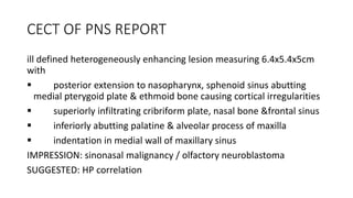
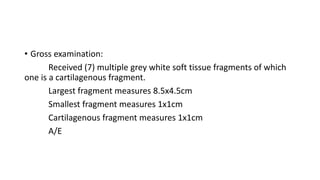
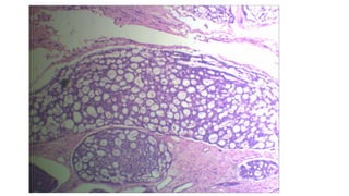
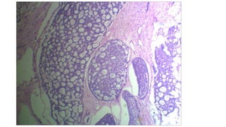
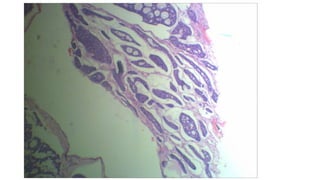
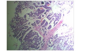
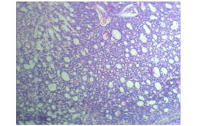
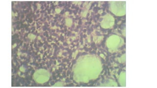
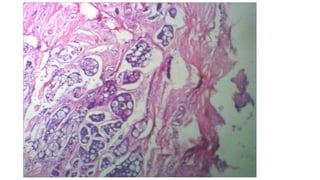
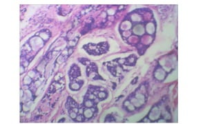
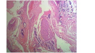
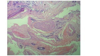
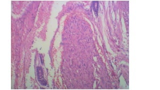
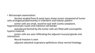
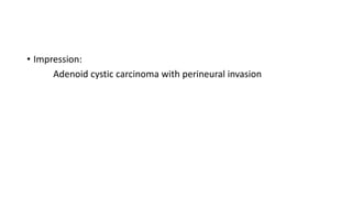
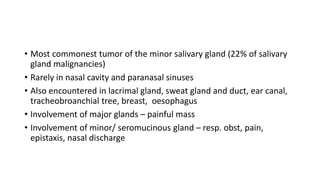
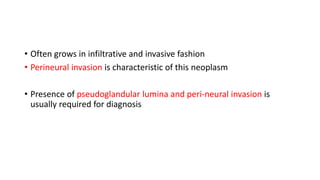
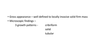
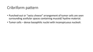
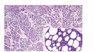
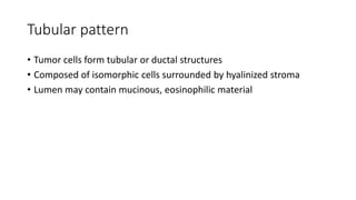
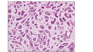
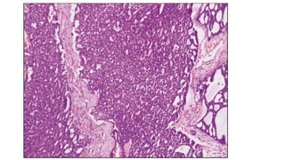
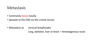
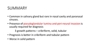
ACADEMIC REVIEW
Case details
Ms Aameena
29/F
C/O – bilateral nasal obstruction for 6 months
O/E – A smooth swelling filled the right nasal cavity and pushed the
nasal septum to the left side
Clinical diagnosis – Rt sinonasal mass for evaluation
? Adenocarcinoma
? Squamous cell carcinoma
? Inverted papilloma
CECT OF PNS REPORT
ill defined heterogeneously enhancing lesion measuring 6.4x5.4x5cm
with
 posterior extension to nasopharynx, sphenoid sinus abutting
medial pterygoid plate & ethmoid bone causing cortical irregularities
 superiorly infiltrating cribriform plate, nasal bone &frontal sinus
 inferiorly abutting palatine & alveolar process of maxilla
 indentation in medial wall of maxillary sinus
IMPRESSION: sinonasal malignancy / olfactory neuroblastoma
SUGGESTED: HP correlation
• Gross examination:
Received (7) multiple grey white soft tissue fragments of which
one is a cartilagenous fragment.
Largest fragment measures 8.5x4.5cm
Smallest fragment measures 1x1cm
Cartilagenous fragment measures 1x1cm
A/E
• Microscopic examination:
Section studied from R nasal mass shows tumor composed of tumor
cells arranged predominantly in cribriform and tubular pattern
tumor cells are small, round to oval with scanty cytoplasm,
hyperchromatic nuclei and indistinct cell border
pseudocyst formed by the tumor cells are filled with eosinophilic
hyaline material.
tumor cells are seen infiltrating the adjacent mucosal glands and
interstitium.
Neural invasion is seen
adjacent attached respiratory epithelium show normal histology
• Impression:
Adenoid cystic carcinoma with perineural invasion
Discussion
• Most commonest tumor of the minor salivary gland (22% of salivary
gland malignancies)
• Rarely in nasal cavity and paranasal sinuses
• Also encountered in lacrimal gland, sweat gland and duct, ear canal,
tracheobroanchial tree, breast, oesophagus
• Involvement of major glands – painful mass
• Involvement of minor/ seromucinous gland – resp. obst, pain,
epistaxis, nasal discharge
Incidence
• Occurs in 5th to 6th decades
• Male predominance
• Slow growing, indolent but aggressive
• Often grows in infiltrative and invasive fashion
• Perineural invasion is characteristic of this neoplasm
• Presence of pseudoglandular lumina and peri-neural invasion is
usually required for diagnosis
• Gross appearance – well defined to locally invasive solid firm mass
• Microscopic findings –
3 growth patterns - cribriform
solid
tubular
Cribriform pattern
• Punched-out or “swiss cheese” arrangement of tumor cells are seen
surrounding acellular spaces containing mucoid/ hyaline material.
• Tumor cells – dense basophilic nuclei with inconspicuous nucleoli.
Tubular pattern
• Tumor cells form tubular or ductal structures
• Composed of isomorphic cells surrounded by hyalinized stroma
• Lumen may contain mucinous, eosinophilic material
Solid pattern
• Large masses of tumor cells
• May contain occasional tubular &/or cribriform pattern
• Area of necrosis may be prominent in the central portion
IHC
• POSITIVE -
cytokeratin,
CEA,
vimentin,
actin,
S100,
CD-117,
c-kit.
GRADING
Differential diagnosis
• Polymorphous low grade adenocarcinoma
• Basal cell adenocarcinoma
• Pleomorphic adenoma
• Polymorphous low grade adenocarcinoma:
very rare in major salivary glands,
Differentiating with ACC:
bland uniform cells,
CD117 negative,
s100 positive,
negative for smooth muscle markers.
• Basal cell adenocarcinoma: Low grade malignant counterpart of basal cell
adenoma
Resembles basaloid (cloacogenic) carcinoma of anal canal or upper
aerodigestive tract
Also called basaloid carcinoma
Similarity with ACC:
infiltrative with perineurial invasion
Differentiating with ACC:
absence of peripheral palisading nuclei, lack of continuity with
epidermis/hair sheath, negative to CEA, S-100, Amylase.
• Pleomorphic adenoma:
Most common tumor of salivary glands
Painless, slow growing tumor, composed of biphasic population of
epithelial and mesenchymal cells
Also called benign mixed tumor
Differentiating with ACC:
mesenchyme-like areas, no invasion, no perineurial invasion
Treatment
• Wide surgical excision + radiotherapy
• Prognosis is better in cribriform and tubular pattern
• Worse in solid pattern
Metastasis
• Commonly recurs locally
• Spreads to the CNS via the cranial nerves
• Metastasis to cervical lymphnodes
lung, skeleton, liver or brain – hematogenous route
survival rate
• 5 year survival rate – 60-70%
• 10 year survival rate – 30%
• 15 year survival rate – 15%
SUMMARY
• Common in salivary gland but rare in nasal cavity and paranasal
sinuses.
• Presence of pseudoglandular lumina and peri-neural invasion is
usually required for diagnosis
3 growth patterns – cribriform, solid, tubular
• Prognosis is better in cribriform and tubular pattern
• Worse in solid pattern
REFERENCE
• Robbins Pathologic basis of disease
• Lever’s textbook
• Washington’s manual of surgical pathology
• http://pathologyoutlines.com/topic/salivaryglandsadenoidcystic.html
• Thankyou

Adenoid cystic carcinoma

  • 1.
  • 2.
    Case details Ms Aameena 29/F C/O– bilateral nasal obstruction for 6 months O/E – A smooth swelling filled the right nasal cavity and pushed the nasal septum to the left side Clinical diagnosis – Rt sinonasal mass for evaluation ? Adenocarcinoma ? Squamous cell carcinoma ? Inverted papilloma
  • 3.
    CECT OF PNSREPORT ill defined heterogeneously enhancing lesion measuring 6.4x5.4x5cm with  posterior extension to nasopharynx, sphenoid sinus abutting medial pterygoid plate & ethmoid bone causing cortical irregularities  superiorly infiltrating cribriform plate, nasal bone &frontal sinus  inferiorly abutting palatine & alveolar process of maxilla  indentation in medial wall of maxillary sinus IMPRESSION: sinonasal malignancy / olfactory neuroblastoma SUGGESTED: HP correlation
  • 4.
    • Gross examination: Received(7) multiple grey white soft tissue fragments of which one is a cartilagenous fragment. Largest fragment measures 8.5x4.5cm Smallest fragment measures 1x1cm Cartilagenous fragment measures 1x1cm A/E
  • 16.
    • Microscopic examination: Sectionstudied from R nasal mass shows tumor composed of tumor cells arranged predominantly in cribriform and tubular pattern tumor cells are small, round to oval with scanty cytoplasm, hyperchromatic nuclei and indistinct cell border pseudocyst formed by the tumor cells are filled with eosinophilic hyaline material. tumor cells are seen infiltrating the adjacent mucosal glands and interstitium. Neural invasion is seen adjacent attached respiratory epithelium show normal histology
  • 17.
    • Impression: Adenoid cysticcarcinoma with perineural invasion
  • 18.
  • 19.
    • Most commonesttumor of the minor salivary gland (22% of salivary gland malignancies) • Rarely in nasal cavity and paranasal sinuses • Also encountered in lacrimal gland, sweat gland and duct, ear canal, tracheobroanchial tree, breast, oesophagus • Involvement of major glands – painful mass • Involvement of minor/ seromucinous gland – resp. obst, pain, epistaxis, nasal discharge
  • 20.
    Incidence • Occurs in5th to 6th decades • Male predominance • Slow growing, indolent but aggressive
  • 21.
    • Often growsin infiltrative and invasive fashion • Perineural invasion is characteristic of this neoplasm • Presence of pseudoglandular lumina and peri-neural invasion is usually required for diagnosis
  • 22.
    • Gross appearance– well defined to locally invasive solid firm mass • Microscopic findings – 3 growth patterns - cribriform solid tubular
  • 23.
    Cribriform pattern • Punched-outor “swiss cheese” arrangement of tumor cells are seen surrounding acellular spaces containing mucoid/ hyaline material. • Tumor cells – dense basophilic nuclei with inconspicuous nucleoli.
  • 25.
    Tubular pattern • Tumorcells form tubular or ductal structures • Composed of isomorphic cells surrounded by hyalinized stroma • Lumen may contain mucinous, eosinophilic material
  • 27.
    Solid pattern • Largemasses of tumor cells • May contain occasional tubular &/or cribriform pattern • Area of necrosis may be prominent in the central portion
  • 29.
  • 30.
  • 31.
    Differential diagnosis • Polymorphouslow grade adenocarcinoma • Basal cell adenocarcinoma • Pleomorphic adenoma
  • 32.
    • Polymorphous lowgrade adenocarcinoma: very rare in major salivary glands, Differentiating with ACC: bland uniform cells, CD117 negative, s100 positive, negative for smooth muscle markers.
  • 33.
    • Basal celladenocarcinoma: Low grade malignant counterpart of basal cell adenoma Resembles basaloid (cloacogenic) carcinoma of anal canal or upper aerodigestive tract Also called basaloid carcinoma Similarity with ACC: infiltrative with perineurial invasion Differentiating with ACC: absence of peripheral palisading nuclei, lack of continuity with epidermis/hair sheath, negative to CEA, S-100, Amylase.
  • 34.
    • Pleomorphic adenoma: Mostcommon tumor of salivary glands Painless, slow growing tumor, composed of biphasic population of epithelial and mesenchymal cells Also called benign mixed tumor Differentiating with ACC: mesenchyme-like areas, no invasion, no perineurial invasion
  • 35.
    Treatment • Wide surgicalexcision + radiotherapy • Prognosis is better in cribriform and tubular pattern • Worse in solid pattern
  • 36.
    Metastasis • Commonly recurslocally • Spreads to the CNS via the cranial nerves • Metastasis to cervical lymphnodes lung, skeleton, liver or brain – hematogenous route
  • 37.
    survival rate • 5year survival rate – 60-70% • 10 year survival rate – 30% • 15 year survival rate – 15%
  • 38.
    SUMMARY • Common insalivary gland but rare in nasal cavity and paranasal sinuses. • Presence of pseudoglandular lumina and peri-neural invasion is usually required for diagnosis 3 growth patterns – cribriform, solid, tubular • Prognosis is better in cribriform and tubular pattern • Worse in solid pattern
  • 39.
    REFERENCE • Robbins Pathologicbasis of disease • Lever’s textbook • Washington’s manual of surgical pathology • http://pathologyoutlines.com/topic/salivaryglandsadenoidcystic.html
  • 40.