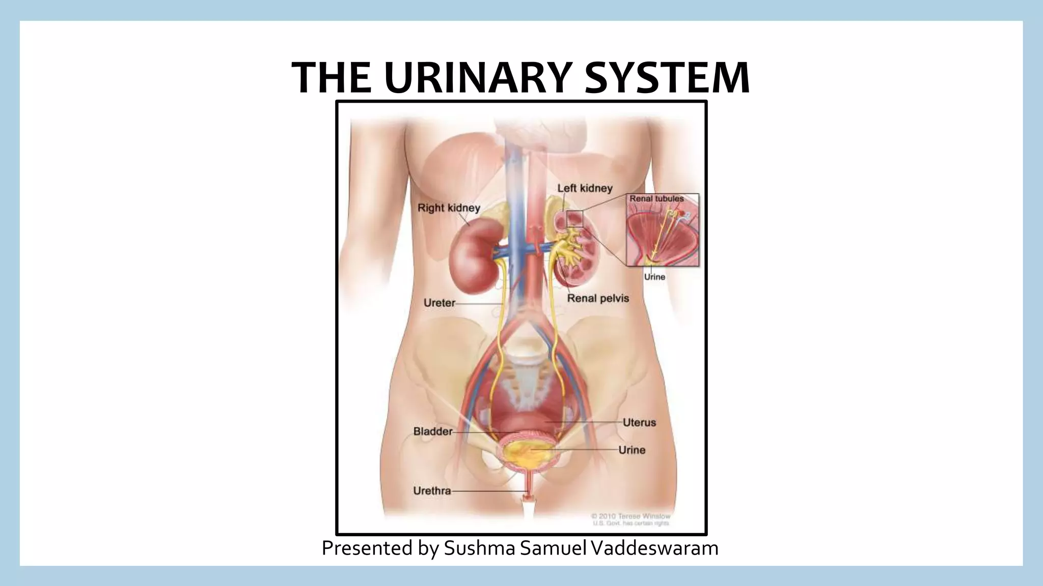
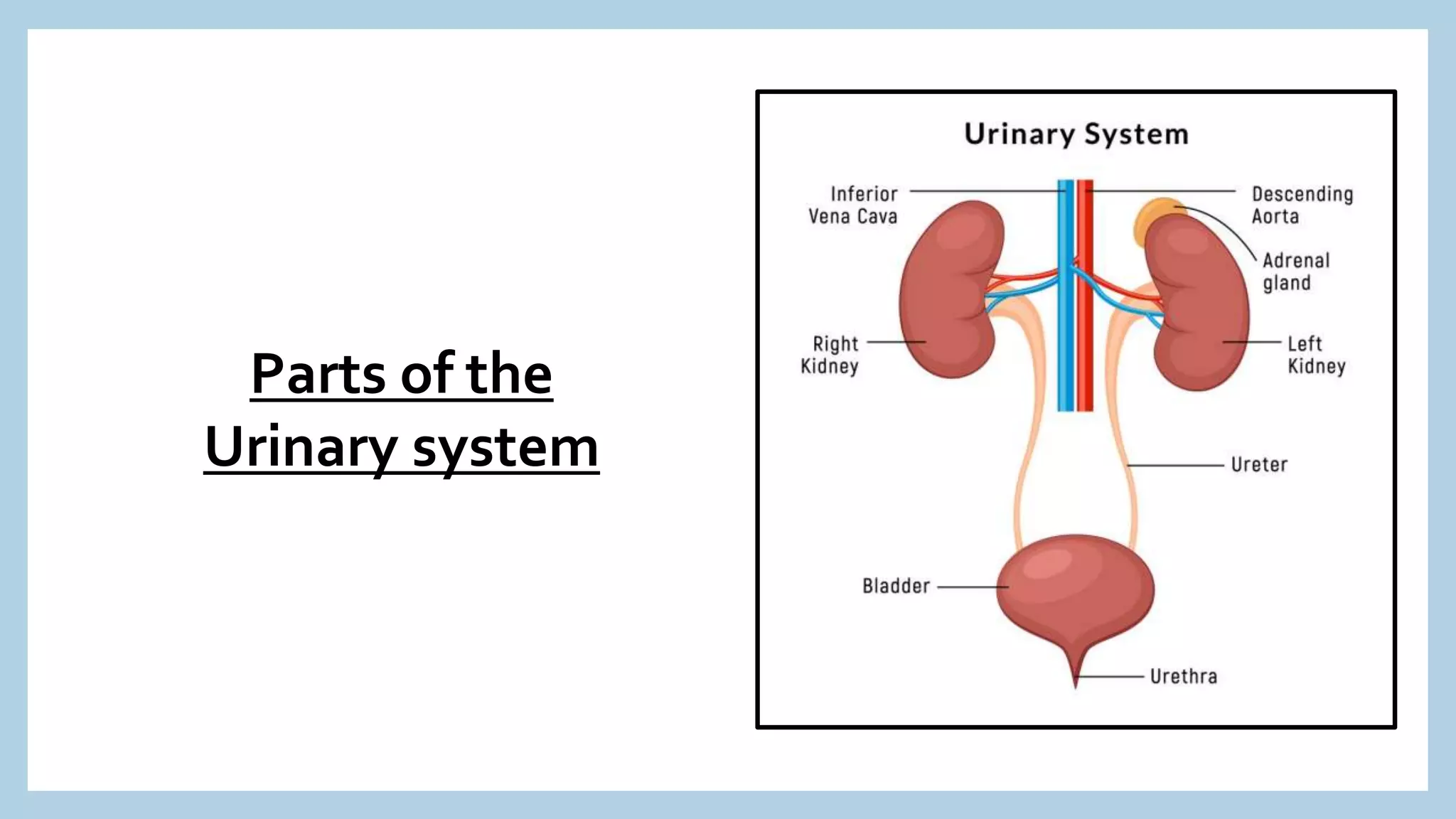
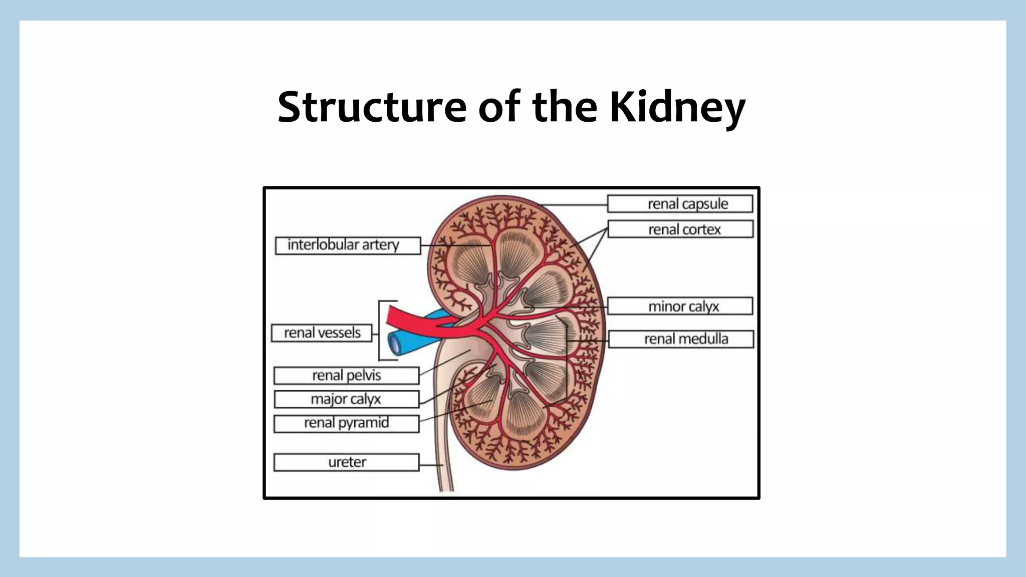
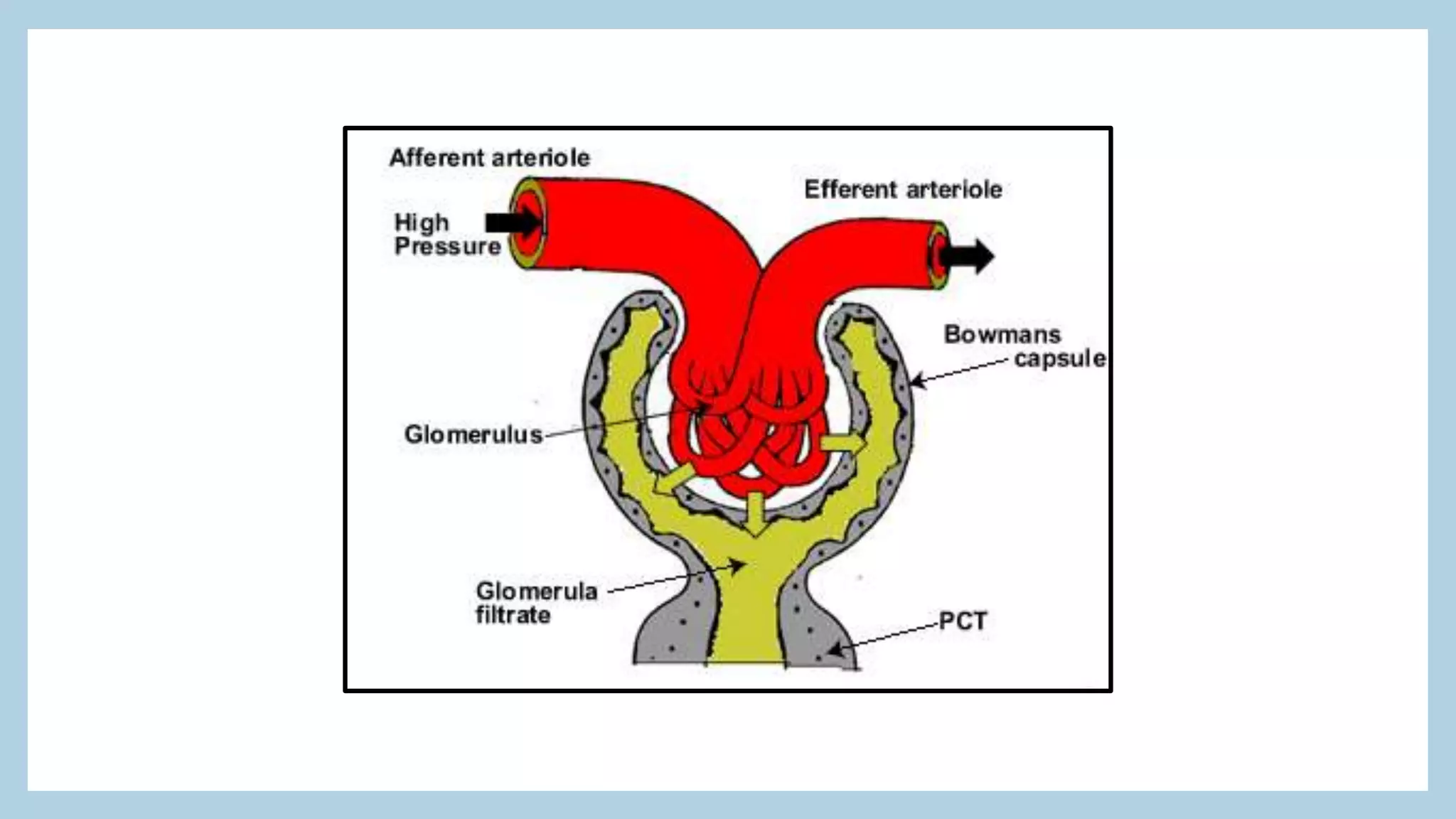
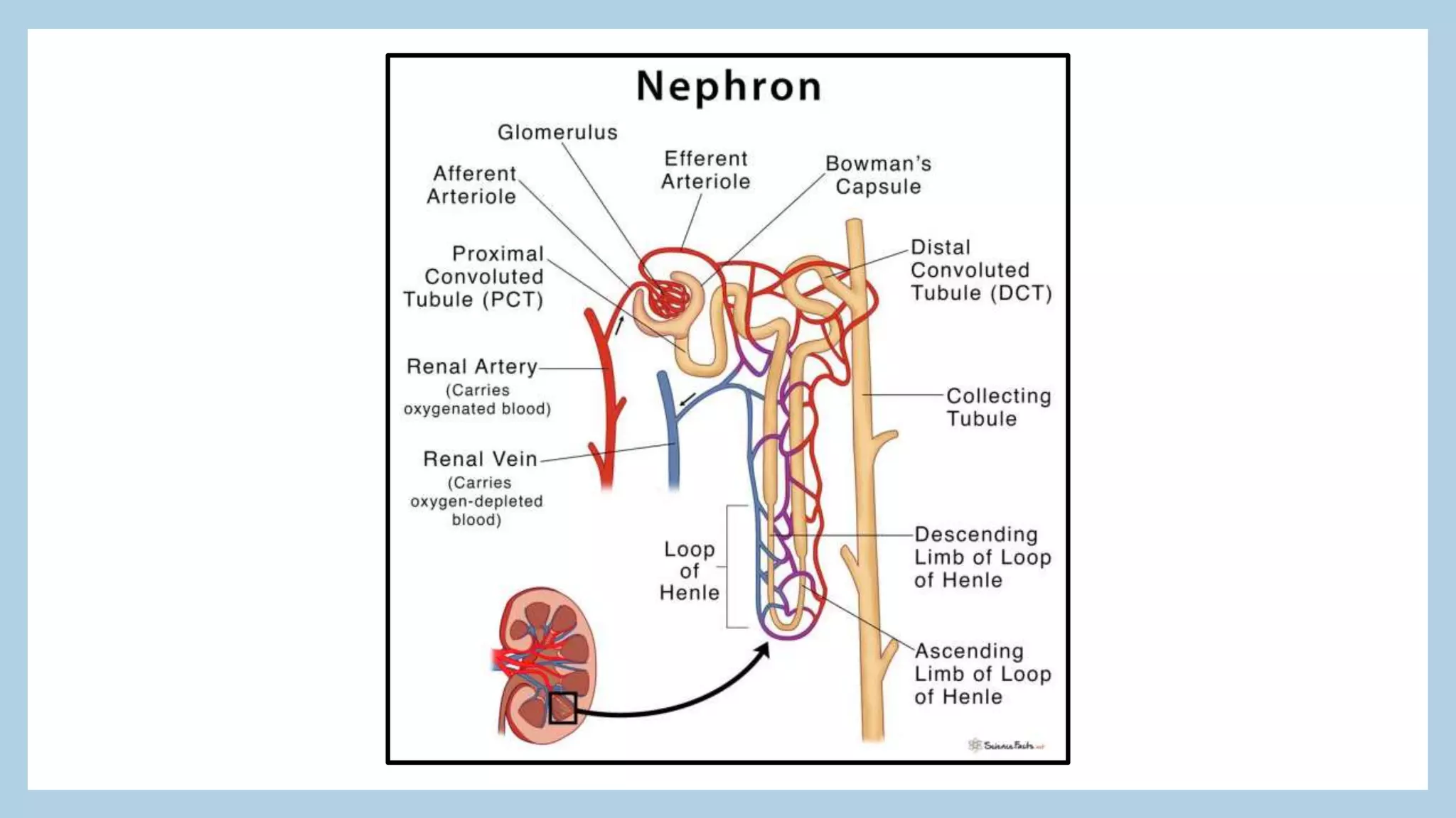
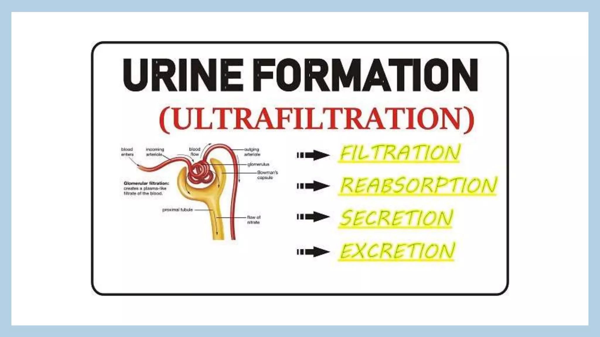
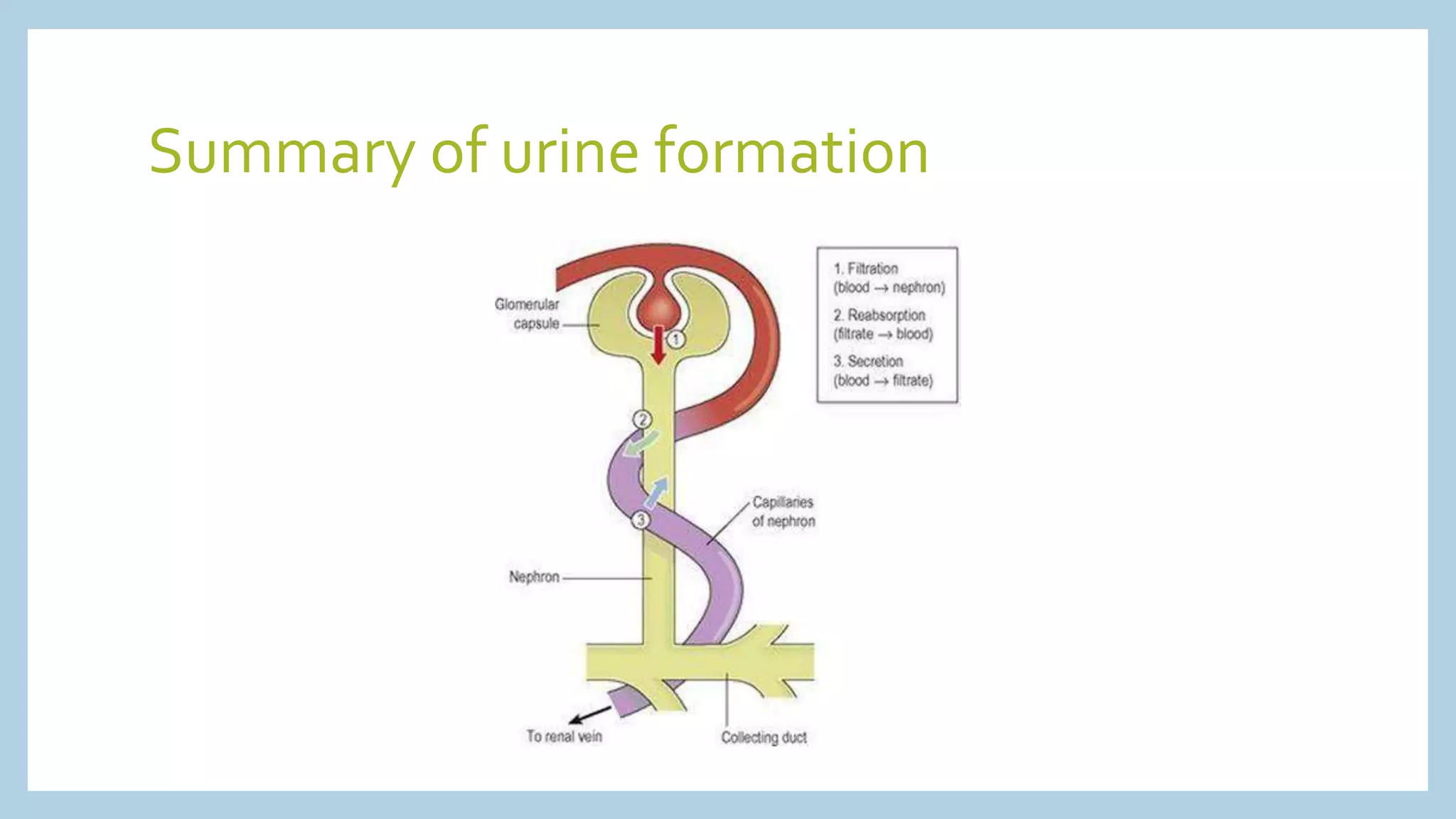
THE URINARY SYSTEM
Presented by Sushma SamuelVaddeswaram
The urinary system is the main excretory system and consists of
the following structures:
• 2 kidneys, which secrete urine
• 2 ureters, which convey the urine from the kidneys to the urinary
bladder
• the urinary bladder where urine collects and is temporarily
stored
• the urethra through which the urine passes from the urinary
bladder to the exterior.
Urine is stored in the bladder and excreted by the process
of micturition.
Introduction
Parts of the
Urinary system
• The kidneys
• lie on the posterior abdominal wall, one on each side of the
vertebral column, behind the peritoneum and below the
diaphragm.
• They extend from the level of the 12th thoracic vertebra to the 3rd
lumbar vertebra, receiving some protection from the lower rib
cage. The right kidney is usually slightly lower than the left,
probably because of the considerable space occupied by the liver.
• Kidneys are bean-shaped organs, about 11 cm long, 6 cm wide, 3
cm thick and weigh 150 g. They are embedded in, and held in
position by, a mass of fat.
Organs associated with the kidneys:
• Right kidney
• Superiorly – the right adrenal gland
• Anteriorly – the right lobe of the liver, the duodenum and the hepatic
flexure of the colon
• Posteriorly – the diaphragm, and muscles of the posterior abdominal wall
• Left kidney
• Superiorly – the left adrenal gland
• Anteriorly – the spleen, stomach, pancreas, jejunum and splenic flexure
of the colon
• Posteriorly – the diaphragm and muscles of the posterior abdominal wall
Gross Structure of the Kidney:
• There are three areas of tissue that can be distinguished when a
longitudinal section of the kidney is viewed with the naked eye.
• • an outer fibrous capsule, surrounding the kidney
• • the cortex, a reddish-brown layer of tissue immediately below
the capsule and outside the pyramids
• • the medulla, the innermost layer, consisting of pale conical-
shaped striations, the renal pyramids.
Structure of the Kidney
Gross structure of the kidney
The hilum is the concave medial border of the kidney where the renal blood and lymph
vessels, the ureter and nerves enter.
The renal pelvis is the funnel-shaped structure that collects urine formed by the kidney. Urine
formed in the kidney passes through a renal papilla at the apex of a pyramid into a minor
calyx, then into a major calyx before passing through the renal pelvis into the ureter.
The walls of the pelvis contain smooth muscle and are lined with transitional epithelium.
Peristalsis of the smooth muscle originating in pacemaker cells in the walls of the calyces
propels urine through the renal pelvis and ureters to the bladder.
This is an intrinsic property of the smooth muscle, and is not under nerve control.
Microscopic Structure
• The kidney is composed of about 1–2 million functional units, the nephrons, and
a smaller number of collecting ducts.
• The nephron
• The nephron consists of a tubule closed at one end, the other end opening into a
collecting tubule.
• The closed or blind end is indented to form the cup-shaped glomerular
capsule (Bowman’s capsule), which almost completely encloses a network of tiny
arterial capillaries, the glomerulus. These resemble a coiled tuft. Continuing from
the glomerular capsule, the remainder of the nephron is about 3 cm long and is
described in three parts:
• • the proximal convoluted tubule
• • the medullary loop (loop of Henle)
• • the distal convoluted tubule, leading into a collecting duct.
NEPHRON (Contd..)
• The walls of the glomerulus and the glomerular capsule consist of a
single layer of flattened epithelial cells. The glomerular walls are more
permeable than those of other capillaries. The remainder of the nephron
and the collecting duct are formed by a single layer of simple squamous
epithelium.
• The blood vessel leading away from the glomerulus is the efferent
arteriole. The afferent arteriole has a larger diameter than the efferent
arteriole, which increases pressure inside the glomerulus and drives
filtration across the glomerular capillary walls .
• The efferent arteriole divides into a second peritubular (meaning ‘around
tubules’) capillary network, which wraps around the remainder of the
tubule, allowing exchange between the fluid in the tubule and the
bloodstream. This maintains the supply of oxygen and nutrients to the
local tissues and removes waste products. Venous blood drained from
this capillary bed eventually leaves the kidney in the renal vein, which
empties into the inferior vena cava.
Function of kidney in urine formation
• The kidneys form urine, which passes through the ureters to the bladder for storage prior
to excretion. Waste products of protein metabolism are excreted, electrolyte levels are
controlled and pH (acid–base balance) is maintained by excretion of hydrogen ions.
There are three processes involved in the formation of urine:
• • filtration
• • selective reabsorption
• • secretion.
• FILTRATION
• This takes place through the semipermeable walls of the glomerulus and glomerular
capsule. Water and other small molecules pass through, although some are reabsorbed
later.
• The filtrate in the glomerulus is very similar in composition to plasma with the important
exceptions of plasma proteins and blood cells.
•
Glomerular filtrate
Blood constituents in glomerular filtrate Blood constituents remaining in glomerular
capillaries
Water Leukocytes
Mineral salts Erythrocytes
Amino acids Platelets
Ketoacids Plasma proteins
Glucose Some drugs (large molecules)
Some hormones
Creatinine
Urea
Uric acid
Some drugs (small molecules)
• The volume of filtrate formed by both kidneys each minute is
called the glomerular filtration rate (GFR). In a healthy adult the
GFR is about 125 ml/min, i.e. 180 litres of filtrate are formed each
day by the two kidneys. Nearly all of the filtrate is later
reabsorbed from the kidney tubules with less than 1%, i.e. 1 to
1.5 litres, excreted as urine.
• In severe shock, when the systolic blood pressure falls below 80
mmHg, autoregulation fails and renal blood flow and the
hydrostatic pressure decrease, impairing filtration within the
nephrons.
Selective Reabsorption
• Most reabsorption from the filtrate back into the blood takes place in the
proximal convoluted tubule. Materials essential to the body are
reabsorbed here, including some water, electrolytes and organic nutrients
such as glucose.
• Only 60–70% of filtrate reaches the loop of the nephron. Much of this,
especially water, sodium and chloride, is reabsorbed in the loop, so only
15–20% of the original filtrate reaches the distal convoluted tubule, and
the composition of the filtrate is now very different from its starting
values.
• More electrolytes are reabsorbed here, especially sodium, so the filtrate
entering the collecting ducts is actually quite dilute. The main function of
the collecting ducts therefore is to reabsorb as much water as the body
needs.
•
Hormones
• Antidiuretic hormone
• Also known as ADH, this is secreted by the posterior lobe of the
pituitary gland and increases the permeability of the distal
convoluted tubules and collecting tubules, increasing water
reabsorption.
• Aldosterone
• Secreted by the adrenal cortex, this hormone increases the
reabsorption of sodium and water, and the excretion of potassium.
Secretion is regulated through a negative feedback system.
• Parathyroid Hormone
Tubular Secretion
• Filtration occurs as the blood flows through the glomerulus.
Substances not required and foreign materials, e.g. drugs
including penicillin and aspirin, may not be cleared from the blood
by filtration because of the short time it remains in the
glomerulus. Such substances are cleared by secretion from the
peritubular capillaries into the convoluted tubules and excreted
from the body in the urine.
• Tubular secretion of hydrogen ions (H+) is important in
maintaining normal blood pH.
Summary of urine formation

urinary system.pptx

  • 1.
    THE URINARY SYSTEM Presentedby Sushma SamuelVaddeswaram
  • 3.
    The urinary systemis the main excretory system and consists of the following structures: • 2 kidneys, which secrete urine • 2 ureters, which convey the urine from the kidneys to the urinary bladder • the urinary bladder where urine collects and is temporarily stored • the urethra through which the urine passes from the urinary bladder to the exterior. Urine is stored in the bladder and excreted by the process of micturition. Introduction
  • 4.
  • 5.
    • The kidneys •lie on the posterior abdominal wall, one on each side of the vertebral column, behind the peritoneum and below the diaphragm. • They extend from the level of the 12th thoracic vertebra to the 3rd lumbar vertebra, receiving some protection from the lower rib cage. The right kidney is usually slightly lower than the left, probably because of the considerable space occupied by the liver. • Kidneys are bean-shaped organs, about 11 cm long, 6 cm wide, 3 cm thick and weigh 150 g. They are embedded in, and held in position by, a mass of fat.
  • 6.
    Organs associated withthe kidneys: • Right kidney • Superiorly – the right adrenal gland • Anteriorly – the right lobe of the liver, the duodenum and the hepatic flexure of the colon • Posteriorly – the diaphragm, and muscles of the posterior abdominal wall • Left kidney • Superiorly – the left adrenal gland • Anteriorly – the spleen, stomach, pancreas, jejunum and splenic flexure of the colon • Posteriorly – the diaphragm and muscles of the posterior abdominal wall
  • 7.
    Gross Structure ofthe Kidney: • There are three areas of tissue that can be distinguished when a longitudinal section of the kidney is viewed with the naked eye. • • an outer fibrous capsule, surrounding the kidney • • the cortex, a reddish-brown layer of tissue immediately below the capsule and outside the pyramids • • the medulla, the innermost layer, consisting of pale conical- shaped striations, the renal pyramids.
  • 8.
  • 9.
    Gross structure ofthe kidney The hilum is the concave medial border of the kidney where the renal blood and lymph vessels, the ureter and nerves enter. The renal pelvis is the funnel-shaped structure that collects urine formed by the kidney. Urine formed in the kidney passes through a renal papilla at the apex of a pyramid into a minor calyx, then into a major calyx before passing through the renal pelvis into the ureter. The walls of the pelvis contain smooth muscle and are lined with transitional epithelium. Peristalsis of the smooth muscle originating in pacemaker cells in the walls of the calyces propels urine through the renal pelvis and ureters to the bladder. This is an intrinsic property of the smooth muscle, and is not under nerve control.
  • 12.
    Microscopic Structure • Thekidney is composed of about 1–2 million functional units, the nephrons, and a smaller number of collecting ducts. • The nephron • The nephron consists of a tubule closed at one end, the other end opening into a collecting tubule. • The closed or blind end is indented to form the cup-shaped glomerular capsule (Bowman’s capsule), which almost completely encloses a network of tiny arterial capillaries, the glomerulus. These resemble a coiled tuft. Continuing from the glomerular capsule, the remainder of the nephron is about 3 cm long and is described in three parts: • • the proximal convoluted tubule • • the medullary loop (loop of Henle) • • the distal convoluted tubule, leading into a collecting duct.
  • 15.
    NEPHRON (Contd..) • Thewalls of the glomerulus and the glomerular capsule consist of a single layer of flattened epithelial cells. The glomerular walls are more permeable than those of other capillaries. The remainder of the nephron and the collecting duct are formed by a single layer of simple squamous epithelium. • The blood vessel leading away from the glomerulus is the efferent arteriole. The afferent arteriole has a larger diameter than the efferent arteriole, which increases pressure inside the glomerulus and drives filtration across the glomerular capillary walls . • The efferent arteriole divides into a second peritubular (meaning ‘around tubules’) capillary network, which wraps around the remainder of the tubule, allowing exchange between the fluid in the tubule and the bloodstream. This maintains the supply of oxygen and nutrients to the local tissues and removes waste products. Venous blood drained from this capillary bed eventually leaves the kidney in the renal vein, which empties into the inferior vena cava.
  • 17.
    Function of kidneyin urine formation • The kidneys form urine, which passes through the ureters to the bladder for storage prior to excretion. Waste products of protein metabolism are excreted, electrolyte levels are controlled and pH (acid–base balance) is maintained by excretion of hydrogen ions. There are three processes involved in the formation of urine: • • filtration • • selective reabsorption • • secretion. • FILTRATION • This takes place through the semipermeable walls of the glomerulus and glomerular capsule. Water and other small molecules pass through, although some are reabsorbed later. • The filtrate in the glomerulus is very similar in composition to plasma with the important exceptions of plasma proteins and blood cells. •
  • 18.
    Glomerular filtrate Blood constituentsin glomerular filtrate Blood constituents remaining in glomerular capillaries Water Leukocytes Mineral salts Erythrocytes Amino acids Platelets Ketoacids Plasma proteins Glucose Some drugs (large molecules) Some hormones Creatinine Urea Uric acid Some drugs (small molecules)
  • 19.
    • The volumeof filtrate formed by both kidneys each minute is called the glomerular filtration rate (GFR). In a healthy adult the GFR is about 125 ml/min, i.e. 180 litres of filtrate are formed each day by the two kidneys. Nearly all of the filtrate is later reabsorbed from the kidney tubules with less than 1%, i.e. 1 to 1.5 litres, excreted as urine. • In severe shock, when the systolic blood pressure falls below 80 mmHg, autoregulation fails and renal blood flow and the hydrostatic pressure decrease, impairing filtration within the nephrons.
  • 20.
    Selective Reabsorption • Mostreabsorption from the filtrate back into the blood takes place in the proximal convoluted tubule. Materials essential to the body are reabsorbed here, including some water, electrolytes and organic nutrients such as glucose. • Only 60–70% of filtrate reaches the loop of the nephron. Much of this, especially water, sodium and chloride, is reabsorbed in the loop, so only 15–20% of the original filtrate reaches the distal convoluted tubule, and the composition of the filtrate is now very different from its starting values. • More electrolytes are reabsorbed here, especially sodium, so the filtrate entering the collecting ducts is actually quite dilute. The main function of the collecting ducts therefore is to reabsorb as much water as the body needs. •
  • 21.
    Hormones • Antidiuretic hormone •Also known as ADH, this is secreted by the posterior lobe of the pituitary gland and increases the permeability of the distal convoluted tubules and collecting tubules, increasing water reabsorption. • Aldosterone • Secreted by the adrenal cortex, this hormone increases the reabsorption of sodium and water, and the excretion of potassium. Secretion is regulated through a negative feedback system. • Parathyroid Hormone
  • 22.
    Tubular Secretion • Filtrationoccurs as the blood flows through the glomerulus. Substances not required and foreign materials, e.g. drugs including penicillin and aspirin, may not be cleared from the blood by filtration because of the short time it remains in the glomerulus. Such substances are cleared by secretion from the peritubular capillaries into the convoluted tubules and excreted from the body in the urine. • Tubular secretion of hydrogen ions (H+) is important in maintaining normal blood pH.
  • 23.