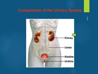
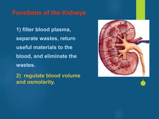
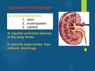
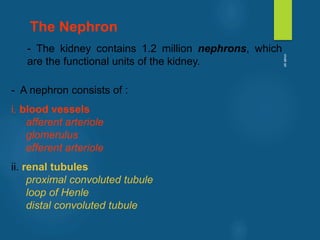
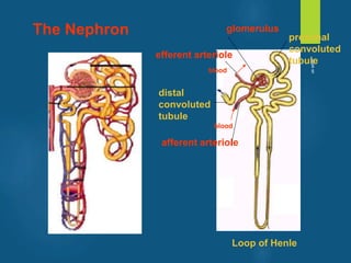
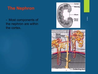
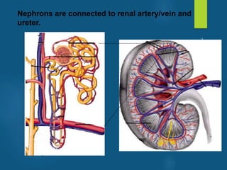
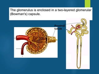
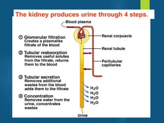
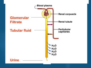
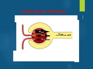
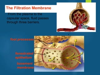
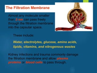
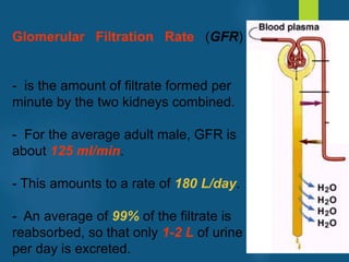
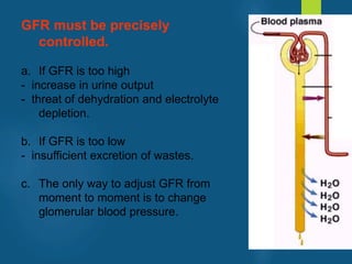
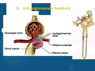
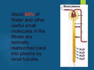
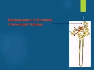
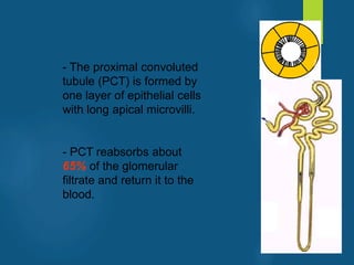
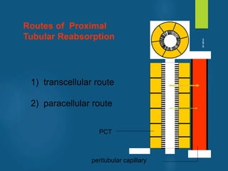
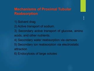
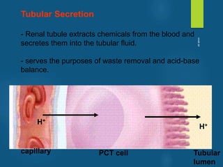
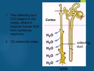
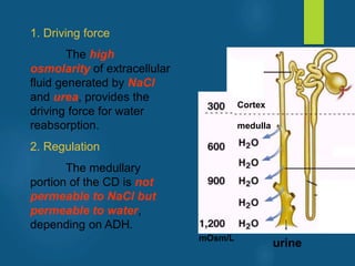
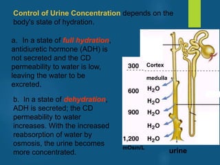
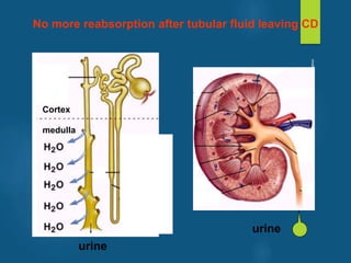
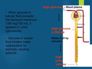
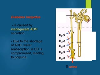
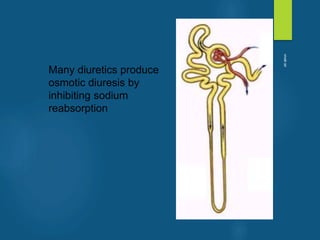
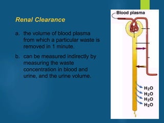
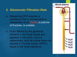
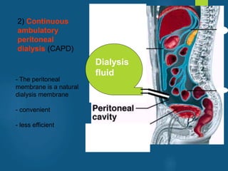
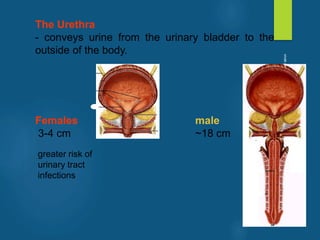
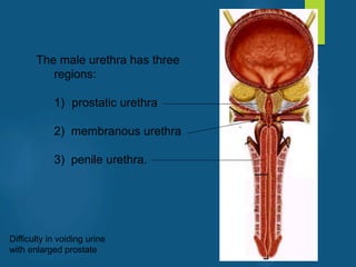
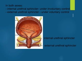
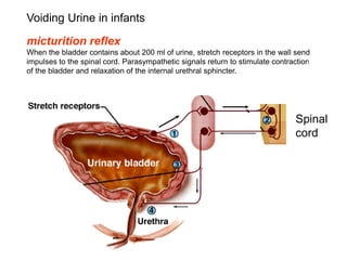
The Urinary System
Prepared by: vivek sir
vivek
sir
TABLE OF CONTENT
1) General Introduction
2) Anatomy of Urinary System
3) Urine Formation
4) Urine Storage and Elimination
vivek
sir
Composition of the Urinary System
vivek
sir
Functions of the Kidneys
1) filter blood plasma, separate
wastes, return useful materials
to the blood, and eliminate the
wastes.
Toxic nitrogenous wastes
- ammonia, urea, uric acid, creatine, and
creatinine
- cause diarrhea, vomiting, and cardiac arrhythmia,
convulsions, coma, and death.
vivek
sir
Functions of the Kidneys
1) filter blood plasma,
separate wastes, return
useful materials to the
blood, and eliminate the
wastes.
2) regulate blood volume
and osmolarity.
vivek
sir
3) produce hormones
1. renin
2. erythropoietin
3. calcitrol
Functions of the Kidneys
4) regulate acid-base balance
of the body fluids.
5) detoxify superoxides, free
radicals, and drugs.
vivek
sir
ANATOMY OF THE KIDNEYS
vivek
sir
- The kidneys lie along the posterior abdominal wall
vivek
sir
- The medial surface of the kidney is concave with
a hilum carrying renal nerves and blood vessels.
The renal parenchyma is divided into an outer
cortex and inner medulla.
vivek
sir
Extensions of the cortex
(renal columns) project
toward the sinus, dividing
the medulla into 6-10
renal pyramids. Each
pyramid is conical with a
blunt point called the
papilla facing the sinus.
vivek
sir
The papilla is nestled into
a cup called a minor calyx,
which collects its urine.
Two or three minor calyces
merge to form a major
calyx. The major calyces
merge to form the renal
pelvis.
vivek
sir
- The kidney contains 1.2 million nephrons, which
are the functional units of the kidney.
- A nephron consists of :
i. blood vessels
afferent arteriole
glomerulus
efferent arteriole
ii. renal tubules
proximal convoluted tubule
loop of Henle
distal convoluted tubule
The Nephron
vivek
sir
afferent arteriole
glomerulus
efferent arteriole
proximal
convoluted
tubule
distal
convoluted
tubule
Loop of Henle
blood
blood
The Nephron
vivek
sir
- Most components of
the nephron are within
the cortex.
The Nephron
vivek
sir
Nephrons are connected to renal artery/vein and
ureter.
vivek
sir
The glomerulus is enclosed in a two-layered glomerular
(Bowman's) capsule.
Proximal
tubule
vivek
sir
URINE FORMATION
vivek
sir
The kidney produces urine through 4 steps.
vivek
sir
Glomerular
Filtrate
Tubular fluid
Urine
vivek
sir
1) Glomerular Filtration
vivek
sir
The Filtration Membrane
From the plasma to the
capsular space, fluid passes
through three barriers.
fenestrated
epithelium
basement
membrane
foot processes
vivek
sir
The Filtration Membrane
Almost any molecule smaller
than 3 nm can pass freely
through the filtration membrane
into the capsular space.
These include:
Water, electrolytes, glucose, amino acids,
lipids, vitamins, and nitrogenous wastes
Kidney infections and trauma commonly damage
the filtration membrane and allow plasma
proteins or blood cells to pass through.
vivek
sir
Blood cells
in urine
Plasma proteins
vivek
sir
Filtration Pressure
Glomerular filtration follows the same principles
that govern filtration in other capillaries.
vivek
sir
Glomerular Filtration Rate (GFR)
- is the amount of filtrate formed per
minute by the two kidneys combined.
- For the average adult male, GFR is
about 125 ml/min.
- This amounts to a rate of 180 L/day.
- An average of 99% of the filtrate is
reabsorbed, so that only 1-2 L of urine
per day is excreted.
vivek
sir
GFR must be precisely
controlled.
a. If GFR is too high
- increase in urine output
- threat of dehydration and electrolyte
depletion.
b. If GFR is too low
- insufficient excretion of wastes.
c. The only way to adjust GFR from
moment to moment is to change
glomerular blood pressure.
vivek
sir
Renal Autoregulation
- the ability of the kidneys to maintain a
relatively stable GFR in spite of the
changes (75 - 175 mmHg) in arterial
blood pressure.
vivek
sir
The nephron has two ways to
prevent drastic changes in GFR
when blood pressure rises:
1) Constriction of the afferent
arteriole to reduce blood flow into
the glomerulus
2) Dilation of the efferent arteriole
to allow the blood to flow out
more easily.
Change in an opposite direction if blood pressure falls
vivek
sir
Mechanisms of Renal Autoregulation
1) myogenic response
2) tubuloglomerular feedback
vivek
sir
1) myogenic response
vivek
sir
2) tubuloglomerular feedback
vivek
sir
1) Glomerular Filtration
2) Tubular Reabsorption
3) Tubular Secretion
4) Concentrating Urine by Collecting Duct
vivek
sir
About 99% of
Water and other
useful small
molecules in the
filtrate are
normally
reabsorbed back
into plasma by
renal tubules.
vivek
sir
Reabsorption in Proximal
Convoluted Tubules
vivek
sir
- The proximal convoluted
tubule (PCT) is formed by
one layer of epithelial cells
with long apical microvilli.
- PCT reabsorbs about
65% of the glomerular
filtrate and return it to the
blood.
vivek
sir
1) transcellular route
2) paracellular route
Routes of Proximal
Tubular Reabsorption
PCT
peritubular capillary
vivek
sir
Mechanisms of Proximal Tubular
Reabsorption
1) Solvent drag
2) Active transport of sodium.
3) Secondary active transport of glucose, amino
acids, and other nutrients.
4) Secondary water reabsorption via osmosis
5) Secondary ion reabsorption via electrostatic
attraction
6) Endocytosis of large solutes
vivek
sir
Osmosis
Water moves from a compartment of low osmolarity
to the compartment of high osmolarity.
low osmolarity
( high H2O conc.)
high osmolarity
( low H2O conc.)
H2O
vivek
sir
1) Solvent drag
- driven by high
colloid osmotic
pressure (COP) in the
peritubular capillaries
- Water is reabsorbed
by osmosis and
carries all other
solutes along.
- Both routes are
involved.
Proteins stay
H2O
Proteins
vivek
sir
2) Active transport of sodium
Sodium pumps (Na-K ATPase) in basolateral
membranes transport sodium out of the cells against its
concentration gradient using ATP.
capillary PCT cell Tubular
lumen
Na+
K+
Na+
vivek
sir
There are also pumps for other ions
capillary PCT cell Tubular
lumen
Ca++
Ca++
vivek
sir
- Various cotransporters can carry both Na+ and
other solutes. For example, the sodium-
dependent glucose transporter (SDGT) can
carry both Na+ and glucose.
3) Secondary active transport of glucose, amino
acids, and other nutrients
Na+
Glucose
capillary PCT cell
Na+
K+
vivek
sir
Amino acids and many other nutrients are
reabsorbed by their specific cotransporters
with sodium.
Na+
capillary PCT cell
Na+
K+
amino acids
3) Secondary active transport of glucose, amino
acids, and other nutrients
vivek
sir
Sodium reabsorption makes both
intracellular and extracellular fluid hypertonic
to the tubular fluid. Water follows sodium into
the peritubular capillaries.
4) Secondary water reabsorption via osmosis
H2O
Na+
Na+
capillary PCT cell Tubular
lumen
vivek
sir
Negative ions tend to follow the positive
sodium ions by electrostatic attraction.
5) Secondary ion reabsorption via
electrostatic attraction
Na Na+
Cl-
capillary PCT cell Tubular
lumen
vivek
sir
The glomerulus filters a small amount of protein
from the blood. The PCT reclaims it by endocytosis,
hydrolzes it to amino acids, and releases these to the
ECF by facilitated diffusion.
6) Endocytosis of large solutes
protein
capillary PCT cell Tubular
lumen
amino acids
vivek
sir
The Transport Maximum
- There is a limit to the amount of solute that the renal
tubule can reabsorb because there are limited numbers of
transport proteins in the plasma membranes.
- If all the transporters are occupied as solute molecules
pass through, some solute will remain in the tubular fluid
and appear in the urine.
Na+
Glucose
Example of diabetes
vivek
sir
Glucose in urine
high glucose in blood
high glucose in filtrate
Exceeds Tm for glucose
vivek
sir
Reabsorption in the
Nephron Loop
vivek
sir
- The primary purpose
is to establish a high
extracellular osmotic
concentration.
- The thick ascending
limb reabsorbs solutes
but is impermeable to
water. Thus, the tubular
fluid becomes very
diluted while
extracellular fluid
becomes very
concentrated with
solutes.
mOsm/L
vivek
sir
The high osmolarity enables the collecting duct
to concentrate the urine later.
vivek
sir
Reabsorption in Distal
Convoluted Tubules
vivek
sir
- Fluid arriving in the
DCT still contains about
20% of the water and
10% of the salts of the
glomerular filtrate.
- A distinguishing feature
of these parts of the renal
tubule is that they are
subject to hormonal
control.
vivek
sir
Aldosterone
a. secreted from adrenal
gland in response to a 
Na+ or a  K+ in blood
b. to increase Na+ absorption
and K+ secretion in the DCT
and cortical portion of the
collecting duct.
c. helps to maintain blood
volume and pressure.
vivek
sir
Atrial Natriuretic Factor
- secreted by the atrial
myocardium in response to
high blood pressure.
- It inhibits sodium and
water reabsorption,
increases the output of both
in the urine, and thus
reduces blood volume and
pressure.
vivek
sir
1) Glomerular Filtration
2) Tubular Reabsorption
3) Tubular Secretion
4) Concentrating Urine by Collecting Duct
vivek
sir
Tubular Secretion
- Renal tubule extracts chemicals from the blood and
secretes them into the tubular fluid.
- serves the purposes of waste removal and acid-base
balance.
H+
H+
capillary PCT cell Tubular
lumen
vivek
sir
1) Glomerular Filtration
2) Tubular Reabsorption
3) Tubular Secretion
4) Concentrating Urine by Collecting Duct
vivek
sir
1. The collecting duct
(CD) begins in the
cortex, where it
receives tubular fluid
from numerous
nephrons.
2. CD reabsorbs water.
Cortex
collecting
duct
urine
vivek
sir
1. Driving force
The high
osmolarity of extracellular
fluid generated by NaCl
and urea, provides the
driving force for water
reabsorption.
2. Regulation
The medullary
portion of the CD is not
permeable to NaCl but
permeable to water,
depending on ADH.
Cortex
mOsm/L
medulla
urine
vivek
sir
a. In a state of full hydration,
antidiuretic hormone (ADH) is
not secreted and the CD
permeability to water is low,
leaving the water to be
excreted.
Cortex
mOsm/L
medulla
Control of Urine Concentration depends on the
body's state of hydration.
urine
b. In a state of dehydration,
ADH is secreted; the CD
permeability to water
increases. With the increased
reabsorption of water by
osmosis, the urine becomes
more concentrated.
vivek
sir
Cortex
medulla
urine
No more reabsorption after tubular fluid leaving CD
urine
vivek
sir
Composition and Properties of Urine
Fresh urine is clear, containing no blood
cells and little proteins. If cloudy, it could
indicate the presence of bacteria, semen,
blood, or menstrual fluid.
Urine Properties
vivek
sir
Substance Blood Plasma
(total amount)
Urine
(amount per day)
Urea 4.8 g 25 g
Uric acid 0.15 g 0.8 g
Creatinine 0.03 g 1.6 g
Potassium 0.5 g 2.0 g
Chloride 10.7 g 6.3 g
Sodium 9.7 g 4.6 g
Protein 200 g 0.1 g
HCO3
- 4.6 g 0 g
Glucose 3 g 0 g
vivek
sir
Urine Volume
An average adult produces 1-2 L of urine per
day.
a. Excessive urine output is called
polyuria.
b. Scanty urine output is oliguria. An
output of less than 400 mL/day is
insufficient to excrete toxic wastes.
vivek
sir
Diabetes
- is chronic polyuria resulting from various
metabolic disorders, including Diabetes
mellitus and Diabetes insipidus
vivek
sir
Diabetes mellitus
- caused by either
1) deficiency of insulin
(Type I) or
2) deficiency of insulin
receptors (Type II).
- Diabetes mellitus features
high glucose in the blood
(hyperglycemia)
glucose
cell
cell
blood
insulin
receptors
insulin
pancreatic  cell
glycogen
vivek
sir
- When glucose in
tubular fluid exceeds
the transport maximum
(180 mg/100 ml), it
appears in urine
(glycosuria).
- Glucose in tubular
fluid hinders water
reabsorption by
osmosis, causing
polyuria.
high
urine
volume
high glucose
high glucose
in filtrate
Retain H2O by
osmosis
vivek
sir
Diabetes insipidus
- is caused by
inadequeate ADH
secretion.
- Due to the shortage
of ADH, water
reabsorption in CD is
compromised, leading
to polyuria.
 urine
vivek
sir
Diuresis
refers to excretion of large amount of urine.
Natriuresis
refers to enhanced urinary excretion of sodium
vivek
sir
Diuretics
- are chemicals that increase
urine volume. They are used for
treating hypertension and
congestive heart failure
because they reduce overall
fluid volume.
- work by either increasing
glomerular filtration or reducing
tubular reabsorption. Caffeine
falls into the former category;
alcohol into the latter (alcohol
suppresses the release of
ADH).
vivek
sir
Many diuretics produce
osmotic diuresis by
inhibiting sodium
reabsorption
vivek
sir
Renal Function Tests
vivek
sir
Renal Clearance
a. the volume of blood plasma
from which a particular waste is
removed in 1 minute.
b. can be measured indirectly by
measuring the waste
concentration in blood and
urine, and the urine volume.
vivek
sir
2. Glomerular Filtration Rate
a. Measuring GFR requires a
substance that is not secreted or
reabsorbed at all. Inulin, a polymer
of fructose, is suitable.
b. Inulin filtered by the glomeruli
remains in the renal tubule and
appears in the urine; none is
reabsorbed, and the tubule does not
secrete it. For this solute, GFR is
equal to the renal clearance.
vivek
sir
Hemodialysis
artificially clearing wastes from the blood
vivek
sir
1) Dialysis
machine
- efficient
- inconvenient
vivek
sir
2) Continuous
ambulatory
peritoneal
dialysis (CAPD)
Dialysis
fluid
- The peritoneal
membrane is a natural
dialysis membrane
- convenient
- less efficient
vivek
sir
Urine Storage and Elimination
vivek
sir
The Ureters
The ureters are muscular tubes leading from the
renal pelvis to the lower bladder.
vivek
sir
The Urinary Bladder
- is a muscular sac on the floor of the pelvic cavity.
- is highly distensible and expands superiorly.
vivek
sir
The openings of the two ureters and the urethra mark a
triangular area called the trigone on the bladder floor.
vivek
sir
The Urethra
- conveys urine from the urinary bladder to the
outside of the body.
Females male
3-4 cm ~18 cm
greater risk of
urinary tract
infections
vivek
sir
The male urethra has three
regions:
1) prostatic urethra
2) membranous urethra
3) penile urethra.
Difficulty in voiding urine
with enlarged prostate
vivek
sir
In both sexes:
- internal urethral sphincter- under involuntary control.
- external urethral sphincter - under voluntary control
internal urethral sphincter
external urethral sphincter
vivek
sir
Spinal
cord
Voiding Urine in infants
micturition reflex
When the bladder contains about 200 ml of urine, stretch receptors in the wall send
impulses to the spinal cord. Parasympathetic signals return to stimulate contraction
of the bladder and relaxation of the internal urethral sphincter.
vivek
sir
2. Once voluntary control has developed, emptying of the bladder
is controlled predominantly by a micturition center in the pons. This
center receives signals from stretch receptors and integrates this
information with cortical input concerning the appropriateness of
urinating at the moment. It sends back impulses to stimulate
relaxation of the external sphincter.
Once voluntary control has developed, emptying of the
bladder is controlled predominantly by a micturition center
in the pons. This center receives signals from stretch
receptors and integrates this information with cortical input
concerning the appropriateness of urinating at the
moment. It sends back impulses to stimulate relaxation of
the external sphincter.
Voiding Urine in adults
Voluntary
control
vivek
sir
SUMMARY
1) General Introduction
2) Anatomy of Urinary System
3) Urine Formation
4) Urine Storage and Elimination
vivek
sir

Urinary system anatomy ppt

  • 1.
    The Urinary System Preparedby: vivek sir vivek sir
  • 2.
    TABLE OF CONTENT 1)General Introduction 2) Anatomy of Urinary System 3) Urine Formation 4) Urine Storage and Elimination vivek sir
  • 3.
    Composition of theUrinary System vivek sir
  • 4.
    Functions of theKidneys 1) filter blood plasma, separate wastes, return useful materials to the blood, and eliminate the wastes. Toxic nitrogenous wastes - ammonia, urea, uric acid, creatine, and creatinine - cause diarrhea, vomiting, and cardiac arrhythmia, convulsions, coma, and death. vivek sir
  • 5.
    Functions of theKidneys 1) filter blood plasma, separate wastes, return useful materials to the blood, and eliminate the wastes. 2) regulate blood volume and osmolarity. vivek sir
  • 6.
    3) produce hormones 1.renin 2. erythropoietin 3. calcitrol Functions of the Kidneys 4) regulate acid-base balance of the body fluids. 5) detoxify superoxides, free radicals, and drugs. vivek sir
  • 7.
    ANATOMY OF THEKIDNEYS vivek sir
  • 8.
    - The kidneyslie along the posterior abdominal wall vivek sir
  • 9.
    - The medialsurface of the kidney is concave with a hilum carrying renal nerves and blood vessels. The renal parenchyma is divided into an outer cortex and inner medulla. vivek sir
  • 10.
    Extensions of thecortex (renal columns) project toward the sinus, dividing the medulla into 6-10 renal pyramids. Each pyramid is conical with a blunt point called the papilla facing the sinus. vivek sir
  • 11.
    The papilla isnestled into a cup called a minor calyx, which collects its urine. Two or three minor calyces merge to form a major calyx. The major calyces merge to form the renal pelvis. vivek sir
  • 12.
    - The kidneycontains 1.2 million nephrons, which are the functional units of the kidney. - A nephron consists of : i. blood vessels afferent arteriole glomerulus efferent arteriole ii. renal tubules proximal convoluted tubule loop of Henle distal convoluted tubule The Nephron vivek sir
  • 13.
  • 14.
    - Most componentsof the nephron are within the cortex. The Nephron vivek sir
  • 15.
    Nephrons are connectedto renal artery/vein and ureter. vivek sir
  • 16.
    The glomerulus isenclosed in a two-layered glomerular (Bowman's) capsule. Proximal tubule vivek sir
  • 17.
  • 18.
    The kidney producesurine through 4 steps. vivek sir
  • 19.
  • 20.
  • 21.
    The Filtration Membrane Fromthe plasma to the capsular space, fluid passes through three barriers. fenestrated epithelium basement membrane foot processes vivek sir
  • 22.
    The Filtration Membrane Almostany molecule smaller than 3 nm can pass freely through the filtration membrane into the capsular space. These include: Water, electrolytes, glucose, amino acids, lipids, vitamins, and nitrogenous wastes Kidney infections and trauma commonly damage the filtration membrane and allow plasma proteins or blood cells to pass through. vivek sir
  • 23.
    Blood cells in urine Plasmaproteins vivek sir
  • 24.
    Filtration Pressure Glomerular filtrationfollows the same principles that govern filtration in other capillaries. vivek sir
  • 25.
    Glomerular Filtration Rate(GFR) - is the amount of filtrate formed per minute by the two kidneys combined. - For the average adult male, GFR is about 125 ml/min. - This amounts to a rate of 180 L/day. - An average of 99% of the filtrate is reabsorbed, so that only 1-2 L of urine per day is excreted. vivek sir
  • 26.
    GFR must beprecisely controlled. a. If GFR is too high - increase in urine output - threat of dehydration and electrolyte depletion. b. If GFR is too low - insufficient excretion of wastes. c. The only way to adjust GFR from moment to moment is to change glomerular blood pressure. vivek sir
  • 27.
    Renal Autoregulation - theability of the kidneys to maintain a relatively stable GFR in spite of the changes (75 - 175 mmHg) in arterial blood pressure. vivek sir
  • 28.
    The nephron hastwo ways to prevent drastic changes in GFR when blood pressure rises: 1) Constriction of the afferent arteriole to reduce blood flow into the glomerulus 2) Dilation of the efferent arteriole to allow the blood to flow out more easily. Change in an opposite direction if blood pressure falls vivek sir
  • 29.
    Mechanisms of RenalAutoregulation 1) myogenic response 2) tubuloglomerular feedback vivek sir
  • 30.
  • 31.
  • 32.
    1) Glomerular Filtration 2)Tubular Reabsorption 3) Tubular Secretion 4) Concentrating Urine by Collecting Duct vivek sir
  • 33.
    About 99% of Waterand other useful small molecules in the filtrate are normally reabsorbed back into plasma by renal tubules. vivek sir
  • 34.
  • 35.
    - The proximalconvoluted tubule (PCT) is formed by one layer of epithelial cells with long apical microvilli. - PCT reabsorbs about 65% of the glomerular filtrate and return it to the blood. vivek sir
  • 36.
    1) transcellular route 2)paracellular route Routes of Proximal Tubular Reabsorption PCT peritubular capillary vivek sir
  • 37.
    Mechanisms of ProximalTubular Reabsorption 1) Solvent drag 2) Active transport of sodium. 3) Secondary active transport of glucose, amino acids, and other nutrients. 4) Secondary water reabsorption via osmosis 5) Secondary ion reabsorption via electrostatic attraction 6) Endocytosis of large solutes vivek sir
  • 38.
    Osmosis Water moves froma compartment of low osmolarity to the compartment of high osmolarity. low osmolarity ( high H2O conc.) high osmolarity ( low H2O conc.) H2O vivek sir
  • 39.
    1) Solvent drag -driven by high colloid osmotic pressure (COP) in the peritubular capillaries - Water is reabsorbed by osmosis and carries all other solutes along. - Both routes are involved. Proteins stay H2O Proteins vivek sir
  • 40.
    2) Active transportof sodium Sodium pumps (Na-K ATPase) in basolateral membranes transport sodium out of the cells against its concentration gradient using ATP. capillary PCT cell Tubular lumen Na+ K+ Na+ vivek sir
  • 41.
    There are alsopumps for other ions capillary PCT cell Tubular lumen Ca++ Ca++ vivek sir
  • 42.
    - Various cotransporterscan carry both Na+ and other solutes. For example, the sodium- dependent glucose transporter (SDGT) can carry both Na+ and glucose. 3) Secondary active transport of glucose, amino acids, and other nutrients Na+ Glucose capillary PCT cell Na+ K+ vivek sir
  • 43.
    Amino acids andmany other nutrients are reabsorbed by their specific cotransporters with sodium. Na+ capillary PCT cell Na+ K+ amino acids 3) Secondary active transport of glucose, amino acids, and other nutrients vivek sir
  • 44.
    Sodium reabsorption makesboth intracellular and extracellular fluid hypertonic to the tubular fluid. Water follows sodium into the peritubular capillaries. 4) Secondary water reabsorption via osmosis H2O Na+ Na+ capillary PCT cell Tubular lumen vivek sir
  • 45.
    Negative ions tendto follow the positive sodium ions by electrostatic attraction. 5) Secondary ion reabsorption via electrostatic attraction Na Na+ Cl- capillary PCT cell Tubular lumen vivek sir
  • 46.
    The glomerulus filtersa small amount of protein from the blood. The PCT reclaims it by endocytosis, hydrolzes it to amino acids, and releases these to the ECF by facilitated diffusion. 6) Endocytosis of large solutes protein capillary PCT cell Tubular lumen amino acids vivek sir
  • 47.
    The Transport Maximum -There is a limit to the amount of solute that the renal tubule can reabsorb because there are limited numbers of transport proteins in the plasma membranes. - If all the transporters are occupied as solute molecules pass through, some solute will remain in the tubular fluid and appear in the urine. Na+ Glucose Example of diabetes vivek sir
  • 48.
    Glucose in urine highglucose in blood high glucose in filtrate Exceeds Tm for glucose vivek sir
  • 49.
  • 50.
    - The primarypurpose is to establish a high extracellular osmotic concentration. - The thick ascending limb reabsorbs solutes but is impermeable to water. Thus, the tubular fluid becomes very diluted while extracellular fluid becomes very concentrated with solutes. mOsm/L vivek sir
  • 51.
    The high osmolarityenables the collecting duct to concentrate the urine later. vivek sir
  • 52.
  • 53.
    - Fluid arrivingin the DCT still contains about 20% of the water and 10% of the salts of the glomerular filtrate. - A distinguishing feature of these parts of the renal tubule is that they are subject to hormonal control. vivek sir
  • 54.
    Aldosterone a. secreted fromadrenal gland in response to a  Na+ or a  K+ in blood b. to increase Na+ absorption and K+ secretion in the DCT and cortical portion of the collecting duct. c. helps to maintain blood volume and pressure. vivek sir
  • 55.
    Atrial Natriuretic Factor -secreted by the atrial myocardium in response to high blood pressure. - It inhibits sodium and water reabsorption, increases the output of both in the urine, and thus reduces blood volume and pressure. vivek sir
  • 56.
    1) Glomerular Filtration 2)Tubular Reabsorption 3) Tubular Secretion 4) Concentrating Urine by Collecting Duct vivek sir
  • 57.
    Tubular Secretion - Renaltubule extracts chemicals from the blood and secretes them into the tubular fluid. - serves the purposes of waste removal and acid-base balance. H+ H+ capillary PCT cell Tubular lumen vivek sir
  • 58.
    1) Glomerular Filtration 2)Tubular Reabsorption 3) Tubular Secretion 4) Concentrating Urine by Collecting Duct vivek sir
  • 59.
    1. The collectingduct (CD) begins in the cortex, where it receives tubular fluid from numerous nephrons. 2. CD reabsorbs water. Cortex collecting duct urine vivek sir
  • 60.
    1. Driving force Thehigh osmolarity of extracellular fluid generated by NaCl and urea, provides the driving force for water reabsorption. 2. Regulation The medullary portion of the CD is not permeable to NaCl but permeable to water, depending on ADH. Cortex mOsm/L medulla urine vivek sir
  • 61.
    a. In astate of full hydration, antidiuretic hormone (ADH) is not secreted and the CD permeability to water is low, leaving the water to be excreted. Cortex mOsm/L medulla Control of Urine Concentration depends on the body's state of hydration. urine b. In a state of dehydration, ADH is secreted; the CD permeability to water increases. With the increased reabsorption of water by osmosis, the urine becomes more concentrated. vivek sir
  • 62.
    Cortex medulla urine No more reabsorptionafter tubular fluid leaving CD urine vivek sir
  • 63.
    Composition and Propertiesof Urine Fresh urine is clear, containing no blood cells and little proteins. If cloudy, it could indicate the presence of bacteria, semen, blood, or menstrual fluid. Urine Properties vivek sir
  • 64.
    Substance Blood Plasma (totalamount) Urine (amount per day) Urea 4.8 g 25 g Uric acid 0.15 g 0.8 g Creatinine 0.03 g 1.6 g Potassium 0.5 g 2.0 g Chloride 10.7 g 6.3 g Sodium 9.7 g 4.6 g Protein 200 g 0.1 g HCO3 - 4.6 g 0 g Glucose 3 g 0 g vivek sir
  • 65.
    Urine Volume An averageadult produces 1-2 L of urine per day. a. Excessive urine output is called polyuria. b. Scanty urine output is oliguria. An output of less than 400 mL/day is insufficient to excrete toxic wastes. vivek sir
  • 66.
    Diabetes - is chronicpolyuria resulting from various metabolic disorders, including Diabetes mellitus and Diabetes insipidus vivek sir
  • 67.
    Diabetes mellitus - causedby either 1) deficiency of insulin (Type I) or 2) deficiency of insulin receptors (Type II). - Diabetes mellitus features high glucose in the blood (hyperglycemia) glucose cell cell blood insulin receptors insulin pancreatic  cell glycogen vivek sir
  • 68.
    - When glucosein tubular fluid exceeds the transport maximum (180 mg/100 ml), it appears in urine (glycosuria). - Glucose in tubular fluid hinders water reabsorption by osmosis, causing polyuria. high urine volume high glucose high glucose in filtrate Retain H2O by osmosis vivek sir
  • 69.
    Diabetes insipidus - iscaused by inadequeate ADH secretion. - Due to the shortage of ADH, water reabsorption in CD is compromised, leading to polyuria.  urine vivek sir
  • 70.
    Diuresis refers to excretionof large amount of urine. Natriuresis refers to enhanced urinary excretion of sodium vivek sir
  • 71.
    Diuretics - are chemicalsthat increase urine volume. They are used for treating hypertension and congestive heart failure because they reduce overall fluid volume. - work by either increasing glomerular filtration or reducing tubular reabsorption. Caffeine falls into the former category; alcohol into the latter (alcohol suppresses the release of ADH). vivek sir
  • 72.
    Many diuretics produce osmoticdiuresis by inhibiting sodium reabsorption vivek sir
  • 73.
  • 74.
    Renal Clearance a. thevolume of blood plasma from which a particular waste is removed in 1 minute. b. can be measured indirectly by measuring the waste concentration in blood and urine, and the urine volume. vivek sir
  • 75.
    2. Glomerular FiltrationRate a. Measuring GFR requires a substance that is not secreted or reabsorbed at all. Inulin, a polymer of fructose, is suitable. b. Inulin filtered by the glomeruli remains in the renal tubule and appears in the urine; none is reabsorbed, and the tubule does not secrete it. For this solute, GFR is equal to the renal clearance. vivek sir
  • 76.
  • 77.
    1) Dialysis machine - efficient -inconvenient vivek sir
  • 78.
    2) Continuous ambulatory peritoneal dialysis (CAPD) Dialysis fluid -The peritoneal membrane is a natural dialysis membrane - convenient - less efficient vivek sir
  • 79.
    Urine Storage andElimination vivek sir
  • 80.
    The Ureters The uretersare muscular tubes leading from the renal pelvis to the lower bladder. vivek sir
  • 81.
    The Urinary Bladder -is a muscular sac on the floor of the pelvic cavity. - is highly distensible and expands superiorly. vivek sir
  • 82.
    The openings ofthe two ureters and the urethra mark a triangular area called the trigone on the bladder floor. vivek sir
  • 83.
    The Urethra - conveysurine from the urinary bladder to the outside of the body. Females male 3-4 cm ~18 cm greater risk of urinary tract infections vivek sir
  • 84.
    The male urethrahas three regions: 1) prostatic urethra 2) membranous urethra 3) penile urethra. Difficulty in voiding urine with enlarged prostate vivek sir
  • 85.
    In both sexes: -internal urethral sphincter- under involuntary control. - external urethral sphincter - under voluntary control internal urethral sphincter external urethral sphincter vivek sir
  • 86.
    Spinal cord Voiding Urine ininfants micturition reflex When the bladder contains about 200 ml of urine, stretch receptors in the wall send impulses to the spinal cord. Parasympathetic signals return to stimulate contraction of the bladder and relaxation of the internal urethral sphincter. vivek sir
  • 87.
    2. Once voluntarycontrol has developed, emptying of the bladder is controlled predominantly by a micturition center in the pons. This center receives signals from stretch receptors and integrates this information with cortical input concerning the appropriateness of urinating at the moment. It sends back impulses to stimulate relaxation of the external sphincter. Once voluntary control has developed, emptying of the bladder is controlled predominantly by a micturition center in the pons. This center receives signals from stretch receptors and integrates this information with cortical input concerning the appropriateness of urinating at the moment. It sends back impulses to stimulate relaxation of the external sphincter. Voiding Urine in adults Voluntary control vivek sir
  • 88.
    SUMMARY 1) General Introduction 2)Anatomy of Urinary System 3) Urine Formation 4) Urine Storage and Elimination vivek sir