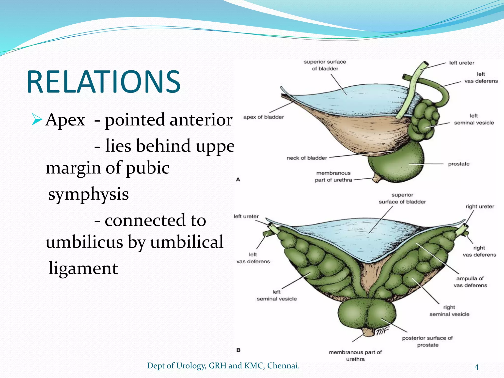
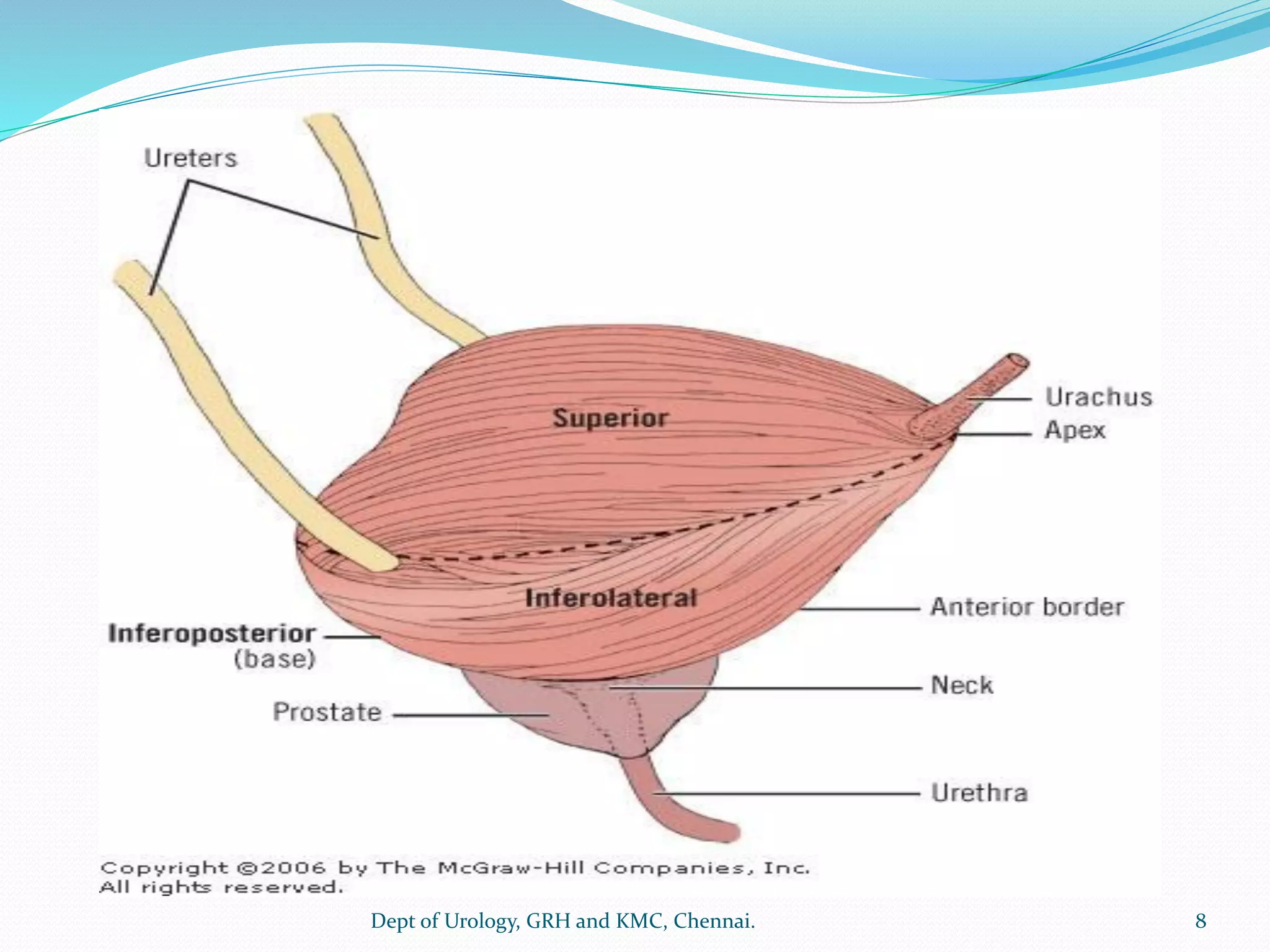
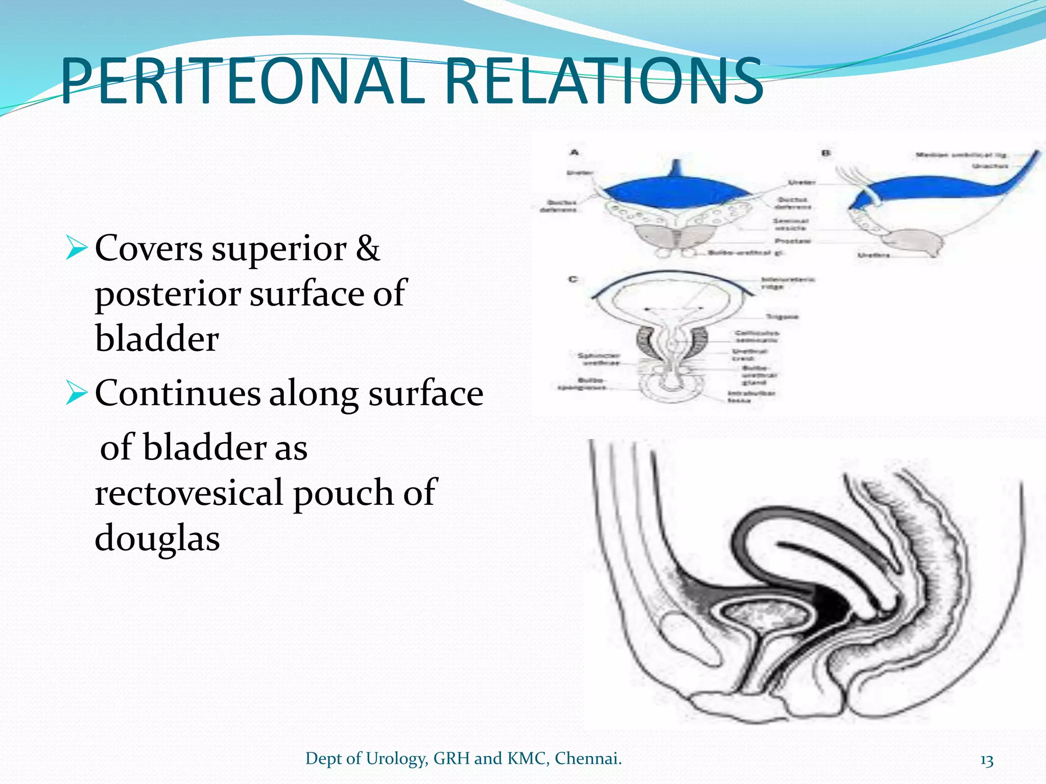
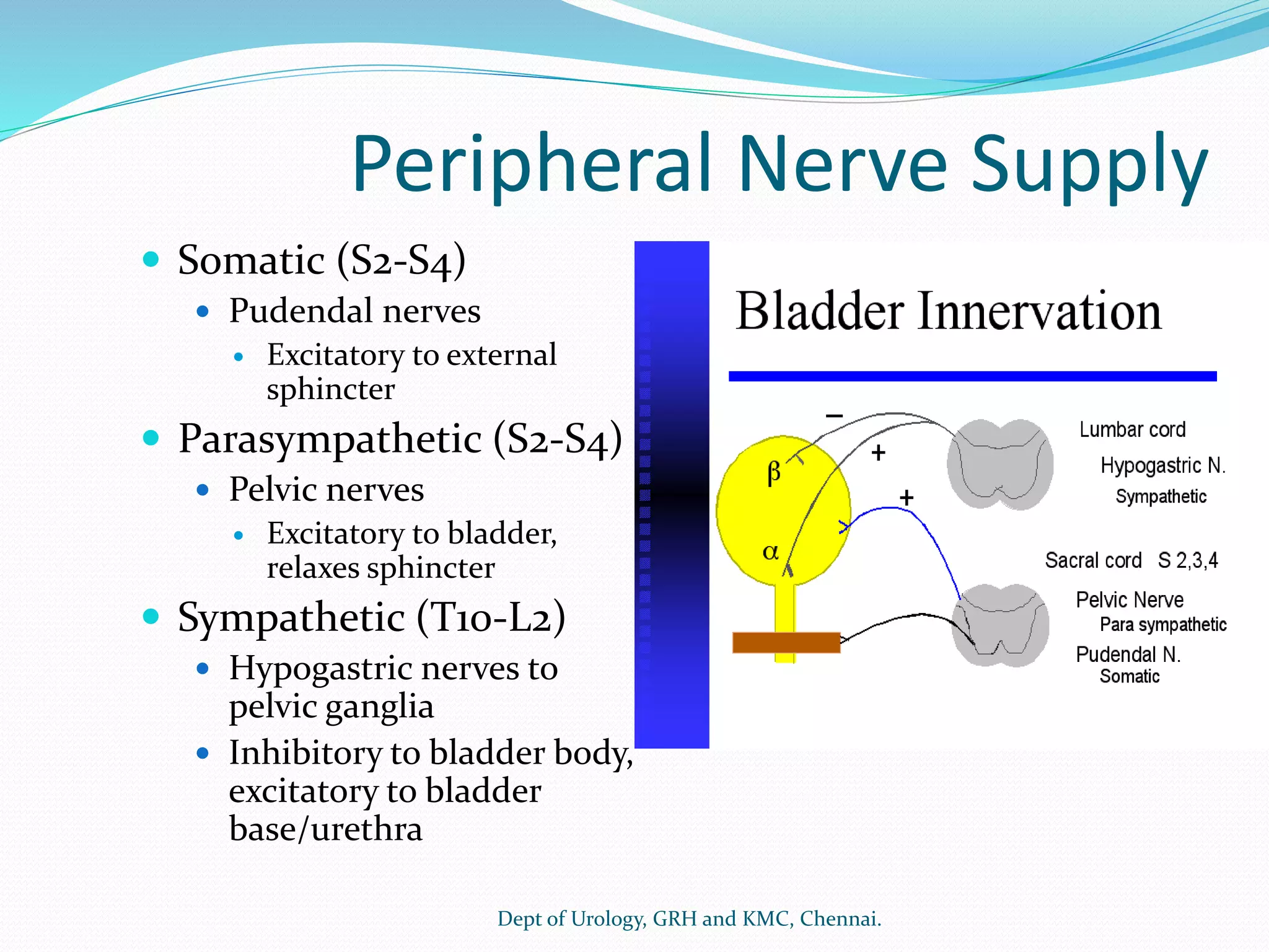
Dept of Urology
Govt Royapettah Hospital and Kilpauk Medical College
Chennai
1
Moderators:
Professors:
 Prof. Dr. G. Sivasankar, M.S., M.Ch.,
 Prof. Dr. A. Senthilvel, M.S., M.Ch.,
Asst Professors:
 Dr. J. Sivabalan, M.S., M.Ch.,
 Dr. R. Bhargavi, M.S., M.Ch.,
 Dr. S. Raju, M.S., M.Ch.,
 Dr. K. Muthurathinam, M.S., M.Ch.,
 Dr. D. Tamilselvan, M.S., M.Ch.,
 Dr. K. Senthilkumar, M.S., M.Ch.
Dept of Urology, GRH and KMC, Chennai. 2
➢Hollow, retroperitoneal organ
➢Pyramidal in shape
➢Capacity about 400-500ml
➢When empty bladder, tetrahedral in shape
➢Apex, base, superior, two inferolateral surface, &
neck
3
Dept of Urology, GRH and KMC, Chennai.
RELATIONS
➢Apex - pointed anterior
- lies behind upper
margin of pubic
symphysis
- connected to
umbilicus by umbilical
ligament
4
Dept of Urology, GRH and KMC, Chennai.
➢Base (posterior) -triangular
- superolaterally joins by the ureter
- inferior angle rise to urethra
- two vas lie side by side on posterior
- upper part- covered by peritoneum, forms
ant wall of rectovesical pouch
- lower part – seperates from rectum by
seminal vesicles & recto vesical fascia
5
Dept of Urology, GRH and KMC, Chennai.
6
Dept of Urology, GRH and KMC, Chennai.
➢Superior ;
covered with peritoneum
when bladder fills , bladder becomes
contact with anterior abdominal wall
➢ Inferolateral-
related to front of retro pubic, perivesical pad of
fat & loose connective tissue- SPACE OF RETZIUS
7
Dept of Urology, GRH and KMC, Chennai.
8
Dept of Urology, GRH and KMC, Chennai.
➢Neck-
lies inferiorly
rests on prostate
held in positions – puboprostatic ligament
➢ Interior-
mucous membrane are thrown into folds-
empty bladder,
disappears on full bladder
9
Dept of Urology, GRH and KMC, Chennai.
LIGAMENTS OF BLADDER
➢TRUE LIGAMENTS
1. Median umbilical ligament( Urachus) -
dome of bladder to umbilicus
2. Lateral umbilical ligament - lateral wall of
bladder to tendinous arch of pelvic fascia
3. Medial umbilical ligament – inguinal ligament
( OBLITERATED UMBILICAL ARTERY)
4. Medial & lateral Pubo prostatic ligament -
pelvic wall to prostate gland
10
Dept of Urology, GRH and KMC, Chennai.
11
Dept of Urology, GRH and KMC, Chennai.
➢False ligaments
Superior false ligament – course the urachus
Lateral false ligament - bladder to wall of
pelvis
Lateral superior ligament – covers the medial
umbilical ligaments
Posterior ligament - around rectum to ant
aspect of sacrum
12
Dept of Urology, GRH and KMC, Chennai.
PERITEONAL RELATIONS
➢Covers superior &
posterior surface of
bladder
➢Continues along surface
of bladder as
rectovesical pouch of
douglas
13
Dept of Urology, GRH and KMC, Chennai.
TRIGONE
➢ Triangle of smooth urothelium between two
ureteric orifice and internal urethral meatus
➢ Firmly adherent to muscular coat
➢ superior angle- corresponds to ureteric orifice
➢ inferior angle – internal urethral orifice
➢ muscular thickened between ureteric orifice –
INTERURETERIC CREST or MERCIER BAR
➢ between ureters and internal urethral meatus
BELL MUSCLE
14
Dept of Urology, GRH and KMC, Chennai.
➢ Three distinct layers
➢ SUPERFICIAL-
derived from longitudinal
layers of ureteric smooth
muscle and inserts to
verumontenum.
➢ DEEP –
Continues from waldeyer’s
sheath(fibroelastic tissue)
which inserts into bladder
neck.
15
Dept of Urology, GRH and KMC, Chennai.
➢ Detrussor layers –
from outer longitudinal
& middle circular and inner longitudinal.
These fibers form a network that enables coor-dinated
emptying of the bladder.
Longitudinal fibers are continuous with the prostatic
urethra.
The circular fibers form the internal urethral sphincter.
16
Dept of Urology, GRH and KMC, Chennai.
URETEROVESICAL JUNCTION
➢ Spiral fibres of ureter becomes longitudinally
& enchased in waldeyer’s sheath
➢ enters the bladder postero inferiorly course about
2 cm as intramural ureter & terminates at ureteric
orifice
➢ unique anatomy of intramural ureter & trigone
contributes intrisinic continence, prevents UV reflex
17
Dept of Urology, GRH and KMC, Chennai.
BLOOD SUPPLY
 ARTERIAL
- Superior & inferior vesical artery
through 1. Lateral
2. Posterior pedicle
- branch of internal iliac artery
 multiple other branches arising from the hypogastric
artery also contribute to the vascular pedicles of the
bladder,
18
Dept of Urology, GRH and KMC, Chennai.
19
Dept of Urology, GRH and KMC, Chennai.
1.Superior vesical artery :
-supplies the superior part of bladder
2.Inferior vesicle artery:
-supplies the lower ureter ,bladder base , prostate
and the seminal vesical in male
-in female supply the ureter ,bladder base and
vagina
3.Trigone is mainly supplied by
-vesiculo-deferential artery in male
-uterine artery in female
20
Dept of Urology, GRH and KMC, Chennai.
VENOUS
-The veins form a plexus within these pedicles and drain
to the internal iliac vein
LYMPHATIC
-lymphatics from the bladder start in the lamina propria
layer and then drain largely to the external iliac lymph
nodes, with some drainage to the internal iliac and
obturator lymph nodes as well.
significant cross-drainage of lymphatics from the
bladder, with drainage to both sides of the pelvis .
21
Dept of Urology, GRH and KMC, Chennai.
Peripheral Nerve Supply
 Somatic (S2-S4)
 Pudendal nerves
 Excitatory to external
sphincter
 Parasympathetic (S2-S4)
 Pelvic nerves
 Excitatory to bladder,
relaxes sphincter
 Sympathetic (T10-L2)
 Hypogastric nerves to
pelvic ganglia
 Inhibitory to bladder body,
excitatory to bladder
base/urethra
Dept of Urology, GRH and KMC, Chennai.
 Efferent fibers come from the anterior portion of the pelvic plexus.
 The bladder has a high density of parasympathetic cholinergic nerve
endings, with relatively little sympathetic innervation.
 There are also nonadrenergic, noncholinergic (NANC) fibers that
innervate the bladder and are thought to use purines as
neurotransmitters (Yoshida et al., 2001).
 The bladder neck has dense alpha 1-receptors in males, enabling
closure of the bladder neck for antegrade ejaculation and aiding
continence.
 Nitric oxide synthase containing neurons can also be found in the
bladder neck and trigone, which may promote relaxation during
micturition.
 The afferent nerves from the bladder travel with the hypogastric
plexus to reach the dorsal root ganglia in the spine.
23
Dept of Urology, GRH and KMC, Chennai.
HISTOLOGICAL STRUCTURE
➢Urothelium
- lined by transitional epithelium
- characterized by outer layer of umbrella
cells sealed closely together communicate
via tight junctions
➢ Lamina propria
- connective tissue
24
Dept of Urology, GRH and KMC, Chennai.
25
Dept of Urology, GRH and KMC, Chennai.
➢Muscularis propria
detrusor smooth muscle course in
outer- longitudinal, middle – circular
inner – longitudinal
- prominent at bladder neck
- middle layer continues as preprostatic sphincter
➢ adventitia
26
Dept of Urology, GRH and KMC, Chennai.
27
Dept of Urology, GRH and KMC, Chennai.
Normal LUT function
 Two-Phase Concept:
Filling/Storage
Emptying/Voiding
28
Dept of Urology, GRH and KMC, Chennai.
29
Dept of Urology, GRH and KMC, Chennai.
 Bladder compliance (C) is defined as the change in
volume (V) relative to the corresponding change in
intravesical pressure (P).
C = ΔV/ΔP
 When there is decreased compliance of the bladder
(steep filling curve), it may be the result of multiple
factors including (1) fast filling rate; (2) change in
composition of the bladder wall (e.g., more
collagen, less elastin); (3) hyperactivity of the
smooth muscle; and (4) a combination of any of
these factors.
30
Dept of Urology, GRH and KMC, Chennai.
Neural pathway
 Pelvic parasympathetic nerves arise at the sacral level
of the spinal cord, excite the bladder, and relax the
urethra.
 Lumbar sympathetic nerves inhibit the bladder body
and excite the bladder base and urethra.
 Pudendal nerves excite the EUS.
 Afferent fibers innervate the LUT via pelvic,
hypogastric (lumber splanchnic), and pudendal nerves
31
Dept of Urology, GRH and KMC, Chennai.
32
Dept of Urology, GRH and KMC, Chennai.
33
Dept of Urology, GRH and KMC, Chennai.
STORAGE PHASE VOIDING PHASE
34
Dept of Urology, GRH and KMC, Chennai.
35
Dept of Urology, GRH and KMC, Chennai.
36
Dept of Urology, GRH and KMC, Chennai.
Bladder in Filling Phase
 Bladder accommodation –
passive
dependent on visco-elastic property
 Increase in collagen leads to decreased compliance
37
Dept of Urology, GRH and KMC, Chennai.
Outlet in Filling phase
 “Guarding reflex”
 Urethral wall tension is not only a product of Smooth
and striated muscle but also of the elastic, collagenous,
and vascular components of the urethral wall
 soft or plastic inner layer capable of being compressed
to a closed configuration
 “Mucosal seal mechanism”
38
Dept of Urology, GRH and KMC, Chennai.
Bladder in Voiding phase
 inhibition of the spinal somatic and sympathetic
reflexes
 activation of the vesical parasympathetic pathways
 the organizational center is in the rostral brainstem
 shaping or funneling of the relaxed outlet- smooth
muscle continuity between the bladder base and the
proximal urethra
39
Dept of Urology, GRH and KMC, Chennai.
Bladder Compartments
 Urothelium
 Lamina propria and vasculature
 Stroma – Collagen
Elastin and Matrix
 Smooth muscle
40
Dept of Urology, GRH and KMC, Chennai.
Smooth muscle - Detrusor
 Detrusor cells contraction- “crossbridge cycling”
between the thick and thin filaments
 Thick filaments (15nm diameter) – myosin. Thin
filaments (6 to 8nm diameter) - actin.
41
Dept of Urology, GRH and KMC, Chennai.
Action potential
Rise in intracellular Calcium
Calcium binds to CaM
Activation of MLCK
MLCK phosphorylates MLC20
Phosphorylated MLC20 forms crossbridges
42
Dept of Urology, GRH and KMC, Chennai.
43
Dept of Urology, GRH and KMC, Chennai.
• Phase 1 fast upstroke of the
AP is composed of a Ca2+
inward current
• Phases 2 repolarization and
3 hyperpolarization of AP
are the result of a K+
outward current
• Blockage or inhibition of any
of these K+ channels would
promote myocyte
contractility and increase the
propensity of spontaneous
myocye activity. 44
Dept of Urology, GRH and KMC, Chennai.
Propagation of Electrical Responses
 Specialized proteins called connexin 43 (gap-junction
proteins) are expressed between the membranes of
connected smooth muscle cells.
 Detrusor is less well coupled electrically than other
smooth muscles.
 Poor coupling could be a feature of a normal detrusor
that prevents synchronous activation of the smooth
muscle cells during bladder filling.
45
Dept of Urology, GRH and KMC, Chennai.
46
Dept of Urology, GRH and KMC, Chennai.
Key Points
 Muscarinic receptors induce detrusor contraction, in
response to ACh released from parasympathetic nerve
terminals, by calcium entry through Ca2+ channels
 The contractile response is slower and longer lasting
than that of skeletal and cardiac muscle
 Interstitial cells or myofibroblasts- pacemaking role in
spontaneous activity of the bladder.
47
Dept of Urology, GRH and KMC, Chennai.
 In addition to smooth muscle, the human bladder is
composed of roughly 50% collagen and 2% elastin.
 With injury, obstruction, or denervation, collagen content
increases
 When collagen levels increase, compliance falls.
 Bladder wall thinning during filling is the result of a
rearrangement of the muscle bundles and also alteration of
collagen coil structure
 During filling, the detrusor reorganizes and muscle
bundles shift position from a top-to-bottom to a side-to-
side configuration
48
Dept of Urology, GRH and KMC, Chennai.
External urethral sphincter
 Twitch type – slow twitch and fast twitch
 Slow-twitch fibers -maintaining sphincter tone for
prolonged periods
 Fast-twitch- add to sphincter tone rapidly to maintain
continence when intra-abdominal pressure is abruptly
increased.
49
Dept of Urology, GRH and KMC, Chennai.
 Male - 35% fast-twitch and 65% slow-twitch fibers.
 Female - 87% slow-twitch and 13% fast-twitch fibers.
50
Dept of Urology, GRH and KMC, Chennai.
`
THANK YOU.
51
Dept of Urology, GRH and KMC, Chennai.

urinary Bladder anatomy 2

  • 1.
    Dept of Urology GovtRoyapettah Hospital and Kilpauk Medical College Chennai 1
  • 2.
    Moderators: Professors:  Prof. Dr.G. Sivasankar, M.S., M.Ch.,  Prof. Dr. A. Senthilvel, M.S., M.Ch., Asst Professors:  Dr. J. Sivabalan, M.S., M.Ch.,  Dr. R. Bhargavi, M.S., M.Ch.,  Dr. S. Raju, M.S., M.Ch.,  Dr. K. Muthurathinam, M.S., M.Ch.,  Dr. D. Tamilselvan, M.S., M.Ch.,  Dr. K. Senthilkumar, M.S., M.Ch. Dept of Urology, GRH and KMC, Chennai. 2
  • 3.
    ➢Hollow, retroperitoneal organ ➢Pyramidalin shape ➢Capacity about 400-500ml ➢When empty bladder, tetrahedral in shape ➢Apex, base, superior, two inferolateral surface, & neck 3 Dept of Urology, GRH and KMC, Chennai.
  • 4.
    RELATIONS ➢Apex - pointedanterior - lies behind upper margin of pubic symphysis - connected to umbilicus by umbilical ligament 4 Dept of Urology, GRH and KMC, Chennai.
  • 5.
    ➢Base (posterior) -triangular -superolaterally joins by the ureter - inferior angle rise to urethra - two vas lie side by side on posterior - upper part- covered by peritoneum, forms ant wall of rectovesical pouch - lower part – seperates from rectum by seminal vesicles & recto vesical fascia 5 Dept of Urology, GRH and KMC, Chennai.
  • 6.
    6 Dept of Urology,GRH and KMC, Chennai.
  • 7.
    ➢Superior ; covered withperitoneum when bladder fills , bladder becomes contact with anterior abdominal wall ➢ Inferolateral- related to front of retro pubic, perivesical pad of fat & loose connective tissue- SPACE OF RETZIUS 7 Dept of Urology, GRH and KMC, Chennai.
  • 8.
    8 Dept of Urology,GRH and KMC, Chennai.
  • 9.
    ➢Neck- lies inferiorly rests onprostate held in positions – puboprostatic ligament ➢ Interior- mucous membrane are thrown into folds- empty bladder, disappears on full bladder 9 Dept of Urology, GRH and KMC, Chennai.
  • 10.
    LIGAMENTS OF BLADDER ➢TRUELIGAMENTS 1. Median umbilical ligament( Urachus) - dome of bladder to umbilicus 2. Lateral umbilical ligament - lateral wall of bladder to tendinous arch of pelvic fascia 3. Medial umbilical ligament – inguinal ligament ( OBLITERATED UMBILICAL ARTERY) 4. Medial & lateral Pubo prostatic ligament - pelvic wall to prostate gland 10 Dept of Urology, GRH and KMC, Chennai.
  • 11.
    11 Dept of Urology,GRH and KMC, Chennai.
  • 12.
    ➢False ligaments Superior falseligament – course the urachus Lateral false ligament - bladder to wall of pelvis Lateral superior ligament – covers the medial umbilical ligaments Posterior ligament - around rectum to ant aspect of sacrum 12 Dept of Urology, GRH and KMC, Chennai.
  • 13.
    PERITEONAL RELATIONS ➢Covers superior& posterior surface of bladder ➢Continues along surface of bladder as rectovesical pouch of douglas 13 Dept of Urology, GRH and KMC, Chennai.
  • 14.
    TRIGONE ➢ Triangle ofsmooth urothelium between two ureteric orifice and internal urethral meatus ➢ Firmly adherent to muscular coat ➢ superior angle- corresponds to ureteric orifice ➢ inferior angle – internal urethral orifice ➢ muscular thickened between ureteric orifice – INTERURETERIC CREST or MERCIER BAR ➢ between ureters and internal urethral meatus BELL MUSCLE 14 Dept of Urology, GRH and KMC, Chennai.
  • 15.
    ➢ Three distinctlayers ➢ SUPERFICIAL- derived from longitudinal layers of ureteric smooth muscle and inserts to verumontenum. ➢ DEEP – Continues from waldeyer’s sheath(fibroelastic tissue) which inserts into bladder neck. 15 Dept of Urology, GRH and KMC, Chennai.
  • 16.
    ➢ Detrussor layers– from outer longitudinal & middle circular and inner longitudinal. These fibers form a network that enables coor-dinated emptying of the bladder. Longitudinal fibers are continuous with the prostatic urethra. The circular fibers form the internal urethral sphincter. 16 Dept of Urology, GRH and KMC, Chennai.
  • 17.
    URETEROVESICAL JUNCTION ➢ Spiralfibres of ureter becomes longitudinally & enchased in waldeyer’s sheath ➢ enters the bladder postero inferiorly course about 2 cm as intramural ureter & terminates at ureteric orifice ➢ unique anatomy of intramural ureter & trigone contributes intrisinic continence, prevents UV reflex 17 Dept of Urology, GRH and KMC, Chennai.
  • 18.
    BLOOD SUPPLY  ARTERIAL -Superior & inferior vesical artery through 1. Lateral 2. Posterior pedicle - branch of internal iliac artery  multiple other branches arising from the hypogastric artery also contribute to the vascular pedicles of the bladder, 18 Dept of Urology, GRH and KMC, Chennai.
  • 19.
    19 Dept of Urology,GRH and KMC, Chennai.
  • 20.
    1.Superior vesical artery: -supplies the superior part of bladder 2.Inferior vesicle artery: -supplies the lower ureter ,bladder base , prostate and the seminal vesical in male -in female supply the ureter ,bladder base and vagina 3.Trigone is mainly supplied by -vesiculo-deferential artery in male -uterine artery in female 20 Dept of Urology, GRH and KMC, Chennai.
  • 21.
    VENOUS -The veins forma plexus within these pedicles and drain to the internal iliac vein LYMPHATIC -lymphatics from the bladder start in the lamina propria layer and then drain largely to the external iliac lymph nodes, with some drainage to the internal iliac and obturator lymph nodes as well. significant cross-drainage of lymphatics from the bladder, with drainage to both sides of the pelvis . 21 Dept of Urology, GRH and KMC, Chennai.
  • 22.
    Peripheral Nerve Supply Somatic (S2-S4)  Pudendal nerves  Excitatory to external sphincter  Parasympathetic (S2-S4)  Pelvic nerves  Excitatory to bladder, relaxes sphincter  Sympathetic (T10-L2)  Hypogastric nerves to pelvic ganglia  Inhibitory to bladder body, excitatory to bladder base/urethra Dept of Urology, GRH and KMC, Chennai.
  • 23.
     Efferent fiberscome from the anterior portion of the pelvic plexus.  The bladder has a high density of parasympathetic cholinergic nerve endings, with relatively little sympathetic innervation.  There are also nonadrenergic, noncholinergic (NANC) fibers that innervate the bladder and are thought to use purines as neurotransmitters (Yoshida et al., 2001).  The bladder neck has dense alpha 1-receptors in males, enabling closure of the bladder neck for antegrade ejaculation and aiding continence.  Nitric oxide synthase containing neurons can also be found in the bladder neck and trigone, which may promote relaxation during micturition.  The afferent nerves from the bladder travel with the hypogastric plexus to reach the dorsal root ganglia in the spine. 23 Dept of Urology, GRH and KMC, Chennai.
  • 24.
    HISTOLOGICAL STRUCTURE ➢Urothelium - linedby transitional epithelium - characterized by outer layer of umbrella cells sealed closely together communicate via tight junctions ➢ Lamina propria - connective tissue 24 Dept of Urology, GRH and KMC, Chennai.
  • 25.
    25 Dept of Urology,GRH and KMC, Chennai.
  • 26.
    ➢Muscularis propria detrusor smoothmuscle course in outer- longitudinal, middle – circular inner – longitudinal - prominent at bladder neck - middle layer continues as preprostatic sphincter ➢ adventitia 26 Dept of Urology, GRH and KMC, Chennai.
  • 27.
    27 Dept of Urology,GRH and KMC, Chennai.
  • 28.
    Normal LUT function Two-Phase Concept: Filling/Storage Emptying/Voiding 28 Dept of Urology, GRH and KMC, Chennai.
  • 29.
    29 Dept of Urology,GRH and KMC, Chennai.
  • 30.
     Bladder compliance(C) is defined as the change in volume (V) relative to the corresponding change in intravesical pressure (P). C = ΔV/ΔP  When there is decreased compliance of the bladder (steep filling curve), it may be the result of multiple factors including (1) fast filling rate; (2) change in composition of the bladder wall (e.g., more collagen, less elastin); (3) hyperactivity of the smooth muscle; and (4) a combination of any of these factors. 30 Dept of Urology, GRH and KMC, Chennai.
  • 31.
    Neural pathway  Pelvicparasympathetic nerves arise at the sacral level of the spinal cord, excite the bladder, and relax the urethra.  Lumbar sympathetic nerves inhibit the bladder body and excite the bladder base and urethra.  Pudendal nerves excite the EUS.  Afferent fibers innervate the LUT via pelvic, hypogastric (lumber splanchnic), and pudendal nerves 31 Dept of Urology, GRH and KMC, Chennai.
  • 32.
    32 Dept of Urology,GRH and KMC, Chennai.
  • 33.
    33 Dept of Urology,GRH and KMC, Chennai.
  • 34.
    STORAGE PHASE VOIDINGPHASE 34 Dept of Urology, GRH and KMC, Chennai.
  • 35.
    35 Dept of Urology,GRH and KMC, Chennai.
  • 36.
    36 Dept of Urology,GRH and KMC, Chennai.
  • 37.
    Bladder in FillingPhase  Bladder accommodation – passive dependent on visco-elastic property  Increase in collagen leads to decreased compliance 37 Dept of Urology, GRH and KMC, Chennai.
  • 38.
    Outlet in Fillingphase  “Guarding reflex”  Urethral wall tension is not only a product of Smooth and striated muscle but also of the elastic, collagenous, and vascular components of the urethral wall  soft or plastic inner layer capable of being compressed to a closed configuration  “Mucosal seal mechanism” 38 Dept of Urology, GRH and KMC, Chennai.
  • 39.
    Bladder in Voidingphase  inhibition of the spinal somatic and sympathetic reflexes  activation of the vesical parasympathetic pathways  the organizational center is in the rostral brainstem  shaping or funneling of the relaxed outlet- smooth muscle continuity between the bladder base and the proximal urethra 39 Dept of Urology, GRH and KMC, Chennai.
  • 40.
    Bladder Compartments  Urothelium Lamina propria and vasculature  Stroma – Collagen Elastin and Matrix  Smooth muscle 40 Dept of Urology, GRH and KMC, Chennai.
  • 41.
    Smooth muscle -Detrusor  Detrusor cells contraction- “crossbridge cycling” between the thick and thin filaments  Thick filaments (15nm diameter) – myosin. Thin filaments (6 to 8nm diameter) - actin. 41 Dept of Urology, GRH and KMC, Chennai.
  • 42.
    Action potential Rise inintracellular Calcium Calcium binds to CaM Activation of MLCK MLCK phosphorylates MLC20 Phosphorylated MLC20 forms crossbridges 42 Dept of Urology, GRH and KMC, Chennai.
  • 43.
    43 Dept of Urology,GRH and KMC, Chennai.
  • 44.
    • Phase 1fast upstroke of the AP is composed of a Ca2+ inward current • Phases 2 repolarization and 3 hyperpolarization of AP are the result of a K+ outward current • Blockage or inhibition of any of these K+ channels would promote myocyte contractility and increase the propensity of spontaneous myocye activity. 44 Dept of Urology, GRH and KMC, Chennai.
  • 45.
    Propagation of ElectricalResponses  Specialized proteins called connexin 43 (gap-junction proteins) are expressed between the membranes of connected smooth muscle cells.  Detrusor is less well coupled electrically than other smooth muscles.  Poor coupling could be a feature of a normal detrusor that prevents synchronous activation of the smooth muscle cells during bladder filling. 45 Dept of Urology, GRH and KMC, Chennai.
  • 46.
    46 Dept of Urology,GRH and KMC, Chennai.
  • 47.
    Key Points  Muscarinicreceptors induce detrusor contraction, in response to ACh released from parasympathetic nerve terminals, by calcium entry through Ca2+ channels  The contractile response is slower and longer lasting than that of skeletal and cardiac muscle  Interstitial cells or myofibroblasts- pacemaking role in spontaneous activity of the bladder. 47 Dept of Urology, GRH and KMC, Chennai.
  • 48.
     In additionto smooth muscle, the human bladder is composed of roughly 50% collagen and 2% elastin.  With injury, obstruction, or denervation, collagen content increases  When collagen levels increase, compliance falls.  Bladder wall thinning during filling is the result of a rearrangement of the muscle bundles and also alteration of collagen coil structure  During filling, the detrusor reorganizes and muscle bundles shift position from a top-to-bottom to a side-to- side configuration 48 Dept of Urology, GRH and KMC, Chennai.
  • 49.
    External urethral sphincter Twitch type – slow twitch and fast twitch  Slow-twitch fibers -maintaining sphincter tone for prolonged periods  Fast-twitch- add to sphincter tone rapidly to maintain continence when intra-abdominal pressure is abruptly increased. 49 Dept of Urology, GRH and KMC, Chennai.
  • 50.
     Male -35% fast-twitch and 65% slow-twitch fibers.  Female - 87% slow-twitch and 13% fast-twitch fibers. 50 Dept of Urology, GRH and KMC, Chennai.
  • 51.
    ` THANK YOU. 51 Dept ofUrology, GRH and KMC, Chennai.