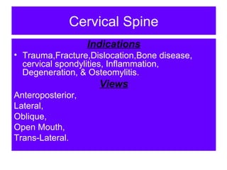
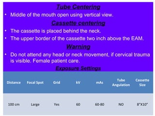
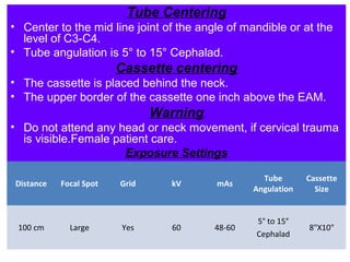
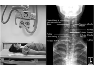
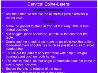
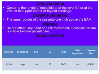
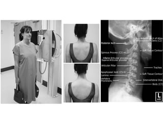
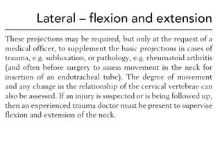
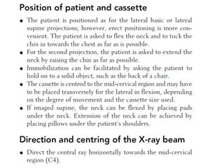
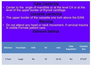
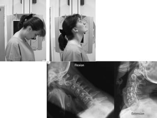
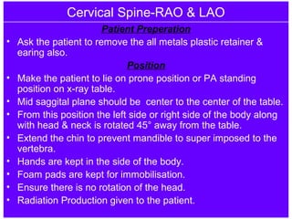
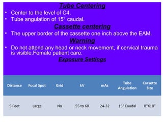
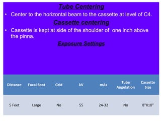
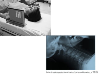
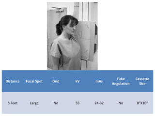
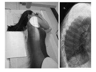
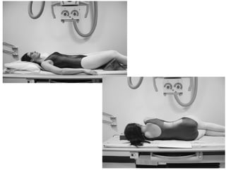
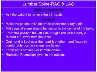
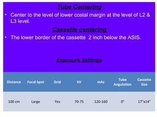
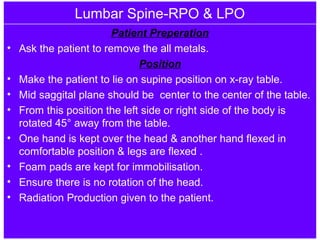
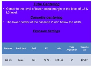
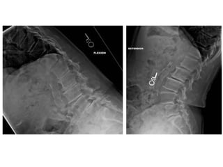
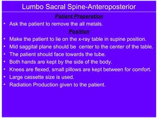
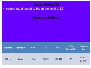
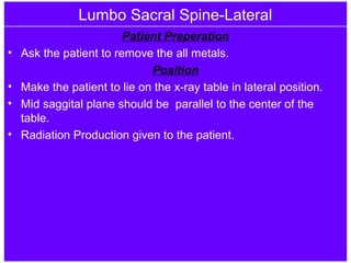
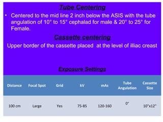
SlideShare a Scribd company logo
V.Deepak Prasath, B.Sc RIT.
K.G.Hospital & Post Graduate Medical Institute,
Coimbatore.
POSITIONING TERMINOLOGY
• Radiographic positioning refers to the study of
patient positioning performed for radiographic
demonstration or visualization of specific body
parts on image receptors.
• Each person who plans to work as a radiologic
technologist must clearly understand the
correct use of positioning terminology.
Cervical Spine
Indications
• Trauma,Fracture,Dislocation,Bone disease,
cervical spondylities, Inflammation,
Degeneration, & Osteomylitis.
Views
Anteroposterior,
Lateral,
Oblique,
Open Mouth,
Trans-Lateral.
Cervical Spine-Open Mouth
Patient Preperation
• Ask the patient to remove the all metals
plastic retainer & earing also.
Position
• Make the patient to lie on the x-ray table or sit.
• Mid saggital plane should be center to the
center of the table.
• The patient should face towards the tube.
• The chin is extented.
• Ensure the mouth is wide open & there is no
rotation of head & neck.
• Radiation Production given to the patient.
Tube Centering
• Middle of the mouth open using vertical view.
Cassette centering
• The cassette is placed behind the neck.
• The upper border of the cassette two inch above the EAM.
Warning
• Do not attend any head or neck movement, if cervical trauma
is visible. Female patient care.
Exposure Settings
Distance Focal Spot Grid kV mAs
Tube
Angulation
Cassette
Size
100 cm Large Yes 60 60-80 NO 8"X10"
Cervical Spine-Anteroposterior
Patient Preperation
• Ask the patient to remove the all metals plastic retainer &
earing also.
Position
• Make the patient to lie on the x-ray table.
• Mid saggital plane should be center to the center of the
table.
• The patient should face towards the tube.
• The chin is raised, so that the mandible is superimposedon
the occipital.(The line from the tip of the mandible to the base
of the skull should be parallel to the center.)
• Ensure there is no rotation of the head.
• Radiation Production given to the patient.
Tube Centering
• Center to the mid line joint of the angle of mandible or at the
level of C3-C4.
• Tube angulation is 5° to 15° Cephalad.
Cassette centering
• The cassette is placed behind the neck.
• The upper border of the cassette one inch above the EAM.
Warning
• Do not attend any head or neck movement, if cervical trauma
is visible.Female patient care.
Exposure Settings
Distance Focal Spot Grid kV mAs
Tube
Angulation
Cassette
Size
100 cm Large Yes 60 48-60
5° to 15°
Cephalad
8"X10"
Cervical Spine-Lateral
Patient Preperation
• Ask the patient to remove the all metals plastic retainer &
earing also.
Position
• Make the patient to stand in front of the x-ray table in true
lateral position.
• Mid saggital plane should be parallel to the center of the
table.
• Depressed the shoulder as much as possible.Ask the patient
to lowered there shoulder as much as possible so as to avoid
overlapping.
• To pull out the patient shoulder more with help of weight
bearing(sand bag) on both hands.
• The chin is raised, so that angle of mandible dose not come in
way to upper c-spine.
• Ensure there is no rotation of the head.
Tube Centering
• Center to the angle of mandible or at the level C4 or at the
level of the upper border of thyroid cardilage.
Cassette centering
• The upper border of the cassette one inch above the EAM.
Warning
• Do not attend any head or neck movement, if cervical trauma
is visible.Female patient care.
Exposure Settings
Distance Focal Spot Grid kV mAs
Tube
Angulation
Cassette
Size
5 Feet Large No 55 24-32 No 8"X10"
Cervical Spine-RAO & LAO
Patient Preperation
• Ask the patient to remove the all metals plastic retainer &
earing also.
Position
• Make the patient to lie on prone position or PA standing
position on x-ray table.
• Mid saggital plane should be center to the center of the table.
• From this position the left side or right side of the body along
with head & neck is rotated 45° away from the table.
• Extend the chin to prevent mandible to super imposed to the
vertebra.
• Hands are kept in the side of the body.
• Foam pads are kept for immobilisation.
• Ensure there is no rotation of the head.
• Radiation Production given to the patient.
Tube Centering
• Center to the level of C4.
• Tube angulation of 15° caudal.
Cassette centering
• The upper border of the cassette one inch above the EAM.
Warning
• Do not attend any head or neck movement, if cervical trauma
is visible.Female patient care.
Exposure Settings
Distance Focal Spot Grid kV mAs
Tube
Angulation
Cassette
Size
5 Feet Large No 55 to 60 24-32 15° Caudal 8"X10"
Cervical Spine-RPO & LPO
Patient Preperation
• Ask the patient to remove the all metals plastic retainer &
earing also.
Position
• Make the patient to lie on supine position or AP standing
position on x-ray table.
• Mid saggital plane should be center to the center of the table.
• From this position the left side or right side of the body along
with head & neck is rotated 45° away from the table.
• Extend the chin to prevent mandible to super imposed to the
vertebra.
• Hands are kept in the side of the body.
• Foam pads are kept for immobilisation.
• Ensure there is no rotation of the head.
• Radiation Production given to the patient.
Tube Centering
• Center to the level of C4.
• Tube angulation of 15° caudal.
Cassette centering
• The upper border of the cassette one inch above the EAM.
Anatomical Structure
• Intervertebral foramen, pedicle.
Exposure Settings
Distance Focal Spot Grid kV mAs
Tube
Angulation
Cassette
Size
5 Feet Large No 55 to 60 24-32 15° Caudal 8"X10"
Cervical Spine-Trans-Lateral
Patient Preperation
• Ask the patient to remove the all metals plastic retainer & earing
also.
Position
• Make the patient to lie on the x-ray table in supine position.
• Hands are kept at side of the body.
• Ensure there is no rotation of the head & neck without much
pressure to the injury neck & foam pads are kept for immobilization.
• Radiation Production given to the patient.
Tube Centering
• Center to the horizontal beam to the cassette at level of C4.
Cassette centering
• Cassette is kept at side of the shoulder of one inch above
the pinna.
Exposure Settings
Distance Focal Spot Grid kV mAs
Tube
Angulation
Cassette
Size
5 Feet Large No 55 24-32 No 8"X10"
Swimmers View
Distance Focal Spot Grid kV mAs
Tube
Angulation
Cassette
Size
5 Feet Large No 55 24-32 No 8"X10"
Thoracic Spine
Indications
• Trauma,Fracture,Dislocation,Bone disease,
Inflammation, Degeneration, & Kyphosis,
skoliosis.
Views
Anteroposterior,
Lateral.
Thoracic Spine-Anteroposterior
Patient Preperation
• Ask the patient to remove the all metals plastic retainer.
Position
• Make the patient to lie on the x-ray table in supine position.
• Mid saggital plane should be center to the center of the table.
• The patient should face towards the tube.
• Knees are flexed, small pillows are kept between for comfort.
• Large cassette size is used.
• Radiation Production given to the patient.
Tube Centering
• Direct the central ray at right-angles to the cassette and
towards a point 2.5 cm below the sternal angle.
• Collimate tightly to the spine.
Cassette centering
• The upper border of the cassette is kept above spinoces
process of the 7th c-spine vertebra.
Exposure Settings
Distance Focal Spot Grid kV mAs
Tube
Angulation
Cassette
Size
100 cm Large Yes 65-70 80-120 0° 12"X15"
Thoracic Spine-Lateral
Patient Preperation
• Ask the patient to remove the all metals plastic retainer.
Position
• Make the patient to lie or stand in front of the x-ray table in
lateral position.
• Mid saggital plane should be parallel to the center of the
table.
• Arm should be raised well above the head.
• The head can be supported by pillow & pads may be placed
between the knee for the patients comfort.
• Radiation Production given to the patient.
Tube Centering
• The tube center at the level of 1" below the axila T7 & T8.
Cassette centering
• The upper border of the cassette is kept 3 to 4 cm above the
spinous process of the C7.
Exposure Settings
Distance Focal Spot Grid kV mAs
Tube
Angulation
Cassette
Size
100 cm Large Yes 65-70 100-120 0° 12"X15"
Lumbar Spine
Indications
• Trauma,Fracture,Dislocation,Bone disease,
Inflammation, Degeneration, & Kyphosis,
skoliosis.
Views
Anteroposterior,
Lateral,
Oblique.
Lumbar Spine-Anteroposterior
Patient Preperation
• Ask the patient to remove the all metals.
Position
• Make the patient to lie on the x-ray table in supine position.
• Mid saggital plane should be center to the center of the table.
• The patient should face towards the tube.
• Both hands are kept by the side of the body.
• Knees are flexed, small pillows are kept between for comfort.
• Large cassette size is used.
• Radiation Production given to the patient.
Tube Centering
• Direct the central ray towards the midline at the level of the
lower costal margin (L3).
Cassette centering
• The lower border of the cassette 2 inch below the ASIS.
Exposure Settings
Distance Focal Spot Grid kV mAs
Tube
Angulation
Cassette
Size
100 cm Large Yes 65-75 100-120 0°
10"x12"
12"x15"
Lumbar Spine-Lateral
Patient Preperation
• Ask the patient to remove the all metals.
Position
• Make the patient to lie on the x-ray table in lateral position.
• Mid saggital plane should be parallel to the center of the
table.
• Hands are fold & kept on the head & knees are flex & small
foam pads are kept to avoid movement.
• Small foam pad are kept under the lumbar spine to reduce the
lumbar arch & lumbar lordorsis.
• Radiation Production given to the patient.
Tube Centering
• Center to the level of lower costal margin at the level of L2 &
L3 level.
Cassette centering
• The lower border of the cassette 2 inch below the ASIS.
Exposure Settings
Distance Focal Spot Grid kV mAs
Tube
Angulation
Cassette
Size
100 cm Large Yes 70-75 120-160 0° 12"x15"
Lumbar Spine-RAO & LAO
Patient Preperation
• Ask the patient to remove the all metals.
Position
• Make the patient to lie on prone positionon x-ray table.
• Mid saggital plane should be center to the center of the table.
• From this position the left side or right side of the body is
rotated 45° away from the table.
• One hand is kept over the head & another hand flexed in
comfortable position & legs are flexed .
• Foam pads are kept for immobilisation.
• Radiation Production given to the patient.
Tube Centering
• Center to the level of lower costal margin at the level of L2 &
L3 level.
Cassette centering
• The lower border of the cassette 2 inch below the ASIS.
Exposure Settings
Distance Focal Spot Grid kV mAs
Tube
Angulation
Cassette
Size
100 cm Large Yes 70-75 120-160 0° 17"x14"
Lumbar Spine-RPO & LPO
Patient Preperation
• Ask the patient to remove the all metals.
Position
• Make the patient to lie on supine position on x-ray table.
• Mid saggital plane should be center to the center of the table.
• From this position the left side or right side of the body is
rotated 45° away from the table.
• One hand is kept over the head & another hand flexed in
comfortable position & legs are flexed .
• Foam pads are kept for immobilisation.
• Ensure there is no rotation of the head.
• Radiation Production given to the patient.
Tube Centering
• Center to the level of lower costal margin at the level of L2 &
L3 level.
Cassette centering
• The lower border of the cassette 2 inch below the ASIS.
Exposure Settings
Distance Focal Spot Grid kV mAs
Tube
Angulation
Cassette
Size
100 cm Large Yes 70-75 120-160 0° 17"x14"
Lumbo Sacral Spine-Anteroposterior
Patient Preperation
• Ask the patient to remove the all metals.
Position
• Make the patient to lie on the x-ray table in supine position.
• Mid saggital plane should be center to the center of the table.
• The patient should face towards the tube.
• Both hands are kept by the side of the body.
• Knees are flexed, small pillows are kept between for comfort.
• Large cassette size is used.
• Radiation Production given to the patient.
Tube Centering
• central ray directed to the at the level of L5.
Exposure Settings
Distance Focal Spot Grid kV mAs
Tube
Angulation
Cassette
Size
100 cm Large Yes 70-75 100-120 0°
10"x12"
12"x15"
Lumbo Sacral Spine-Lateral
Patient Preperation
• Ask the patient to remove the all metals.
Position
• Make the patient to lie on the x-ray table in lateral position.
• Mid saggital plane should be parallel to the center of the
table.
• Radiation Production given to the patient.
Tube Centering
• Central ray directed to the at the level of L5.
Exposure Settings
Distance Focal Spot Grid kV mAs
Tube
Angulation
Cassette
Size
100 cm Large Yes 75-85 120-160 0° 12"x15"
Sacrum-Anteroposterior
Patient Preperation
• Ask the patient to remove the all metals.
Position
• Make the patient to lie on the x-ray table in supine position.
• Mid saggital plane should be center to the center of the table.
• The patient should face towards the tube.
• Both hands are kept by the side of the body.
• Knees are flexed, small pillows are kept between for comfort.
• Radiation Production given to the patient.
Tube Centering
• Centered to the mid line 2 inch below the ASIS with the tube
angulation of 10° to 15° cephalad for male & 20° to 25° for
Female.
Cassette centering
Upper border of the cassette placed at the level of illiac creast
Exposure Settings
Distance Focal Spot Grid kV mAs
Tube
Angulation
Cassette
Size
100 cm Large Yes 70-75 80-100
10° to 15°
for male
20° to 25°
for Female
cephalad
10"x12"
Sacrum-Lateral
Patient Preperation
• Ask the patient to remove the all metals.
Position
• Make the patient to lie on the x-ray table in lateral position.
• Mid saggital plane should be parallel to the center of the
table.
• Knees are flexed, small pillows are kept between for comfort.
• Radiation Production given to the patient.
Tube Centering
• Centered to the mid line 2 inch below the ASIS with the tube
angulation of 10° to 15° cephalad for male & 20° to 25° for
Female.
Cassette centering
Upper border of the cassette placed at the level of illiac creast
Exposure Settings
Distance Focal Spot Grid kV mAs
Tube
Angulation
Cassette
Size
100 cm Large Yes 75-85 120-160
0°
10"x12"
Coccyx-Anteroposterior
Patient Preperation
• Ask the patient to remove the all metals.
Position
• Make the patient to lie on the x-ray table in supine position.
• Mid saggital plane should be center to the center of the table.
• The patient should face towards the tube.
• Both hands are kept by the side of the body.
• Knees are flexed, small pillows are kept between for comfort.
• Radiation Production given to the patient.
Tube Centering
• Centered to the mid line 2 inch below the ASIS with the tube
angulation of 10° to 15° cephalad for male & 20° to 25° for
Female.
Cassette centering
Upper border of the cassette placed at the level of illiac creast
Exposure Settings
Distance Focal Spot Grid kV mAs
Tube
Angulation
Cassette
Size
100 cm Large Yes 70-75 80-100
10° to 15°
for male
20° to 25°
for Female
cephalad
10"x12"
Coccyx-Lateral
Patient Preperation
• Ask the patient to remove the all metals.
Position
• Make the patient to lie on the x-ray table in lateral position.
• Mid saggital plane should be parallel to the center of the
table.
• Knees are flexed, small pillows are kept between for comfort.
• Radiation Production given to the patient.
Tube Centering
• Centered to the mid line 2 inch below the ASIS with the tube
angulation of 10° to 15° cephalad for male & 20° to 25° for
Female.
Cassette centering
Upper border of the cassette placed at the level of illiac creast
Exposure Settings
Distance Focal Spot Grid kV mAs
Tube
Angulation
Cassette
Size
100 cm Large Yes 75-85 120-160
0°
10"x12"
Radiography Positioning Spine

More Related Content

What's hot

X ray imaging of forearm and elbow
X ray imaging of forearm and elbowX ray imaging of forearm and elbow
X ray imaging of forearm and elbow
Maajid Mohi ud din
 
Mobile & portable radiography
Mobile & portable radiographyMobile & portable radiography
Mobile & portable radiography
Priyanka Parimala
 
Radiograpic views for shoulder joint
Radiograpic views  for shoulder jointRadiograpic views  for shoulder joint
Radiograpic views for shoulder joint
Ganesan Yogananthem
 
Upper limb
Upper limbUpper limb
Upper limb
Rituraj Mishra
 
9. radiography of shoulder
9. radiography of shoulder9. radiography of shoulder
9. radiography of shoulder
drdharmendra8648
 
X-RAY PELVIS PROJECTIONS
X-RAY PELVIS PROJECTIONSX-RAY PELVIS PROJECTIONS
X-RAY PELVIS PROJECTIONS
Jai Kumar
 
Upper extremity anatomy & positioning
Upper extremity anatomy & positioningUpper extremity anatomy & positioning
Upper extremity anatomy & positioning
InfoUtilRT
 
ELBOW JOINT X-RAY PROJECTIONS
ELBOW JOINT X-RAY PROJECTIONSELBOW JOINT X-RAY PROJECTIONS
ELBOW JOINT X-RAY PROJECTIONS
Jai Kumar
 
Radiographic positioning terminology
Radiographic positioning terminology  Radiographic positioning terminology
Radiographic positioning terminology
Muhammad Arif Afridi
 
Chest radiography ppt
Chest radiography pptChest radiography ppt
Chest radiography ppt
Niranjan Jha
 
Magnification(macro and micro radiography), distortion
Magnification(macro and micro radiography), distortionMagnification(macro and micro radiography), distortion
Magnification(macro and micro radiography), distortion
parthajyotidas11
 
Projections of carpal tunnel and wrist
Projections of  carpal tunnel and wristProjections of  carpal tunnel and wrist
Projections of carpal tunnel and wrist
DonBenny2
 
Radiographic positioning of Upper limb (ELBOW & HUMERUS)
Radiographic positioning of Upper limb (ELBOW & HUMERUS)Radiographic positioning of Upper limb (ELBOW & HUMERUS)
Radiographic positioning of Upper limb (ELBOW & HUMERUS)
Nasir Mohiudin
 
RADIOGRAPHIC VIEWS FOR HIP JOINT
RADIOGRAPHIC VIEWS FOR HIP JOINTRADIOGRAPHIC VIEWS FOR HIP JOINT
RADIOGRAPHIC VIEWS FOR HIP JOINT
Ganesan Yogananthem
 
Emergency radiography
Emergency radiography Emergency radiography
Emergency radiography
milan timilsina
 
Chest x ray positioning
Chest x ray  positioningChest x ray  positioning
Chest x ray positioning
airwave12
 
Advanced radiographic positions for the lower extremities
Advanced radiographic positions for the lower extremitiesAdvanced radiographic positions for the lower extremities
Advanced radiographic positions for the lower extremitiesmr_koky
 
Radiographic views of proximal femur and pelvis
Radiographic views of proximal femur and pelvisRadiographic views of proximal femur and pelvis
Radiographic views of proximal femur and pelvis
Chandan Prasad
 
Radiographic technique of skull
Radiographic technique of skullRadiographic technique of skull
Radiographic technique of skull
SaruGosain
 
Portable n mobile unit
Portable n mobile unitPortable n mobile unit
Portable n mobile unitSudil Paudyal
 

What's hot (20)

X ray imaging of forearm and elbow
X ray imaging of forearm and elbowX ray imaging of forearm and elbow
X ray imaging of forearm and elbow
 
Mobile & portable radiography
Mobile & portable radiographyMobile & portable radiography
Mobile & portable radiography
 
Radiograpic views for shoulder joint
Radiograpic views  for shoulder jointRadiograpic views  for shoulder joint
Radiograpic views for shoulder joint
 
Upper limb
Upper limbUpper limb
Upper limb
 
9. radiography of shoulder
9. radiography of shoulder9. radiography of shoulder
9. radiography of shoulder
 
X-RAY PELVIS PROJECTIONS
X-RAY PELVIS PROJECTIONSX-RAY PELVIS PROJECTIONS
X-RAY PELVIS PROJECTIONS
 
Upper extremity anatomy & positioning
Upper extremity anatomy & positioningUpper extremity anatomy & positioning
Upper extremity anatomy & positioning
 
ELBOW JOINT X-RAY PROJECTIONS
ELBOW JOINT X-RAY PROJECTIONSELBOW JOINT X-RAY PROJECTIONS
ELBOW JOINT X-RAY PROJECTIONS
 
Radiographic positioning terminology
Radiographic positioning terminology  Radiographic positioning terminology
Radiographic positioning terminology
 
Chest radiography ppt
Chest radiography pptChest radiography ppt
Chest radiography ppt
 
Magnification(macro and micro radiography), distortion
Magnification(macro and micro radiography), distortionMagnification(macro and micro radiography), distortion
Magnification(macro and micro radiography), distortion
 
Projections of carpal tunnel and wrist
Projections of  carpal tunnel and wristProjections of  carpal tunnel and wrist
Projections of carpal tunnel and wrist
 
Radiographic positioning of Upper limb (ELBOW & HUMERUS)
Radiographic positioning of Upper limb (ELBOW & HUMERUS)Radiographic positioning of Upper limb (ELBOW & HUMERUS)
Radiographic positioning of Upper limb (ELBOW & HUMERUS)
 
RADIOGRAPHIC VIEWS FOR HIP JOINT
RADIOGRAPHIC VIEWS FOR HIP JOINTRADIOGRAPHIC VIEWS FOR HIP JOINT
RADIOGRAPHIC VIEWS FOR HIP JOINT
 
Emergency radiography
Emergency radiography Emergency radiography
Emergency radiography
 
Chest x ray positioning
Chest x ray  positioningChest x ray  positioning
Chest x ray positioning
 
Advanced radiographic positions for the lower extremities
Advanced radiographic positions for the lower extremitiesAdvanced radiographic positions for the lower extremities
Advanced radiographic positions for the lower extremities
 
Radiographic views of proximal femur and pelvis
Radiographic views of proximal femur and pelvisRadiographic views of proximal femur and pelvis
Radiographic views of proximal femur and pelvis
 
Radiographic technique of skull
Radiographic technique of skullRadiographic technique of skull
Radiographic technique of skull
 
Portable n mobile unit
Portable n mobile unitPortable n mobile unit
Portable n mobile unit
 

Similar to Radiography Positioning Spine

Seminar new
Seminar newSeminar new
Seminar new
Ganesh Gadag
 
X ray c-spine
X ray c-spine X ray c-spine
X ray c-spine
Rajaoct
 
Radiotherapy techniques for Breast Cancer
Radiotherapy techniques for Breast CancerRadiotherapy techniques for Breast Cancer
Radiotherapy techniques for Breast Cancer
Animesh Agrawal
 
Mandible lateral oblique
Mandible lateral obliqueMandible lateral oblique
Mandible lateral oblique
Amila Abeyweera
 
Oesophagus swallow
Oesophagus swallowOesophagus swallow
Oesophagus swallow
mohamedrafi112
 
Extra-oral Radiographic Techniques
Extra-oral Radiographic TechniquesExtra-oral Radiographic Techniques
Extra-oral Radiographic Techniques
Arun Panwar
 
Operative approaches for 4th ventricular tumours
Operative approaches for 4th ventricular tumoursOperative approaches for 4th ventricular tumours
Operative approaches for 4th ventricular tumours
dr. pk gouda
 
Hip surgical approach
Hip surgical approachHip surgical approach
Hip surgical approach
Khadijah Nordin
 
The DECRA trial
The DECRA trialThe DECRA trial
The DECRA trial
joemdas
 
RIBS AND STERNUM RADIO-GRAPHIC PROJECTIONS
RIBS AND STERNUM RADIO-GRAPHIC PROJECTIONSRIBS AND STERNUM RADIO-GRAPHIC PROJECTIONS
RIBS AND STERNUM RADIO-GRAPHIC PROJECTIONS
Jai Kumar
 
ANATOMY AND POSITIONING OF KNEE.pptx
ANATOMY AND POSITIONING OF KNEE.pptxANATOMY AND POSITIONING OF KNEE.pptx
ANATOMY AND POSITIONING OF KNEE.pptx
dypradio
 
Basic Chest X ray Views - AP, PA & Lateral etc . pptx
Basic Chest X ray Views - AP, PA & Lateral etc . pptxBasic Chest X ray Views - AP, PA & Lateral etc . pptx
Basic Chest X ray Views - AP, PA & Lateral etc . pptx
Dr Abna J
 
chest.pptx
chest.pptxchest.pptx
X ray c-spine
X ray c-spineX ray c-spine
X ray c-spine
Milan Silwal
 
Supracondylar humeral fracture
Supracondylar humeral fractureSupracondylar humeral fracture
Supracondylar humeral fracture
MinThu62
 
Incisions and position in general surgery by dr chandrakant sabale
Incisions and position in general surgery by dr chandrakant sabaleIncisions and position in general surgery by dr chandrakant sabale
Incisions and position in general surgery by dr chandrakant sabale
CHANDRAKANT SABALE
 
Craniospinal irradiation
Craniospinal irradiationCraniospinal irradiation
Craniospinal irradiation
Swarnita Sahu
 
Extraoral radiograph lecture
Extraoral radiograph lectureExtraoral radiograph lecture
Extraoral radiograph lecture
Lama K Banna
 

Similar to Radiography Positioning Spine (20)

Seminar new
Seminar newSeminar new
Seminar new
 
Extraoral radiography
Extraoral radiographyExtraoral radiography
Extraoral radiography
 
Occlusal max
Occlusal maxOcclusal max
Occlusal max
 
X ray c-spine
X ray c-spine X ray c-spine
X ray c-spine
 
Radiotherapy techniques for Breast Cancer
Radiotherapy techniques for Breast CancerRadiotherapy techniques for Breast Cancer
Radiotherapy techniques for Breast Cancer
 
Mandible lateral oblique
Mandible lateral obliqueMandible lateral oblique
Mandible lateral oblique
 
Oesophagus swallow
Oesophagus swallowOesophagus swallow
Oesophagus swallow
 
Extra-oral Radiographic Techniques
Extra-oral Radiographic TechniquesExtra-oral Radiographic Techniques
Extra-oral Radiographic Techniques
 
Operative approaches for 4th ventricular tumours
Operative approaches for 4th ventricular tumoursOperative approaches for 4th ventricular tumours
Operative approaches for 4th ventricular tumours
 
Hip surgical approach
Hip surgical approachHip surgical approach
Hip surgical approach
 
The DECRA trial
The DECRA trialThe DECRA trial
The DECRA trial
 
RIBS AND STERNUM RADIO-GRAPHIC PROJECTIONS
RIBS AND STERNUM RADIO-GRAPHIC PROJECTIONSRIBS AND STERNUM RADIO-GRAPHIC PROJECTIONS
RIBS AND STERNUM RADIO-GRAPHIC PROJECTIONS
 
ANATOMY AND POSITIONING OF KNEE.pptx
ANATOMY AND POSITIONING OF KNEE.pptxANATOMY AND POSITIONING OF KNEE.pptx
ANATOMY AND POSITIONING OF KNEE.pptx
 
Basic Chest X ray Views - AP, PA & Lateral etc . pptx
Basic Chest X ray Views - AP, PA & Lateral etc . pptxBasic Chest X ray Views - AP, PA & Lateral etc . pptx
Basic Chest X ray Views - AP, PA & Lateral etc . pptx
 
chest.pptx
chest.pptxchest.pptx
chest.pptx
 
X ray c-spine
X ray c-spineX ray c-spine
X ray c-spine
 
Supracondylar humeral fracture
Supracondylar humeral fractureSupracondylar humeral fracture
Supracondylar humeral fracture
 
Incisions and position in general surgery by dr chandrakant sabale
Incisions and position in general surgery by dr chandrakant sabaleIncisions and position in general surgery by dr chandrakant sabale
Incisions and position in general surgery by dr chandrakant sabale
 
Craniospinal irradiation
Craniospinal irradiationCraniospinal irradiation
Craniospinal irradiation
 
Extraoral radiograph lecture
Extraoral radiograph lectureExtraoral radiograph lecture
Extraoral radiograph lecture
 

Recently uploaded

Surat @ℂall @Girls ꧁❤8527049040❤꧂@ℂall @Girls Service Vip Top Model Safe
Surat @ℂall @Girls ꧁❤8527049040❤꧂@ℂall @Girls Service Vip Top Model SafeSurat @ℂall @Girls ꧁❤8527049040❤꧂@ℂall @Girls Service Vip Top Model Safe
Surat @ℂall @Girls ꧁❤8527049040❤꧂@ℂall @Girls Service Vip Top Model Safe
Savita Shen $i11
 
Report Back from SGO 2024: What’s the Latest in Cervical Cancer?
Report Back from SGO 2024: What’s the Latest in Cervical Cancer?Report Back from SGO 2024: What’s the Latest in Cervical Cancer?
Report Back from SGO 2024: What’s the Latest in Cervical Cancer?
bkling
 
The hemodynamic and autonomic determinants of elevated blood pressure in obes...
The hemodynamic and autonomic determinants of elevated blood pressure in obes...The hemodynamic and autonomic determinants of elevated blood pressure in obes...
The hemodynamic and autonomic determinants of elevated blood pressure in obes...
Catherine Liao
 
Charaka Samhita Sutra sthana Chapter 15 Upakalpaniyaadhyaya
Charaka Samhita Sutra sthana Chapter 15 UpakalpaniyaadhyayaCharaka Samhita Sutra sthana Chapter 15 Upakalpaniyaadhyaya
Charaka Samhita Sutra sthana Chapter 15 Upakalpaniyaadhyaya
Dr KHALID B.M
 
Are There Any Natural Remedies To Treat Syphilis.pdf
Are There Any Natural Remedies To Treat Syphilis.pdfAre There Any Natural Remedies To Treat Syphilis.pdf
Are There Any Natural Remedies To Treat Syphilis.pdf
Little Cross Family Clinic
 
Pulmonary Thromboembolism - etilogy, types, medical- Surgical and nursing man...
Pulmonary Thromboembolism - etilogy, types, medical- Surgical and nursing man...Pulmonary Thromboembolism - etilogy, types, medical- Surgical and nursing man...
Pulmonary Thromboembolism - etilogy, types, medical- Surgical and nursing man...
VarunMahajani
 
Factory Supply Best Quality Pmk Oil CAS 28578–16–7 PMK Powder in Stock
Factory Supply Best Quality Pmk Oil CAS 28578–16–7 PMK Powder in StockFactory Supply Best Quality Pmk Oil CAS 28578–16–7 PMK Powder in Stock
Factory Supply Best Quality Pmk Oil CAS 28578–16–7 PMK Powder in Stock
rebeccabio
 
THOA 2.ppt Human Organ Transplantation Act
THOA 2.ppt Human Organ Transplantation ActTHOA 2.ppt Human Organ Transplantation Act
THOA 2.ppt Human Organ Transplantation Act
DrSathishMS1
 
Couples presenting to the infertility clinic- Do they really have infertility...
Couples presenting to the infertility clinic- Do they really have infertility...Couples presenting to the infertility clinic- Do they really have infertility...
Couples presenting to the infertility clinic- Do they really have infertility...
Sujoy Dasgupta
 
Non-respiratory Functions of the Lungs.pdf
Non-respiratory Functions of the Lungs.pdfNon-respiratory Functions of the Lungs.pdf
Non-respiratory Functions of the Lungs.pdf
MedicoseAcademics
 
BENIGN PROSTATIC HYPERPLASIA.BPH. BPHpdf
BENIGN PROSTATIC HYPERPLASIA.BPH. BPHpdfBENIGN PROSTATIC HYPERPLASIA.BPH. BPHpdf
BENIGN PROSTATIC HYPERPLASIA.BPH. BPHpdf
DR SETH JOTHAM
 
TEST BANK for Operations Management, 14th Edition by William J. Stevenson, Ve...
TEST BANK for Operations Management, 14th Edition by William J. Stevenson, Ve...TEST BANK for Operations Management, 14th Edition by William J. Stevenson, Ve...
TEST BANK for Operations Management, 14th Edition by William J. Stevenson, Ve...
kevinkariuki227
 
Maxilla, Mandible & Hyoid Bone & Clinical Correlations by Dr. RIG.pptx
Maxilla, Mandible & Hyoid Bone & Clinical Correlations by Dr. RIG.pptxMaxilla, Mandible & Hyoid Bone & Clinical Correlations by Dr. RIG.pptx
Maxilla, Mandible & Hyoid Bone & Clinical Correlations by Dr. RIG.pptx
Dr. Rabia Inam Gandapore
 
HOT NEW PRODUCT! BIG SALES FAST SHIPPING NOW FROM CHINA!! EU KU DB BK substit...
HOT NEW PRODUCT! BIG SALES FAST SHIPPING NOW FROM CHINA!! EU KU DB BK substit...HOT NEW PRODUCT! BIG SALES FAST SHIPPING NOW FROM CHINA!! EU KU DB BK substit...
HOT NEW PRODUCT! BIG SALES FAST SHIPPING NOW FROM CHINA!! EU KU DB BK substit...
GL Anaacs
 
POST OPERATIVE OLIGURIA and its management
POST OPERATIVE OLIGURIA and its managementPOST OPERATIVE OLIGURIA and its management
POST OPERATIVE OLIGURIA and its management
touseefaziz1
 
BRACHYTHERAPY OVERVIEW AND APPLICATORS
BRACHYTHERAPY OVERVIEW  AND  APPLICATORSBRACHYTHERAPY OVERVIEW  AND  APPLICATORS
BRACHYTHERAPY OVERVIEW AND APPLICATORS
Krishan Murari
 
Lung Cancer: Artificial Intelligence, Synergetics, Complex System Analysis, S...
Lung Cancer: Artificial Intelligence, Synergetics, Complex System Analysis, S...Lung Cancer: Artificial Intelligence, Synergetics, Complex System Analysis, S...
Lung Cancer: Artificial Intelligence, Synergetics, Complex System Analysis, S...
Oleg Kshivets
 
Prix Galien International 2024 Forum Program
Prix Galien International 2024 Forum ProgramPrix Galien International 2024 Forum Program
Prix Galien International 2024 Forum Program
Levi Shapiro
 
Superficial & Deep Fascia of the NECK.pptx
Superficial & Deep Fascia of the NECK.pptxSuperficial & Deep Fascia of the NECK.pptx
Superficial & Deep Fascia of the NECK.pptx
Dr. Rabia Inam Gandapore
 
Surgical Site Infections, pathophysiology, and prevention.pptx
Surgical Site Infections, pathophysiology, and prevention.pptxSurgical Site Infections, pathophysiology, and prevention.pptx
Surgical Site Infections, pathophysiology, and prevention.pptx
jval Landero
 

Recently uploaded (20)

Surat @ℂall @Girls ꧁❤8527049040❤꧂@ℂall @Girls Service Vip Top Model Safe
Surat @ℂall @Girls ꧁❤8527049040❤꧂@ℂall @Girls Service Vip Top Model SafeSurat @ℂall @Girls ꧁❤8527049040❤꧂@ℂall @Girls Service Vip Top Model Safe
Surat @ℂall @Girls ꧁❤8527049040❤꧂@ℂall @Girls Service Vip Top Model Safe
 
Report Back from SGO 2024: What’s the Latest in Cervical Cancer?
Report Back from SGO 2024: What’s the Latest in Cervical Cancer?Report Back from SGO 2024: What’s the Latest in Cervical Cancer?
Report Back from SGO 2024: What’s the Latest in Cervical Cancer?
 
The hemodynamic and autonomic determinants of elevated blood pressure in obes...
The hemodynamic and autonomic determinants of elevated blood pressure in obes...The hemodynamic and autonomic determinants of elevated blood pressure in obes...
The hemodynamic and autonomic determinants of elevated blood pressure in obes...
 
Charaka Samhita Sutra sthana Chapter 15 Upakalpaniyaadhyaya
Charaka Samhita Sutra sthana Chapter 15 UpakalpaniyaadhyayaCharaka Samhita Sutra sthana Chapter 15 Upakalpaniyaadhyaya
Charaka Samhita Sutra sthana Chapter 15 Upakalpaniyaadhyaya
 
Are There Any Natural Remedies To Treat Syphilis.pdf
Are There Any Natural Remedies To Treat Syphilis.pdfAre There Any Natural Remedies To Treat Syphilis.pdf
Are There Any Natural Remedies To Treat Syphilis.pdf
 
Pulmonary Thromboembolism - etilogy, types, medical- Surgical and nursing man...
Pulmonary Thromboembolism - etilogy, types, medical- Surgical and nursing man...Pulmonary Thromboembolism - etilogy, types, medical- Surgical and nursing man...
Pulmonary Thromboembolism - etilogy, types, medical- Surgical and nursing man...
 
Factory Supply Best Quality Pmk Oil CAS 28578–16–7 PMK Powder in Stock
Factory Supply Best Quality Pmk Oil CAS 28578–16–7 PMK Powder in StockFactory Supply Best Quality Pmk Oil CAS 28578–16–7 PMK Powder in Stock
Factory Supply Best Quality Pmk Oil CAS 28578–16–7 PMK Powder in Stock
 
THOA 2.ppt Human Organ Transplantation Act
THOA 2.ppt Human Organ Transplantation ActTHOA 2.ppt Human Organ Transplantation Act
THOA 2.ppt Human Organ Transplantation Act
 
Couples presenting to the infertility clinic- Do they really have infertility...
Couples presenting to the infertility clinic- Do they really have infertility...Couples presenting to the infertility clinic- Do they really have infertility...
Couples presenting to the infertility clinic- Do they really have infertility...
 
Non-respiratory Functions of the Lungs.pdf
Non-respiratory Functions of the Lungs.pdfNon-respiratory Functions of the Lungs.pdf
Non-respiratory Functions of the Lungs.pdf
 
BENIGN PROSTATIC HYPERPLASIA.BPH. BPHpdf
BENIGN PROSTATIC HYPERPLASIA.BPH. BPHpdfBENIGN PROSTATIC HYPERPLASIA.BPH. BPHpdf
BENIGN PROSTATIC HYPERPLASIA.BPH. BPHpdf
 
TEST BANK for Operations Management, 14th Edition by William J. Stevenson, Ve...
TEST BANK for Operations Management, 14th Edition by William J. Stevenson, Ve...TEST BANK for Operations Management, 14th Edition by William J. Stevenson, Ve...
TEST BANK for Operations Management, 14th Edition by William J. Stevenson, Ve...
 
Maxilla, Mandible & Hyoid Bone & Clinical Correlations by Dr. RIG.pptx
Maxilla, Mandible & Hyoid Bone & Clinical Correlations by Dr. RIG.pptxMaxilla, Mandible & Hyoid Bone & Clinical Correlations by Dr. RIG.pptx
Maxilla, Mandible & Hyoid Bone & Clinical Correlations by Dr. RIG.pptx
 
HOT NEW PRODUCT! BIG SALES FAST SHIPPING NOW FROM CHINA!! EU KU DB BK substit...
HOT NEW PRODUCT! BIG SALES FAST SHIPPING NOW FROM CHINA!! EU KU DB BK substit...HOT NEW PRODUCT! BIG SALES FAST SHIPPING NOW FROM CHINA!! EU KU DB BK substit...
HOT NEW PRODUCT! BIG SALES FAST SHIPPING NOW FROM CHINA!! EU KU DB BK substit...
 
POST OPERATIVE OLIGURIA and its management
POST OPERATIVE OLIGURIA and its managementPOST OPERATIVE OLIGURIA and its management
POST OPERATIVE OLIGURIA and its management
 
BRACHYTHERAPY OVERVIEW AND APPLICATORS
BRACHYTHERAPY OVERVIEW  AND  APPLICATORSBRACHYTHERAPY OVERVIEW  AND  APPLICATORS
BRACHYTHERAPY OVERVIEW AND APPLICATORS
 
Lung Cancer: Artificial Intelligence, Synergetics, Complex System Analysis, S...
Lung Cancer: Artificial Intelligence, Synergetics, Complex System Analysis, S...Lung Cancer: Artificial Intelligence, Synergetics, Complex System Analysis, S...
Lung Cancer: Artificial Intelligence, Synergetics, Complex System Analysis, S...
 
Prix Galien International 2024 Forum Program
Prix Galien International 2024 Forum ProgramPrix Galien International 2024 Forum Program
Prix Galien International 2024 Forum Program
 
Superficial & Deep Fascia of the NECK.pptx
Superficial & Deep Fascia of the NECK.pptxSuperficial & Deep Fascia of the NECK.pptx
Superficial & Deep Fascia of the NECK.pptx
 
Surgical Site Infections, pathophysiology, and prevention.pptx
Surgical Site Infections, pathophysiology, and prevention.pptxSurgical Site Infections, pathophysiology, and prevention.pptx
Surgical Site Infections, pathophysiology, and prevention.pptx
 

Radiography Positioning Spine

  • 1. V.Deepak Prasath, B.Sc RIT. K.G.Hospital & Post Graduate Medical Institute, Coimbatore.
  • 2. POSITIONING TERMINOLOGY • Radiographic positioning refers to the study of patient positioning performed for radiographic demonstration or visualization of specific body parts on image receptors. • Each person who plans to work as a radiologic technologist must clearly understand the correct use of positioning terminology.
  • 3. Cervical Spine Indications • Trauma,Fracture,Dislocation,Bone disease, cervical spondylities, Inflammation, Degeneration, & Osteomylitis. Views Anteroposterior, Lateral, Oblique, Open Mouth, Trans-Lateral.
  • 4. Cervical Spine-Open Mouth Patient Preperation • Ask the patient to remove the all metals plastic retainer & earing also. Position • Make the patient to lie on the x-ray table or sit. • Mid saggital plane should be center to the center of the table. • The patient should face towards the tube. • The chin is extented. • Ensure the mouth is wide open & there is no rotation of head & neck. • Radiation Production given to the patient.
  • 5. Tube Centering • Middle of the mouth open using vertical view. Cassette centering • The cassette is placed behind the neck. • The upper border of the cassette two inch above the EAM. Warning • Do not attend any head or neck movement, if cervical trauma is visible. Female patient care. Exposure Settings Distance Focal Spot Grid kV mAs Tube Angulation Cassette Size 100 cm Large Yes 60 60-80 NO 8"X10"
  • 6.
  • 7. Cervical Spine-Anteroposterior Patient Preperation • Ask the patient to remove the all metals plastic retainer & earing also. Position • Make the patient to lie on the x-ray table. • Mid saggital plane should be center to the center of the table. • The patient should face towards the tube. • The chin is raised, so that the mandible is superimposedon the occipital.(The line from the tip of the mandible to the base of the skull should be parallel to the center.) • Ensure there is no rotation of the head. • Radiation Production given to the patient.
  • 8. Tube Centering • Center to the mid line joint of the angle of mandible or at the level of C3-C4. • Tube angulation is 5° to 15° Cephalad. Cassette centering • The cassette is placed behind the neck. • The upper border of the cassette one inch above the EAM. Warning • Do not attend any head or neck movement, if cervical trauma is visible.Female patient care. Exposure Settings Distance Focal Spot Grid kV mAs Tube Angulation Cassette Size 100 cm Large Yes 60 48-60 5° to 15° Cephalad 8"X10"
  • 9.
  • 10. Cervical Spine-Lateral Patient Preperation • Ask the patient to remove the all metals plastic retainer & earing also. Position • Make the patient to stand in front of the x-ray table in true lateral position. • Mid saggital plane should be parallel to the center of the table. • Depressed the shoulder as much as possible.Ask the patient to lowered there shoulder as much as possible so as to avoid overlapping. • To pull out the patient shoulder more with help of weight bearing(sand bag) on both hands. • The chin is raised, so that angle of mandible dose not come in way to upper c-spine. • Ensure there is no rotation of the head.
  • 11. Tube Centering • Center to the angle of mandible or at the level C4 or at the level of the upper border of thyroid cardilage. Cassette centering • The upper border of the cassette one inch above the EAM. Warning • Do not attend any head or neck movement, if cervical trauma is visible.Female patient care. Exposure Settings Distance Focal Spot Grid kV mAs Tube Angulation Cassette Size 5 Feet Large No 55 24-32 No 8"X10"
  • 12.
  • 13.
  • 14.
  • 15. Tube Centering • Center to the angle of mandible or at the level C4 or at the level of the upper border of thyroid cardilage. Cassette centering • The upper border of the cassette one inch above the EAM. Warning • Do not attend any head or neck movement, if cervical trauma is visible.Female patient care. Exposure Settings Distance Focal Spot Grid kV mAs Tube Angulation Cassette Size 5 Feet Large No 55 24-32 No 8"X10"
  • 16.
  • 17. Cervical Spine-RAO & LAO Patient Preperation • Ask the patient to remove the all metals plastic retainer & earing also. Position • Make the patient to lie on prone position or PA standing position on x-ray table. • Mid saggital plane should be center to the center of the table. • From this position the left side or right side of the body along with head & neck is rotated 45° away from the table. • Extend the chin to prevent mandible to super imposed to the vertebra. • Hands are kept in the side of the body. • Foam pads are kept for immobilisation. • Ensure there is no rotation of the head. • Radiation Production given to the patient.
  • 18. Tube Centering • Center to the level of C4. • Tube angulation of 15° caudal. Cassette centering • The upper border of the cassette one inch above the EAM. Warning • Do not attend any head or neck movement, if cervical trauma is visible.Female patient care. Exposure Settings Distance Focal Spot Grid kV mAs Tube Angulation Cassette Size 5 Feet Large No 55 to 60 24-32 15° Caudal 8"X10"
  • 19.
  • 20. Cervical Spine-RPO & LPO Patient Preperation • Ask the patient to remove the all metals plastic retainer & earing also. Position • Make the patient to lie on supine position or AP standing position on x-ray table. • Mid saggital plane should be center to the center of the table. • From this position the left side or right side of the body along with head & neck is rotated 45° away from the table. • Extend the chin to prevent mandible to super imposed to the vertebra. • Hands are kept in the side of the body. • Foam pads are kept for immobilisation. • Ensure there is no rotation of the head. • Radiation Production given to the patient.
  • 21. Tube Centering • Center to the level of C4. • Tube angulation of 15° caudal. Cassette centering • The upper border of the cassette one inch above the EAM. Anatomical Structure • Intervertebral foramen, pedicle. Exposure Settings Distance Focal Spot Grid kV mAs Tube Angulation Cassette Size 5 Feet Large No 55 to 60 24-32 15° Caudal 8"X10"
  • 22.
  • 23. Cervical Spine-Trans-Lateral Patient Preperation • Ask the patient to remove the all metals plastic retainer & earing also. Position • Make the patient to lie on the x-ray table in supine position. • Hands are kept at side of the body. • Ensure there is no rotation of the head & neck without much pressure to the injury neck & foam pads are kept for immobilization. • Radiation Production given to the patient.
  • 24. Tube Centering • Center to the horizontal beam to the cassette at level of C4. Cassette centering • Cassette is kept at side of the shoulder of one inch above the pinna. Exposure Settings Distance Focal Spot Grid kV mAs Tube Angulation Cassette Size 5 Feet Large No 55 24-32 No 8"X10"
  • 25.
  • 27. Distance Focal Spot Grid kV mAs Tube Angulation Cassette Size 5 Feet Large No 55 24-32 No 8"X10"
  • 28. Thoracic Spine Indications • Trauma,Fracture,Dislocation,Bone disease, Inflammation, Degeneration, & Kyphosis, skoliosis. Views Anteroposterior, Lateral.
  • 29. Thoracic Spine-Anteroposterior Patient Preperation • Ask the patient to remove the all metals plastic retainer. Position • Make the patient to lie on the x-ray table in supine position. • Mid saggital plane should be center to the center of the table. • The patient should face towards the tube. • Knees are flexed, small pillows are kept between for comfort. • Large cassette size is used. • Radiation Production given to the patient.
  • 30. Tube Centering • Direct the central ray at right-angles to the cassette and towards a point 2.5 cm below the sternal angle. • Collimate tightly to the spine. Cassette centering • The upper border of the cassette is kept above spinoces process of the 7th c-spine vertebra. Exposure Settings Distance Focal Spot Grid kV mAs Tube Angulation Cassette Size 100 cm Large Yes 65-70 80-120 0° 12"X15"
  • 31.
  • 32. Thoracic Spine-Lateral Patient Preperation • Ask the patient to remove the all metals plastic retainer. Position • Make the patient to lie or stand in front of the x-ray table in lateral position. • Mid saggital plane should be parallel to the center of the table. • Arm should be raised well above the head. • The head can be supported by pillow & pads may be placed between the knee for the patients comfort. • Radiation Production given to the patient.
  • 33. Tube Centering • The tube center at the level of 1" below the axila T7 & T8. Cassette centering • The upper border of the cassette is kept 3 to 4 cm above the spinous process of the C7. Exposure Settings Distance Focal Spot Grid kV mAs Tube Angulation Cassette Size 100 cm Large Yes 65-70 100-120 0° 12"X15"
  • 34.
  • 35. Lumbar Spine Indications • Trauma,Fracture,Dislocation,Bone disease, Inflammation, Degeneration, & Kyphosis, skoliosis. Views Anteroposterior, Lateral, Oblique.
  • 36.
  • 37. Lumbar Spine-Anteroposterior Patient Preperation • Ask the patient to remove the all metals. Position • Make the patient to lie on the x-ray table in supine position. • Mid saggital plane should be center to the center of the table. • The patient should face towards the tube. • Both hands are kept by the side of the body. • Knees are flexed, small pillows are kept between for comfort. • Large cassette size is used. • Radiation Production given to the patient.
  • 38. Tube Centering • Direct the central ray towards the midline at the level of the lower costal margin (L3). Cassette centering • The lower border of the cassette 2 inch below the ASIS. Exposure Settings Distance Focal Spot Grid kV mAs Tube Angulation Cassette Size 100 cm Large Yes 65-75 100-120 0° 10"x12" 12"x15"
  • 39. Lumbar Spine-Lateral Patient Preperation • Ask the patient to remove the all metals. Position • Make the patient to lie on the x-ray table in lateral position. • Mid saggital plane should be parallel to the center of the table. • Hands are fold & kept on the head & knees are flex & small foam pads are kept to avoid movement. • Small foam pad are kept under the lumbar spine to reduce the lumbar arch & lumbar lordorsis. • Radiation Production given to the patient.
  • 40. Tube Centering • Center to the level of lower costal margin at the level of L2 & L3 level. Cassette centering • The lower border of the cassette 2 inch below the ASIS. Exposure Settings Distance Focal Spot Grid kV mAs Tube Angulation Cassette Size 100 cm Large Yes 70-75 120-160 0° 12"x15"
  • 41. Lumbar Spine-RAO & LAO Patient Preperation • Ask the patient to remove the all metals. Position • Make the patient to lie on prone positionon x-ray table. • Mid saggital plane should be center to the center of the table. • From this position the left side or right side of the body is rotated 45° away from the table. • One hand is kept over the head & another hand flexed in comfortable position & legs are flexed . • Foam pads are kept for immobilisation. • Radiation Production given to the patient.
  • 42. Tube Centering • Center to the level of lower costal margin at the level of L2 & L3 level. Cassette centering • The lower border of the cassette 2 inch below the ASIS. Exposure Settings Distance Focal Spot Grid kV mAs Tube Angulation Cassette Size 100 cm Large Yes 70-75 120-160 0° 17"x14"
  • 43. Lumbar Spine-RPO & LPO Patient Preperation • Ask the patient to remove the all metals. Position • Make the patient to lie on supine position on x-ray table. • Mid saggital plane should be center to the center of the table. • From this position the left side or right side of the body is rotated 45° away from the table. • One hand is kept over the head & another hand flexed in comfortable position & legs are flexed . • Foam pads are kept for immobilisation. • Ensure there is no rotation of the head. • Radiation Production given to the patient.
  • 44. Tube Centering • Center to the level of lower costal margin at the level of L2 & L3 level. Cassette centering • The lower border of the cassette 2 inch below the ASIS. Exposure Settings Distance Focal Spot Grid kV mAs Tube Angulation Cassette Size 100 cm Large Yes 70-75 120-160 0° 17"x14"
  • 45.
  • 46. Lumbo Sacral Spine-Anteroposterior Patient Preperation • Ask the patient to remove the all metals. Position • Make the patient to lie on the x-ray table in supine position. • Mid saggital plane should be center to the center of the table. • The patient should face towards the tube. • Both hands are kept by the side of the body. • Knees are flexed, small pillows are kept between for comfort. • Large cassette size is used. • Radiation Production given to the patient.
  • 47. Tube Centering • central ray directed to the at the level of L5. Exposure Settings Distance Focal Spot Grid kV mAs Tube Angulation Cassette Size 100 cm Large Yes 70-75 100-120 0° 10"x12" 12"x15"
  • 48. Lumbo Sacral Spine-Lateral Patient Preperation • Ask the patient to remove the all metals. Position • Make the patient to lie on the x-ray table in lateral position. • Mid saggital plane should be parallel to the center of the table. • Radiation Production given to the patient.
  • 49. Tube Centering • Central ray directed to the at the level of L5. Exposure Settings Distance Focal Spot Grid kV mAs Tube Angulation Cassette Size 100 cm Large Yes 75-85 120-160 0° 12"x15"
  • 50. Sacrum-Anteroposterior Patient Preperation • Ask the patient to remove the all metals. Position • Make the patient to lie on the x-ray table in supine position. • Mid saggital plane should be center to the center of the table. • The patient should face towards the tube. • Both hands are kept by the side of the body. • Knees are flexed, small pillows are kept between for comfort. • Radiation Production given to the patient.
  • 51. Tube Centering • Centered to the mid line 2 inch below the ASIS with the tube angulation of 10° to 15° cephalad for male & 20° to 25° for Female. Cassette centering Upper border of the cassette placed at the level of illiac creast Exposure Settings Distance Focal Spot Grid kV mAs Tube Angulation Cassette Size 100 cm Large Yes 70-75 80-100 10° to 15° for male 20° to 25° for Female cephalad 10"x12"
  • 52. Sacrum-Lateral Patient Preperation • Ask the patient to remove the all metals. Position • Make the patient to lie on the x-ray table in lateral position. • Mid saggital plane should be parallel to the center of the table. • Knees are flexed, small pillows are kept between for comfort. • Radiation Production given to the patient.
  • 53. Tube Centering • Centered to the mid line 2 inch below the ASIS with the tube angulation of 10° to 15° cephalad for male & 20° to 25° for Female. Cassette centering Upper border of the cassette placed at the level of illiac creast Exposure Settings Distance Focal Spot Grid kV mAs Tube Angulation Cassette Size 100 cm Large Yes 75-85 120-160 0° 10"x12"
  • 54. Coccyx-Anteroposterior Patient Preperation • Ask the patient to remove the all metals. Position • Make the patient to lie on the x-ray table in supine position. • Mid saggital plane should be center to the center of the table. • The patient should face towards the tube. • Both hands are kept by the side of the body. • Knees are flexed, small pillows are kept between for comfort. • Radiation Production given to the patient.
  • 55. Tube Centering • Centered to the mid line 2 inch below the ASIS with the tube angulation of 10° to 15° cephalad for male & 20° to 25° for Female. Cassette centering Upper border of the cassette placed at the level of illiac creast Exposure Settings Distance Focal Spot Grid kV mAs Tube Angulation Cassette Size 100 cm Large Yes 70-75 80-100 10° to 15° for male 20° to 25° for Female cephalad 10"x12"
  • 56. Coccyx-Lateral Patient Preperation • Ask the patient to remove the all metals. Position • Make the patient to lie on the x-ray table in lateral position. • Mid saggital plane should be parallel to the center of the table. • Knees are flexed, small pillows are kept between for comfort. • Radiation Production given to the patient.
  • 57. Tube Centering • Centered to the mid line 2 inch below the ASIS with the tube angulation of 10° to 15° cephalad for male & 20° to 25° for Female. Cassette centering Upper border of the cassette placed at the level of illiac creast Exposure Settings Distance Focal Spot Grid kV mAs Tube Angulation Cassette Size 100 cm Large Yes 75-85 120-160 0° 10"x12"