The nervous system of vertebrates (including humans) is divided into the central nervous system (CNS) and the peripheral nervous system (PNS). The (CNS) is the major division, and consists of the brain and the spinal cord. The spinal canal contains the spinal cord, while the cranial cavity contains the brain.
1 GNM anatomy Unit -11 Central Nervous System CNS.pptxthiru murugan
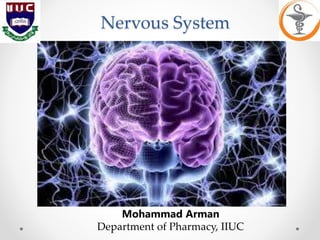
By: M. Thiru murugan
Unit – 11:
Types of nerves- structure and functions
Brain and cranial nerves.
Spinal cord and motor and sensory pathways of the spinal cord, autonomic nervous system.
Nervous system:
Nervous system is one of vital system in our body which control and coordinate all the functions of body parts.
Classification:
Central nervous system (CNS)
Peripheral nervous system (PNS)
1. Central nervous system (CNS): brain and spinal cord
2. Peripheral nervous system (PNS): Somatic nervous System & Autonomic nervous system (ANS)
Central Nervous System (CNS):
The central nervous system (CNS) controls most functions of the body and mind.
It consists of two parts: the brain and the spinal cord.
The brain is the center of our thoughts, the interpreter of our external environment, and the origin of control over body movement.
It interprets information from our special senses, as well as from internal organs
Meninges:
The coverings of brain and spinal cord are called meninge.
There are 3 layers surrounding the brain and spinal cord.
Dura (outer layer)
Arachnoid (middle layer)
Pia matter (inner layer)
Dura mater: The tough outer layer is called the dura mater. protect the central nervous system.
Arachnoid: The middle layer is the arachnoid, It contains cerebrospinal fluid, which acts to cushion the brain
Pia matter: the innermost layer of the meninges, the pia mater closely covers the brain.
Brain:
Introduction:
The brain is a complex organ that controls thought, memory, emotion, touch, motor skills, vision, breathing, temperature, hunger and every process that regulates our body.
the brain and spinal cord Together make up the central nervous system, or CNS
The brain receives information through our five senses: sight, smell, touch, taste, and hearing - often many at one time
Diagram:
Structure:
The brain is composed of the cerebrum, cerebellum, and brainstem
Cerebrum (telencephalon or endbrain): is the largest part of the brain and is composed of right and left hemispheres. It performs higher functions like interpreting touch, vision and hearing, as well as speech, reasoning, emotions, learning, and fine control of movement.
Cerebellum (little brain): is located under the cerebrum. Its function is to coordinate muscle movements, maintain posture, and balance.
Brainstem: consist midbrain, the pons, and the medulla oblongata acts as a relay center connecting the cerebrum and cerebellum to the spinal cord.
Functions such as breathing, heart rate, body temperature, wake and sleep cycles, digestion, sneezing, coughing, vomiting, and swallowing.
Lobes of the brain:
Each hemisphere has 4 lobes:
Frontal lobe
Temporal lobe
Parietal lobe
Occipital lobe
Each lobe may be divided, once again, into areas that serve very specific functions
The cerebral cortex has many folds, called the gyrus (plural: "gyri") and its trough is called a sulcus (plural: sulci)
Deep structure of Brain:
Hypothalamus: is located in the floor of the third ventricle and
Unit-I, Chapter_1 Nervous System Final PPT.pptAudumbar Mali
B. Pharm. Sem:-II,
BP 201T. HUMAN ANATOMY AND PHYSIOLOGY-II (Theory),
Nervous System:
Organization of nervous system, neuron, neuroglia, classification and properties of nerve fibre, electrophysiology, action potential, nerve impulse, receptors, synapse, neurotransmitters. Central nervous system: Meninges, ventricles of brain and
cerebrospinal fluid.structure and functions of brain (cerebrum, brain stem, cerebellum), spinal cord (gross structure, functions of afferent and efferent nerve tracts,reflex activity).
It is the part of central nervous system.
Complex organ that controls every process that regulates human body.
Located in cranium
cranium and bones that protects the brain is called skull
In terms of weight, the average adult human brain weighs in at 1300 to 1400 grams or around 3 pounds
In terms of length, the average brain is around 15 centimeters long.
There are 3 main parts of the brain
Forebrain
Midbrain
Hindbrain
Forebrain is divided into 3 functional parts
Thalamus
Cerebrum
Limbic system
Thalamus : Thalamus carries sensory information to the limbic system and cerebrum. The information includes sensory input from auditory and visual pathways, from the skin and from within the body.
Cerebrum: The largest part of the brain, the cerebrum initiates and coordinates movement and regulates temperature. Other areas of the cerebrum enable speech, judgment, thinking and reasoning, problem-solving, emotions and learning. Other functions relate to vision, hearing, touch and other senses. Further divided into 2 halves:
Right cerebral hemisphere (control the functions of left part of body)
Left cerebral hemisphere (controls the functions of rights part of the body)
Cerebral cortex is the outer layer of cerebrum. This part receives sensory information, processes it, stores some in memory for future use, directs voluntary movements, and is responsible for the poorly understood process that we call thinking.
Lobes of cerebral cortex:
Parietal Lobe Located below the crown of the head Processes sensory information from the whole body (information about pain, touch, and pressure)
Frontal Lobe Located right behind the forehead Responsible for initiating and coordinating motor movements and higher cognitive skills like problem solving and thinking
Occipital Lobe Located in the back of the brain, against the skull Processes all the visual information coming into the brain
Temporal Lobe Located behind the temples and just above the ears In charge of making sense of the information you hear Integrates information from various senses, such as smell and vision
Limbic system: The limbic system is located in an arc between the thalamus and cerebrum. Limbic system works together to produce our most basic and primitive emotions, drives, and behaviors, including fear, rage, tranquility, hunger, thirst, pleasure and sexual responses. Portion of limbic system is also important in the formation of memories. It is further divided into 3 parts:
Amygdala (regulate emotions, such as fear and aggression)
Hippocampus (storage of long term memory)
Hypothalamus (major coordinating center controlling body temperature, hunger, the menstrual cycle, water balance, the sleep-wake cycle through hormone production)
Midbrain is reduced in humans, and it contains auditory relay center and center that controls relax movements of eyes.
Midbrain contains reticular formation, which is a relay center connecting hindbrain with the forebrain.
Reticular formation is very i
The_Body_s_Control_System.pptx;filename= UTF-8''The Body_s Control System.pptxJimbertTingcang2
The system that enables the body to coordinate bodily activities is the nervous system. The nervous system is referred to as the control unit of the body. The main function of the nervous system is to integrate and coordinate bodily activities. It also acts as a storehouse of information. All information outside and inside the body are processed and interpreted by the nervous system.
The basic unit of the nervous system is the nerve cell or the neuron. Its main function is to deliver messages. The nervous system is composed of billions of neurons. Some parts of the body have more neurons than the other parts. Extending from the cell body (nucleus) are filaments called nerve fibers. These include one long fiber, the axon, and many shorter fibers called the dendrites.
The neuron has three main parts, the cell body, dendrite, and axon. They all work together to deliver the message from the sense organ to the brain and then back to the organ which does the action.
The fibers of a neuron are covered with a layer of fatty substances called myelin. This layer acts as an insulator. It is also important in speeding up the rate at which nerve impulses are transmitted.
The cell body is the main component of a neuron. It maintains the health of the neuron.
The dendrites are the short fibers around the cell body, Dendrites carry messages into the nerve cell. Thus, they are the receivers. The axons carry messages away from the nerves. Thus, they are the transmitters.
The long fiber of the neuron is the axon. It carries the message from the cell body to the other neurons.
The central nervous system is composed of the brainand the spinal cord.
The brain is the master control of the nervous system that sends messages to the organism so it can respond properly to different stimuli in the environment. It is protected by a bony structure called skullor cranium.
The brain has three main parts: cerebrum, cerebellum and the brain stem. The cerebrum is responsible for controlling speech, memory and intelligence. The cerebellum controls movement and balance. The brain stem connects the brain to the spinal cord and maintains vital automatic functions that include circulations and breathing.
1.
Cerebrum
2.
Cerebellum
3.
Brain stem
The spinal cord is a cord-like material in the backbone. It extends downward from the medulla oblongata through four fifth of the spinal column. It is protected and encased within a spinal fluid-filled vertebral column/backbone. Like the brain, the spinal cord is a delicate organ. Injuries can cause paralysis or loss of control over voluntary movements.
Science Ideas
•
The nervous system is the body’s internal data processor. Its main parts are the nerves, brains and the spinal cord.
•
The nerve cell is the basic unit of a nervous system.
•
There are two major divisions of the nervous system: the central nervous system and the peripheral nervous system.
•
The brain and the spinal cord comprise the central nervous system, the body’s main con
This slide talks about neuroplasticity, the central nervous system, the brain and its structure, the spinal cord, autonomic nervous system, its functions, nervous system and learning, neurotransmitters, working of neurotransmitters, classification, types of neurotransmitters, neurotransmitters in learning and limbic system in learning.
1 GNM anatomy Unit -11 Central Nervous System CNS.pptxthiru murugan
By: M. Thiru murugan
Unit – 11:
Types of nerves- structure and functions
Brain and cranial nerves.
Spinal cord and motor and sensory pathways of the spinal cord, autonomic nervous system.
Nervous system:
Nervous system is one of vital system in our body which control and coordinate all the functions of body parts.
Classification:
Central nervous system (CNS)
Peripheral nervous system (PNS)
1. Central nervous system (CNS): brain and spinal cord
2. Peripheral nervous system (PNS): Somatic nervous System & Autonomic nervous system (ANS)
Central Nervous System (CNS):
The central nervous system (CNS) controls most functions of the body and mind.
It consists of two parts: the brain and the spinal cord.
The brain is the center of our thoughts, the interpreter of our external environment, and the origin of control over body movement.
It interprets information from our special senses, as well as from internal organs
Meninges:
The coverings of brain and spinal cord are called meninge.
There are 3 layers surrounding the brain and spinal cord.
Dura (outer layer)
Arachnoid (middle layer)
Pia matter (inner layer)
Dura mater: The tough outer layer is called the dura mater. protect the central nervous system.
Arachnoid: The middle layer is the arachnoid, It contains cerebrospinal fluid, which acts to cushion the brain
Pia matter: the innermost layer of the meninges, the pia mater closely covers the brain.
Brain:
Introduction:
The brain is a complex organ that controls thought, memory, emotion, touch, motor skills, vision, breathing, temperature, hunger and every process that regulates our body.
the brain and spinal cord Together make up the central nervous system, or CNS
The brain receives information through our five senses: sight, smell, touch, taste, and hearing - often many at one time
Diagram:
Structure:
The brain is composed of the cerebrum, cerebellum, and brainstem
Cerebrum (telencephalon or endbrain): is the largest part of the brain and is composed of right and left hemispheres. It performs higher functions like interpreting touch, vision and hearing, as well as speech, reasoning, emotions, learning, and fine control of movement.
Cerebellum (little brain): is located under the cerebrum. Its function is to coordinate muscle movements, maintain posture, and balance.
Brainstem: consist midbrain, the pons, and the medulla oblongata acts as a relay center connecting the cerebrum and cerebellum to the spinal cord.
Functions such as breathing, heart rate, body temperature, wake and sleep cycles, digestion, sneezing, coughing, vomiting, and swallowing.
Lobes of the brain:
Each hemisphere has 4 lobes:
Frontal lobe
Temporal lobe
Parietal lobe
Occipital lobe
Each lobe may be divided, once again, into areas that serve very specific functions
The cerebral cortex has many folds, called the gyrus (plural: "gyri") and its trough is called a sulcus (plural: sulci)
Deep structure of Brain:
Hypothalamus: is located in the floor of the third ventricle and
Unit-I, Chapter_1 Nervous System Final PPT.pptAudumbar Mali
B. Pharm. Sem:-II,
BP 201T. HUMAN ANATOMY AND PHYSIOLOGY-II (Theory),
Nervous System:
Organization of nervous system, neuron, neuroglia, classification and properties of nerve fibre, electrophysiology, action potential, nerve impulse, receptors, synapse, neurotransmitters. Central nervous system: Meninges, ventricles of brain and
cerebrospinal fluid.structure and functions of brain (cerebrum, brain stem, cerebellum), spinal cord (gross structure, functions of afferent and efferent nerve tracts,reflex activity).
It is the part of central nervous system.
Complex organ that controls every process that regulates human body.
Located in cranium
cranium and bones that protects the brain is called skull
In terms of weight, the average adult human brain weighs in at 1300 to 1400 grams or around 3 pounds
In terms of length, the average brain is around 15 centimeters long.
There are 3 main parts of the brain
Forebrain
Midbrain
Hindbrain
Forebrain is divided into 3 functional parts
Thalamus
Cerebrum
Limbic system
Thalamus : Thalamus carries sensory information to the limbic system and cerebrum. The information includes sensory input from auditory and visual pathways, from the skin and from within the body.
Cerebrum: The largest part of the brain, the cerebrum initiates and coordinates movement and regulates temperature. Other areas of the cerebrum enable speech, judgment, thinking and reasoning, problem-solving, emotions and learning. Other functions relate to vision, hearing, touch and other senses. Further divided into 2 halves:
Right cerebral hemisphere (control the functions of left part of body)
Left cerebral hemisphere (controls the functions of rights part of the body)
Cerebral cortex is the outer layer of cerebrum. This part receives sensory information, processes it, stores some in memory for future use, directs voluntary movements, and is responsible for the poorly understood process that we call thinking.
Lobes of cerebral cortex:
Parietal Lobe Located below the crown of the head Processes sensory information from the whole body (information about pain, touch, and pressure)
Frontal Lobe Located right behind the forehead Responsible for initiating and coordinating motor movements and higher cognitive skills like problem solving and thinking
Occipital Lobe Located in the back of the brain, against the skull Processes all the visual information coming into the brain
Temporal Lobe Located behind the temples and just above the ears In charge of making sense of the information you hear Integrates information from various senses, such as smell and vision
Limbic system: The limbic system is located in an arc between the thalamus and cerebrum. Limbic system works together to produce our most basic and primitive emotions, drives, and behaviors, including fear, rage, tranquility, hunger, thirst, pleasure and sexual responses. Portion of limbic system is also important in the formation of memories. It is further divided into 3 parts:
Amygdala (regulate emotions, such as fear and aggression)
Hippocampus (storage of long term memory)
Hypothalamus (major coordinating center controlling body temperature, hunger, the menstrual cycle, water balance, the sleep-wake cycle through hormone production)
Midbrain is reduced in humans, and it contains auditory relay center and center that controls relax movements of eyes.
Midbrain contains reticular formation, which is a relay center connecting hindbrain with the forebrain.
Reticular formation is very i
The_Body_s_Control_System.pptx;filename= UTF-8''The Body_s Control System.pptxJimbertTingcang2
The system that enables the body to coordinate bodily activities is the nervous system. The nervous system is referred to as the control unit of the body. The main function of the nervous system is to integrate and coordinate bodily activities. It also acts as a storehouse of information. All information outside and inside the body are processed and interpreted by the nervous system.
The basic unit of the nervous system is the nerve cell or the neuron. Its main function is to deliver messages. The nervous system is composed of billions of neurons. Some parts of the body have more neurons than the other parts. Extending from the cell body (nucleus) are filaments called nerve fibers. These include one long fiber, the axon, and many shorter fibers called the dendrites.
The neuron has three main parts, the cell body, dendrite, and axon. They all work together to deliver the message from the sense organ to the brain and then back to the organ which does the action.
The fibers of a neuron are covered with a layer of fatty substances called myelin. This layer acts as an insulator. It is also important in speeding up the rate at which nerve impulses are transmitted.
The cell body is the main component of a neuron. It maintains the health of the neuron.
The dendrites are the short fibers around the cell body, Dendrites carry messages into the nerve cell. Thus, they are the receivers. The axons carry messages away from the nerves. Thus, they are the transmitters.
The long fiber of the neuron is the axon. It carries the message from the cell body to the other neurons.
The central nervous system is composed of the brainand the spinal cord.
The brain is the master control of the nervous system that sends messages to the organism so it can respond properly to different stimuli in the environment. It is protected by a bony structure called skullor cranium.
The brain has three main parts: cerebrum, cerebellum and the brain stem. The cerebrum is responsible for controlling speech, memory and intelligence. The cerebellum controls movement and balance. The brain stem connects the brain to the spinal cord and maintains vital automatic functions that include circulations and breathing.
1.
Cerebrum
2.
Cerebellum
3.
Brain stem
The spinal cord is a cord-like material in the backbone. It extends downward from the medulla oblongata through four fifth of the spinal column. It is protected and encased within a spinal fluid-filled vertebral column/backbone. Like the brain, the spinal cord is a delicate organ. Injuries can cause paralysis or loss of control over voluntary movements.
Science Ideas
•
The nervous system is the body’s internal data processor. Its main parts are the nerves, brains and the spinal cord.
•
The nerve cell is the basic unit of a nervous system.
•
There are two major divisions of the nervous system: the central nervous system and the peripheral nervous system.
•
The brain and the spinal cord comprise the central nervous system, the body’s main con
This slide talks about neuroplasticity, the central nervous system, the brain and its structure, the spinal cord, autonomic nervous system, its functions, nervous system and learning, neurotransmitters, working of neurotransmitters, classification, types of neurotransmitters, neurotransmitters in learning and limbic system in learning.
TEST BANK for Operations Management, 14th Edition by William J. Stevenson, Ve...kevinkariuki227
TEST BANK for Operations Management, 14th Edition by William J. Stevenson, Verified Chapters 1 - 19, Complete Newest Version.pdf
TEST BANK for Operations Management, 14th Edition by William J. Stevenson, Verified Chapters 1 - 19, Complete Newest Version.pdf
Pulmonary Thromboembolism - etilogy, types, medical- Surgical and nursing man...VarunMahajani
Disruption of blood supply to lung alveoli due to blockage of one or more pulmonary blood vessels is called as Pulmonary thromboembolism. In this presentation we will discuss its causes, types and its management in depth.
Report Back from SGO 2024: What’s the Latest in Cervical Cancer?bkling
Are you curious about what’s new in cervical cancer research or unsure what the findings mean? Join Dr. Emily Ko, a gynecologic oncologist at Penn Medicine, to learn about the latest updates from the Society of Gynecologic Oncology (SGO) 2024 Annual Meeting on Women’s Cancer. Dr. Ko will discuss what the research presented at the conference means for you and answer your questions about the new developments.
Lung Cancer: Artificial Intelligence, Synergetics, Complex System Analysis, S...Oleg Kshivets
RESULTS: Overall life span (LS) was 2252.1±1742.5 days and cumulative 5-year survival (5YS) reached 73.2%, 10 years – 64.8%, 20 years – 42.5%. 513 LCP lived more than 5 years (LS=3124.6±1525.6 days), 148 LCP – more than 10 years (LS=5054.4±1504.1 days).199 LCP died because of LC (LS=562.7±374.5 days). 5YS of LCP after bi/lobectomies was significantly superior in comparison with LCP after pneumonectomies (78.1% vs.63.7%, P=0.00001 by log-rank test). AT significantly improved 5YS (66.3% vs. 34.8%) (P=0.00000 by log-rank test) only for LCP with N1-2. Cox modeling displayed that 5YS of LCP significantly depended on: phase transition (PT) early-invasive LC in terms of synergetics, PT N0—N12, cell ratio factors (ratio between cancer cells- CC and blood cells subpopulations), G1-3, histology, glucose, AT, blood cell circuit, prothrombin index, heparin tolerance, recalcification time (P=0.000-0.038). Neural networks, genetic algorithm selection and bootstrap simulation revealed relationships between 5YS and PT early-invasive LC (rank=1), PT N0—N12 (rank=2), thrombocytes/CC (3), erythrocytes/CC (4), eosinophils/CC (5), healthy cells/CC (6), lymphocytes/CC (7), segmented neutrophils/CC (8), stick neutrophils/CC (9), monocytes/CC (10); leucocytes/CC (11). Correct prediction of 5YS was 100% by neural networks computing (area under ROC curve=1.0; error=0.0).
CONCLUSIONS: 5YS of LCP after radical procedures significantly depended on: 1) PT early-invasive cancer; 2) PT N0--N12; 3) cell ratio factors; 4) blood cell circuit; 5) biochemical factors; 6) hemostasis system; 7) AT; 8) LC characteristics; 9) LC cell dynamics; 10) surgery type: lobectomy/pneumonectomy; 11) anthropometric data. Optimal diagnosis and treatment strategies for LC are: 1) screening and early detection of LC; 2) availability of experienced thoracic surgeons because of complexity of radical procedures; 3) aggressive en block surgery and adequate lymph node dissection for completeness; 4) precise prediction; 5) adjuvant chemoimmunoradiotherapy for LCP with unfavorable prognosis.
- Video recording of this lecture in English language: https://youtu.be/lK81BzxMqdo
- Video recording of this lecture in Arabic language: https://youtu.be/Ve4P0COk9OI
- Link to download the book free: https://nephrotube.blogspot.com/p/nephrotube-nephrology-books.html
- Link to NephroTube website: www.NephroTube.com
- Link to NephroTube social media accounts: https://nephrotube.blogspot.com/p/join-nephrotube-on-social-media.html
Couples presenting to the infertility clinic- Do they really have infertility...Sujoy Dasgupta
Dr Sujoy Dasgupta presented the study on "Couples presenting to the infertility clinic- Do they really have infertility? – The unexplored stories of non-consummation" in the 13th Congress of the Asia Pacific Initiative on Reproduction (ASPIRE 2024) at Manila on 24 May, 2024.
Prix Galien International 2024 Forum ProgramLevi Shapiro
June 20, 2024, Prix Galien International and Jerusalem Ethics Forum in ROME. Detailed agenda including panels:
- ADVANCES IN CARDIOLOGY: A NEW PARADIGM IS COMING
- WOMEN’S HEALTH: FERTILITY PRESERVATION
- WHAT’S NEW IN THE TREATMENT OF INFECTIOUS,
ONCOLOGICAL AND INFLAMMATORY SKIN DISEASES?
- ARTIFICIAL INTELLIGENCE AND ETHICS
- GENE THERAPY
- BEYOND BORDERS: GLOBAL INITIATIVES FOR DEMOCRATIZING LIFE SCIENCE TECHNOLOGIES AND PROMOTING ACCESS TO HEALTHCARE
- ETHICAL CHALLENGES IN LIFE SCIENCES
- Prix Galien International Awards Ceremony
Acute scrotum is a general term referring to an emergency condition affecting the contents or the wall of the scrotum.
There are a number of conditions that present acutely, predominantly with pain and/or swelling
A careful and detailed history and examination, and in some cases, investigations allow differentiation between these diagnoses. A prompt diagnosis is essential as the patient may require urgent surgical intervention
Testicular torsion refers to twisting of the spermatic cord, causing ischaemia of the testicle.
Testicular torsion results from inadequate fixation of the testis to the tunica vaginalis producing ischemia from reduced arterial inflow and venous outflow obstruction.
The prevalence of testicular torsion in adult patients hospitalized with acute scrotal pain is approximately 25 to 50 percent
Title: Sense of Taste
Presenter: Dr. Faiza, Assistant Professor of Physiology
Qualifications:
MBBS (Best Graduate, AIMC Lahore)
FCPS Physiology
ICMT, CHPE, DHPE (STMU)
MPH (GC University, Faisalabad)
MBA (Virtual University of Pakistan)
Learning Objectives:
Describe the structure and function of taste buds.
Describe the relationship between the taste threshold and taste index of common substances.
Explain the chemical basis and signal transduction of taste perception for each type of primary taste sensation.
Recognize different abnormalities of taste perception and their causes.
Key Topics:
Significance of Taste Sensation:
Differentiation between pleasant and harmful food
Influence on behavior
Selection of food based on metabolic needs
Receptors of Taste:
Taste buds on the tongue
Influence of sense of smell, texture of food, and pain stimulation (e.g., by pepper)
Primary and Secondary Taste Sensations:
Primary taste sensations: Sweet, Sour, Salty, Bitter, Umami
Chemical basis and signal transduction mechanisms for each taste
Taste Threshold and Index:
Taste threshold values for Sweet (sucrose), Salty (NaCl), Sour (HCl), and Bitter (Quinine)
Taste index relationship: Inversely proportional to taste threshold
Taste Blindness:
Inability to taste certain substances, particularly thiourea compounds
Example: Phenylthiocarbamide
Structure and Function of Taste Buds:
Composition: Epithelial cells, Sustentacular/Supporting cells, Taste cells, Basal cells
Features: Taste pores, Taste hairs/microvilli, and Taste nerve fibers
Location of Taste Buds:
Found in papillae of the tongue (Fungiform, Circumvallate, Foliate)
Also present on the palate, tonsillar pillars, epiglottis, and proximal esophagus
Mechanism of Taste Stimulation:
Interaction of taste substances with receptors on microvilli
Signal transduction pathways for Umami, Sweet, Bitter, Sour, and Salty tastes
Taste Sensitivity and Adaptation:
Decrease in sensitivity with age
Rapid adaptation of taste sensation
Role of Saliva in Taste:
Dissolution of tastants to reach receptors
Washing away the stimulus
Taste Preferences and Aversions:
Mechanisms behind taste preference and aversion
Influence of receptors and neural pathways
Impact of Sensory Nerve Damage:
Degeneration of taste buds if the sensory nerve fiber is cut
Abnormalities of Taste Detection:
Conditions: Ageusia, Hypogeusia, Dysgeusia (parageusia)
Causes: Nerve damage, neurological disorders, infections, poor oral hygiene, adverse drug effects, deficiencies, aging, tobacco use, altered neurotransmitter levels
Neurotransmitters and Taste Threshold:
Effects of serotonin (5-HT) and norepinephrine (NE) on taste sensitivity
Supertasters:
25% of the population with heightened sensitivity to taste, especially bitterness
Increased number of fungiform papillae
2. Nervous System
• The nervous system of vertebrates (including humans) is
divided into the central nervous system (CNS) and the
peripheral nervous system (PNS). The (CNS) is the major
division, and consists of the brain and the spinal cord. The
spinal canal contains the spinal cord, while the cranial cavity
contains the brain.
• Nervous system is body’s command center. Originating from
brain, it controls movements, thoughts and automatic
responses to the world. It also controls other body systems and
processes, such as digestion, breathing and sexual
development (puberty). Diseases, accidents, toxins and the
natural aging process can damage nervous system.
3. Affects of Nervous System
Nervous system affects every aspect of your health, including
your:
• Thoughts, memory, learning, and feelings.
• Movements, such as balance and coordination.
• Senses, including how your brain interprets what you see,
hear, taste, touch and feel.
• Sleep, healing and aging.
• Heartbeat and breathing patterns.
• Response to stressful situations.
• Digestion, as well as how hungry and thirsty you feel.
• Body processes, such as puberty.
6. Neuron
• Neurons are the building blocks of the nervous system. They
receive and transmit signals to different parts of the body. This
is carried out in both physical and electrical forms. There are
several different types of neurons that facilitate the
transmission of information.
7. Parts of Neuron
Following are the different parts of a neuron:
1. Dendrites: These are branch-like structures that receive messages
from other neurons and allow the transmission of messages to the cell
body.
2. Cell Body: Each neuron has a cell body with a nucleus, Golgi body,
endoplasmic reticulum, mitochondria and other components.
3. Axon: Axon is a tube-like structure that carries electrical impulse
from the cell body to the axon terminals that pass the impulse to
another neuron.
4. Synapse: It is the chemical junction between the terminal of one
neuron and the dendrites of another neuron.
8. Neuron Types
• There are three different types of neurons:
1. Sensory Neurons: The sensory neurons convert signals from the
external environment into corresponding internal stimuli. The
sensory inputs activate the sensory neurons and carry sensory
information to the brain and spinal cord. They are pseudounipolar
in structure.
2. Motor Neurons: These are multipolar and are located in the
central nervous system extending their axons outside the central
nervous system. This is the most common type of neuron and
transmits information from the brain to the muscles of the body.
3. Interneurons: They are multipolar in structure. Their axons
connect only to the nearby sensory and motor neurons. They help
in passing signals between two neurons.
10. Neuroglia
• Neuroglia, also called glial cell or glia, any of several types of
cell that function primarily to support neurons. The term
neuroglia means “nerve glue.” In 1907 Italian biologist Emilio
Lugaro suggested that neuroglial cells exchange substances
with the extracellular fluid and in this way exert control on the
neuronal environment.
• Neuroglia are a large class of neural cells of ectodermal
(astroglia, oligodendroglia, and peripheral glial cells) and
mesodermal (microglia) origin. Neuroglial cells provide
homeostatic support, protection, and defense to the nervous
tissue.
13. Central Nervous System
• The central nervous system (CNS) consists of the brain and
spinal cord. It controls things like thought, movement, and
emotion, as well as breathing, heart rate, hormones, and body
temperature.
• Each nerve has a protective outer layer called myelin. Myelin
insulates the nerve and helps the messages get through.
It composed of two major interconnected organ:
1. The brain
2. The spinal cord
15. Brain
• Parts of the body:
Cerebrum: The cerebrum (front of brain) comprises gray matter
(the cerebral cortex) and white matter at its center. The largest
part of the brain, the cerebrum initiates and coordinates
movement and regulates temperature. Other areas of the
cerebrum enable speech, judgment, thinking and reasoning,
problem-solving, emotions and learning. Other functions relate to
vision, hearing, touch and other senses.
Cerebral Cortex: Cortex is Latin for “bark,” and describes the
outer gray matter covering of the cerebrum. The cortex has a
large surface area due to its folds, and comprises about half of the
brain’s weight.
16. Continue…
Brainstem: The brainstem (middle of brain) connects the
cerebrum with the spinal cord. The brainstem includes the
midbrain, the pons and the medulla.
I. Midbrain: The midbrain (or mesencephalon) is a very complex structure
with a range of different neuron clusters (nuclei and colliculi), neural
pathways and other structures. These features facilitate various functions,
from hearing and movement to calculating responses and environmental
changes.
II. Pons: The pons is the origin for four of the 12 cranial nerves, which
enable a range of activities such as tear production, chewing, blinking,
focusing vision, balance, hearing and facial expression.
III. Medulla: At the bottom of the brainstem, the medulla is where the brain
meets the spinal cord. The medulla is essential to survival. Functions of
the medulla regulate many bodily activities, including heart rhythm,
breathing, blood flow, and oxygen and carbon dioxide levels.
17. Continue…
Cerebellum: The cerebellum (“little brain”) is a fist-sized portion
of the brain located at the back of the head, below the temporal
and occipital lobes and above the brainstem. Like the cerebral
cortex, it has two hemispheres. The outer portion contains
neurons, and the inner area communicates with the cerebral
cortex. Its function is to coordinate voluntary muscle movements
and to maintain posture, balance and equilibrium. New studies
are exploring the cerebellum’s roles in thought, emotions and
social behavior, as well as its possible involvement in addiction,
autism and schizophrenia.
18. Gray matter and white matter?
Gray and white matter are two different regions of the central
nervous system. In the brain, gray matter refers to the darker,
outer portion, while white matter describes the lighter, inner
section underneath. In the spinal cord, this order is reversed: The
white matter is on the outside, and the gray matter sits within.
19. Deeper Structures Within the Brain
Pituitary Gland: Sometimes called the “master gland,”. The pituitary
gland governs the function of other glands in the body, regulating the
flow of hormones from the thyroid, adrenals, ovaries and testicles. It
receives chemical signals from the hypothalamus through its stalk and
blood supply.
Hypothalamus: The hypothalamus is located above the pituitary gland
and sends it chemical messages that control its function. It regulates
body temperature, synchronizes sleep patterns, controls hunger and
thirst and also plays a role in some aspects of memory and emotion.
Pineal Gland: The pineal gland is located deep in the brain and
attached by a stalk to the top of the third ventricle. The pineal gland
responds to light and dark and secretes melatonin, which regulates
circadian rhythms and the sleep-wake cycle.
Hippocampus: It supports memory, learning, navigation and
perception of space. It receives information from the cerebral cortex
and may play a role in Alzheimer’s disease.
20. Spinal Cord
• The spinal cord is a long, tube-like band of tissue. It connects
your brain to your lower back. Your spinal cord carries nerve
signals from your brain to your body and vice versa. These
nerve signals help you feel sensations and move your body.
Any damage to your spinal cord can affect your movement or
function.
• In most adults, your spinal cord is about 18 inches (45
centimeters) long.
What is the purpose of the spinal cord?
I. Control body movements and functions.
II. Report senses to brain.
III. Manage your reflexes.
21. Continue…
What are the main parts of the spinal cord?
Spinal cord has three main parts:
1. Cervical (neck).
2. Thoracic (chest).
3. Lumbar (lower back).
What tissues and fluids make up the spinal cord?
Like brain, layers of tissue called meninges cover the spinal cord.
These protective tissues include:
1. Dura mater: The outer layer that protects your spinal cord from
injury.
2. Arachnoid mater: The middle layer between the epidural and
subarachnoid space.
3. Pia mater: The inner layer that covers your spinal cord.
22. Peripheral Nervous System (PNS)
Peripheral nervous system (PNS) is that part of your nervous
system that lies outside brain and spinal cord. It plays key role in
both sending information from different areas of body back to
brain, as well as carrying out commands from your brain to
various parts of your body.
The four primary functions of the PNS are to:
i. Control autonomic body functions
ii. Control motor movements
iii. Digestion
iv. Relay sensory information to the central nervous system
23. Cranial Nerves
The cranial nerves are a set of 12 paired nerves in the back of
your brain. Cranial nerves send electrical signals between your
brain, face, neck and torso. Your cranial nerves help you taste,
smell, hear and feel sensations. They also help you make facial
expressions, blink your eyes and move your tongue.
25. Somatic Nervous System
• The Somatic Nervous System is the part of the peripheral nervous
system that handles voluntary control of body movements
• In somatic nervous system, In the process of voluntary movement,
sensory neurons carry impulses to the brain and the spinal cord.
After processing, a signal is sent back to the skeletal muscles,
organs, and skin by way of the somatic motor neurons. The second
function of the somatic nervous system is the process of the reflex
arc.
26. Autonomic Nervous System
• The Autonomic Nervous System is the part of the peripheral
nervous system that acts as an involuntary control system
(below the level of consciousness), and controls visceral
functions.
• The autonomic nervous system (ANS) is still controlled by the
brain too -- but is not conscious. The brain's hypothalamus is
required to send signals to the heart, your glands, breathing,
digestion etc.