Downloaded 42 times
![4
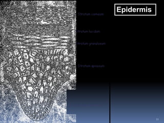
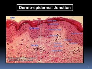
Epidermis
Stratified squamous epithelium
No blood vessels
In palms of hand and soles of feet, epidermis is thicker
Composed of five layers:
[1] Stratum corneum
[2] Stratum lucidum
[3] Stratum granulosum
[4] Stratum spinosum
[5] Stratum basale](https://image.slidesharecdn.com/skinaandplecture12-240511081428-f6bced90/85/Unit-VII-Integumentary-System-Skin-Nail-Hair-4-320.jpg)
![5
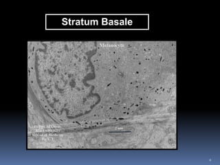
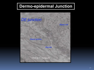
[1] Stratum Basale
Single layer of columnar or cuboidal cells
Lower surface of cells attached to dermis
Receives nutrients from blood in the dermal vessels
Mitosis occurs in this layer
Older cells expelled to outer layer
Melanin is produced in this layer](https://image.slidesharecdn.com/skinaandplecture12-240511081428-f6bced90/85/Unit-VII-Integumentary-System-Skin-Nail-Hair-5-320.jpg)
![7
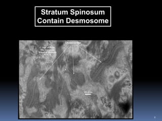
[2] Stratum Spinosum
Prickle cell layer
Several layers of polyhedral (many-sided) cells
Interlocking spine-like projections help binding of this layer
Active protein synthesis takes place (indicates cell growth and
division)
Obtain nutrients through fine elements
Keratinization begins in this layer
Nuclei-containing cells change into flat cells composed of hard
durable protein)](https://image.slidesharecdn.com/skinaandplecture12-240511081428-f6bced90/85/Unit-VII-Integumentary-System-Skin-Nail-Hair-7-320.jpg)
![9
[3] Stratum Granulosum
Granular + 2 to 4 cells thick
Cells contain keratohyaline in granules
Final stages of keratinization occur
Loss of fluid, nucleus disintegrates
[4] Stratum Lucidum
Transparent layer
Flat + translucent dead cells
Protection against UV
Lucidum appears in palm of hands and soles of feet to protect
against sun burn](https://image.slidesharecdn.com/skinaandplecture12-240511081428-f6bced90/85/Unit-VII-Integumentary-System-Skin-Nail-Hair-9-320.jpg)
![10
[5] Stratum Corneum (Horny Layer)
Thick layer of dead cells
Soft keratin (keep skin elastic)
Cells below contain fatty substrate keep skin waterproof +
prevent skin cracking and allowing bacteria inside](https://image.slidesharecdn.com/skinaandplecture12-240511081428-f6bced90/85/Unit-VII-Integumentary-System-Skin-Nail-Hair-10-320.jpg)
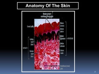
![14
[1] Upper Papillary Layer
Loose connective tissue
Contain protrusions into epidermis called “PAPILLAE”
Fine capillaries to carry waste away + provide nourishment and
oxygen
Nerve endings for heat, pain, cold, pressure and touch
(Meissner’s corpuscles)
Double row of papillae better gripping by hands and feet +
distinctive fingerprint patterns](https://image.slidesharecdn.com/skinaandplecture12-240511081428-f6bced90/85/Unit-VII-Integumentary-System-Skin-Nail-Hair-14-320.jpg)
![15
[2] Reticular Layer
Elastic network of tough collagen fibres interwoven with elastic
fibres
Collagenous fibres arranged in special pattern
Incisions made parallel to these lines during surgery wound
heals faster
Contains sebaceous and sweat glands, arrector pili muscle and
hair follicle
Pacinian corpuscles are distributed through the dermis and
function as pressure receptors
Stretch marks and pregnancy due to breaks in collagen and
elastic fibres](https://image.slidesharecdn.com/skinaandplecture12-240511081428-f6bced90/85/Unit-VII-Integumentary-System-Skin-Nail-Hair-15-320.jpg)
![26
Burns
Seriousness of burns classified according to:
[1] Extent (size of body area involved)
[2] Depth (How many layers of tissue are injured)
First Degree Burn
e.g. Sunburn
Epidermis damaged but not destroyed
Treated by cold water
< 10% of body surface involved](https://image.slidesharecdn.com/skinaandplecture12-240511081428-f6bced90/85/Unit-VII-Integumentary-System-Skin-Nail-Hair-26-320.jpg)

![29
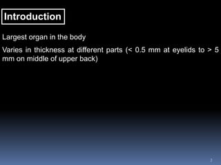
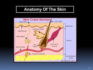
Functions Of The Skin
Non-specific immunity (details to follow in later lectures)
[1] Protection
Anatomical barrier against infection
Melanin = screen out excess UV rays
When melanin is darkened by the tan transferred to outer skin
layers (suntan) skin less sensitive to sunrays
Dark skin due to wider distribution of melanin beyond stratum
basale into higher levels of epidermis](https://image.slidesharecdn.com/skinaandplecture12-240511081428-f6bced90/85/Unit-VII-Integumentary-System-Skin-Nail-Hair-29-320.jpg)
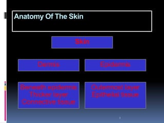
![[2] Thermoregulation
Control of heat production
Shivering
Skeletal muscle
Motor neurons
Control of heat loss
Skin vasocontriction and vasodilation
Skin blood vessels
Sympathetic nervous system
Control of heat loss
Sweating
Sweat glands
Sympathetic nervous system
Hypothalamic thermoregulatory integrating centre
Peripheral Thermoreceptors (Skin)
Skin temperature
30](https://image.slidesharecdn.com/skinaandplecture12-240511081428-f6bced90/85/Unit-VII-Integumentary-System-Skin-Nail-Hair-30-320.jpg)
![31
[3] Sensory Perception
Millions of nerve endings
Receptor for pain, heat and pressure
Therefore, maintain homeostasis
[4] Excretion
Excretion of lactic acid and sodium chloride
Urea (1 g nitrogenous waste eliminated through skin per hour)](https://image.slidesharecdn.com/skinaandplecture12-240511081428-f6bced90/85/Unit-VII-Integumentary-System-Skin-Nail-Hair-31-320.jpg)
![[5] Vitamin D Synthesis
32
Promotes absorption of calcium and phosphate through intestine
Active vitamin D
Modification by liver and kidney enzymes
Vitamin D3 (Cholecalciferol)
Vitamin D precursor (in skin)](https://image.slidesharecdn.com/skinaandplecture12-240511081428-f6bced90/85/Unit-VII-Integumentary-System-Skin-Nail-Hair-32-320.jpg)
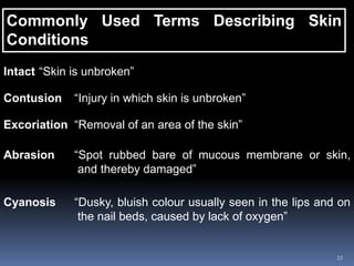

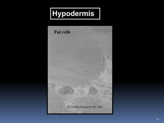
The document provides a detailed overview of the anatomy and physiology of the skin, the largest organ of the body, including its various layers: the epidermis, dermis, and hypodermis, each with specific functions and cell types. It also discusses skin-related glands, the development and functions of the skin, including protection, thermoregulation, sensory perception, excretion, and vitamin D synthesis. Additionally, it covers skin conditions and classifications of burns.


![concept of safety[1].ppt BSN YEAR 1 SEM1](https://cdn.slidesharecdn.com/ss_thumbnails/conceptofsafety1-240509093204-8977567e-thumbnail.jpg?width=640&height=640&fit=bounds)


![CASE_PRESENTATION_ON_subdural_hematoma(SDH)[1 FINAL PPT]-1.pptx](https://cdn.slidesharecdn.com/ss_thumbnails/casepresentationonsubduralhematomasdh1finalppt-1-260129172522-d405d375-thumbnail.jpg?width=640&height=640&fit=bounds)


