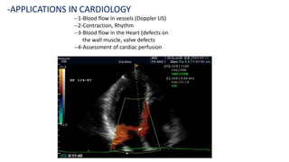
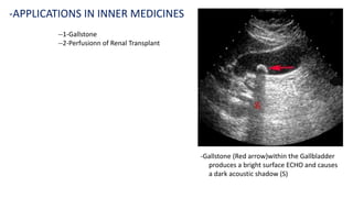
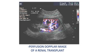
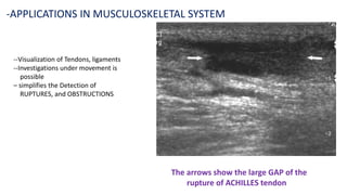
Ultrasound uses high-frequency sound waves to produce images of the inside of the body. It can show internal organs, tissues, and blood flowing through blood vessels in real-time. Ultrasound has many medical uses including examining the heart and blood vessels, liver, gallbladder, spleen, kidneys, uterus and pregnancy, eyes, thyroid, muscles and tendons. It is widely used due to its safety since it does not use ionizing radiation.
ultrasound is not limited to diagnosis, but can also be used in screening for disease and to aid in treatment of diseases . another one is ultrasound is not expose any radiation.
Anaesthetic considerations for Robotic Surgery, What to expect, how to go ahead. An update and incite on the intricacies of Robotic Surgery and Anaesthetic implications.
This presentation informs the procedure of MRI scan.It throws light upon the factors which are important before and after the scan.It also discusses the contraindications before and after the mri scan and which things should be avoided before the scan.
We live in an age of a new unpreceded wonders. The wonders of the world are not seven any more. The inanimate talk to us. We are flying in the air. More than 65,000-Ton can float over the water in an iron vessel. The Robotic Doctor is already a reality. Reviewing the history of mankind's cumulative experience starting with the ancient very primitive trials and ending with the presence of Robotic and Telesurgery
Clearly show that the major and rapid advances in the whole mankind's life occur only in the last few decades especially the last 10 years ? .
• What is Ultrasound imaging?
• Why Ultrasound?
• Common Uses
• History
• Properties of Ultrasound
• Equipment types
• How does the procedure work?
• Physics
• Benefits and Risks etc.
Fetal Studio offers affordable and professional access to prenatal ultrasound, limited medical surveys and level 1 ultrasounds,
also the best 3D & 4D imaging quality available on the market.
Ultrasound is a non-invasive medical imaging modality widely used in various clinical applications.
It is based on the principle of using high-frequency sound waves to create real-time images of internal structures within the human body.
In this presentation, we will discuss into the fundamental principles of ultrasound imaging and its applications in radiology.
Tom Selleck Health: A Comprehensive Look at the Iconic Actor’s Wellness Journeygreendigital
Tom Selleck, an enduring figure in Hollywood. has captivated audiences for decades with his rugged charm, iconic moustache. and memorable roles in television and film. From his breakout role as Thomas Magnum in Magnum P.I. to his current portrayal of Frank Reagan in Blue Bloods. Selleck's career has spanned over 50 years. But beyond his professional achievements. fans have often been curious about Tom Selleck Health. especially as he has aged in the public eye.
Follow us on: Pinterest
Introduction
Many have been interested in Tom Selleck health. not only because of his enduring presence on screen but also because of the challenges. and lifestyle choices he has faced and made over the years. This article delves into the various aspects of Tom Selleck health. exploring his fitness regimen, diet, mental health. and the challenges he has encountered as he ages. We'll look at how he maintains his well-being. the health issues he has faced, and his approach to ageing .
Early Life and Career
Childhood and Athletic Beginnings
Tom Selleck was born on January 29, 1945, in Detroit, Michigan, and grew up in Sherman Oaks, California. From an early age, he was involved in sports, particularly basketball. which played a significant role in his physical development. His athletic pursuits continued into college. where he attended the University of Southern California (USC) on a basketball scholarship. This early involvement in sports laid a strong foundation for his physical health and disciplined lifestyle.
Transition to Acting
Selleck's transition from an athlete to an actor came with its physical demands. His first significant role in "Magnum P.I." required him to perform various stunts and maintain a fit appearance. This role, which he played from 1980 to 1988. necessitated a rigorous fitness routine to meet the show's demands. setting the stage for his long-term commitment to health and wellness.
Fitness Regimen
Workout Routine
Tom Selleck health and fitness regimen has evolved. adapting to his changing roles and age. During his "Magnum, P.I." days. Selleck's workouts were intense and focused on building and maintaining muscle mass. His routine included weightlifting, cardiovascular exercises. and specific training for the stunts he performed on the show.
Selleck adjusted his fitness routine as he aged to suit his body's needs. Today, his workouts focus on maintaining flexibility, strength, and cardiovascular health. He incorporates low-impact exercises such as swimming, walking, and light weightlifting. This balanced approach helps him stay fit without putting undue strain on his joints and muscles.
Importance of Flexibility and Mobility
In recent years, Selleck has emphasized the importance of flexibility and mobility in his fitness regimen. Understanding the natural decline in muscle mass and joint flexibility with age. he includes stretching and yoga in his routine. These practices help prevent injuries, improve posture, and maintain mobilit
- Video recording of this lecture in English language: https://youtu.be/kqbnxVAZs-0
- Video recording of this lecture in Arabic language: https://youtu.be/SINlygW1Mpc
- Link to download the book free: https://nephrotube.blogspot.com/p/nephrotube-nephrology-books.html
- Link to NephroTube website: www.NephroTube.com
- Link to NephroTube social media accounts: https://nephrotube.blogspot.com/p/join-nephrotube-on-social-media.html
Recomendações da OMS sobre cuidados maternos e neonatais para uma experiência pós-natal positiva.
Em consonância com os ODS – Objetivos do Desenvolvimento Sustentável e a Estratégia Global para a Saúde das Mulheres, Crianças e Adolescentes, e aplicando uma abordagem baseada nos direitos humanos, os esforços de cuidados pós-natais devem expandir-se para além da cobertura e da simples sobrevivência, de modo a incluir cuidados de qualidade.
Estas diretrizes visam melhorar a qualidade dos cuidados pós-natais essenciais e de rotina prestados às mulheres e aos recém-nascidos, com o objetivo final de melhorar a saúde e o bem-estar materno e neonatal.
Uma “experiência pós-natal positiva” é um resultado importante para todas as mulheres que dão à luz e para os seus recém-nascidos, estabelecendo as bases para a melhoria da saúde e do bem-estar a curto e longo prazo. Uma experiência pós-natal positiva é definida como aquela em que as mulheres, pessoas que gestam, os recém-nascidos, os casais, os pais, os cuidadores e as famílias recebem informação consistente, garantia e apoio de profissionais de saúde motivados; e onde um sistema de saúde flexível e com recursos reconheça as necessidades das mulheres e dos bebês e respeite o seu contexto cultural.
Estas diretrizes consolidadas apresentam algumas recomendações novas e já bem fundamentadas sobre cuidados pós-natais de rotina para mulheres e neonatos que recebem cuidados no pós-parto em unidades de saúde ou na comunidade, independentemente dos recursos disponíveis.
É fornecido um conjunto abrangente de recomendações para cuidados durante o período puerperal, com ênfase nos cuidados essenciais que todas as mulheres e recém-nascidos devem receber, e com a devida atenção à qualidade dos cuidados; isto é, a entrega e a experiência do cuidado recebido. Estas diretrizes atualizam e ampliam as recomendações da OMS de 2014 sobre cuidados pós-natais da mãe e do recém-nascido e complementam as atuais diretrizes da OMS sobre a gestão de complicações pós-natais.
O estabelecimento da amamentação e o manejo das principais intercorrências é contemplada.
Recomendamos muito.
Vamos discutir essas recomendações no nosso curso de pós-graduação em Aleitamento no Instituto Ciclos.
Esta publicação só está disponível em inglês até o momento.
Prof. Marcus Renato de Carvalho
www.agostodourado.com
These lecture slides, by Dr Sidra Arshad, offer a quick overview of the physiological basis of a normal electrocardiogram.
Learning objectives:
1. Define an electrocardiogram (ECG) and electrocardiography
2. Describe how dipoles generated by the heart produce the waveforms of the ECG
3. Describe the components of a normal electrocardiogram of a typical bipolar lead (limb II)
4. Differentiate between intervals and segments
5. Enlist some common indications for obtaining an ECG
6. Describe the flow of current around the heart during the cardiac cycle
7. Discuss the placement and polarity of the leads of electrocardiograph
8. Describe the normal electrocardiograms recorded from the limb leads and explain the physiological basis of the different records that are obtained
9. Define mean electrical vector (axis) of the heart and give the normal range
10. Define the mean QRS vector
11. Describe the axes of leads (hexagonal reference system)
12. Comprehend the vectorial analysis of the normal ECG
13. Determine the mean electrical axis of the ventricular QRS and appreciate the mean axis deviation
14. Explain the concepts of current of injury, J point, and their significance
Study Resources:
1. Chapter 11, Guyton and Hall Textbook of Medical Physiology, 14th edition
2. Chapter 9, Human Physiology - From Cells to Systems, Lauralee Sherwood, 9th edition
3. Chapter 29, Ganong’s Review of Medical Physiology, 26th edition
4. Electrocardiogram, StatPearls - https://www.ncbi.nlm.nih.gov/books/NBK549803/
5. ECG in Medical Practice by ABM Abdullah, 4th edition
6. Chapter 3, Cardiology Explained, https://www.ncbi.nlm.nih.gov/books/NBK2214/
7. ECG Basics, http://www.nataliescasebook.com/tag/e-c-g-basics
Title: Sense of Taste
Presenter: Dr. Faiza, Assistant Professor of Physiology
Qualifications:
MBBS (Best Graduate, AIMC Lahore)
FCPS Physiology
ICMT, CHPE, DHPE (STMU)
MPH (GC University, Faisalabad)
MBA (Virtual University of Pakistan)
Learning Objectives:
Describe the structure and function of taste buds.
Describe the relationship between the taste threshold and taste index of common substances.
Explain the chemical basis and signal transduction of taste perception for each type of primary taste sensation.
Recognize different abnormalities of taste perception and their causes.
Key Topics:
Significance of Taste Sensation:
Differentiation between pleasant and harmful food
Influence on behavior
Selection of food based on metabolic needs
Receptors of Taste:
Taste buds on the tongue
Influence of sense of smell, texture of food, and pain stimulation (e.g., by pepper)
Primary and Secondary Taste Sensations:
Primary taste sensations: Sweet, Sour, Salty, Bitter, Umami
Chemical basis and signal transduction mechanisms for each taste
Taste Threshold and Index:
Taste threshold values for Sweet (sucrose), Salty (NaCl), Sour (HCl), and Bitter (Quinine)
Taste index relationship: Inversely proportional to taste threshold
Taste Blindness:
Inability to taste certain substances, particularly thiourea compounds
Example: Phenylthiocarbamide
Structure and Function of Taste Buds:
Composition: Epithelial cells, Sustentacular/Supporting cells, Taste cells, Basal cells
Features: Taste pores, Taste hairs/microvilli, and Taste nerve fibers
Location of Taste Buds:
Found in papillae of the tongue (Fungiform, Circumvallate, Foliate)
Also present on the palate, tonsillar pillars, epiglottis, and proximal esophagus
Mechanism of Taste Stimulation:
Interaction of taste substances with receptors on microvilli
Signal transduction pathways for Umami, Sweet, Bitter, Sour, and Salty tastes
Taste Sensitivity and Adaptation:
Decrease in sensitivity with age
Rapid adaptation of taste sensation
Role of Saliva in Taste:
Dissolution of tastants to reach receptors
Washing away the stimulus
Taste Preferences and Aversions:
Mechanisms behind taste preference and aversion
Influence of receptors and neural pathways
Impact of Sensory Nerve Damage:
Degeneration of taste buds if the sensory nerve fiber is cut
Abnormalities of Taste Detection:
Conditions: Ageusia, Hypogeusia, Dysgeusia (parageusia)
Causes: Nerve damage, neurological disorders, infections, poor oral hygiene, adverse drug effects, deficiencies, aging, tobacco use, altered neurotransmitter levels
Neurotransmitters and Taste Threshold:
Effects of serotonin (5-HT) and norepinephrine (NE) on taste sensitivity
Supertasters:
25% of the population with heightened sensitivity to taste, especially bitterness
Increased number of fungiform papillae
Ozempic: Preoperative Management of Patients on GLP-1 Receptor Agonists Saeid Safari
Preoperative Management of Patients on GLP-1 Receptor Agonists like Ozempic and Semiglutide
ASA GUIDELINE
NYSORA Guideline
2 Case Reports of Gastric Ultrasound
Rasamanikya is a excellent preparation in the field of Rasashastra, it is used in various Kushtha Roga, Shwasa, Vicharchika, Bhagandara, Vatarakta, and Phiranga Roga. In this article Preparation& Comparative analytical profile for both Formulationon i.e Rasamanikya prepared by Kushmanda swarasa & Churnodhaka Shodita Haratala. The study aims to provide insights into the comparative efficacy and analytical aspects of these formulations for enhanced therapeutic outcomes.
Adv. biopharm. APPLICATION OF PHARMACOKINETICS : TARGETED DRUG DELIVERY SYSTEMSAkankshaAshtankar
MIP 201T & MPH 202T
ADVANCED BIOPHARMACEUTICS & PHARMACOKINETICS : UNIT 5
APPLICATION OF PHARMACOKINETICS : TARGETED DRUG DELIVERY SYSTEMS By - AKANKSHA ASHTANKAR
Here is the updated list of Top Best Ayurvedic medicine for Gas and Indigestion and those are Gas-O-Go Syp for Dyspepsia | Lavizyme Syrup for Acidity | Yumzyme Hepatoprotective Capsules etc
2. --1-What is the Ultrasound imaging
--2-Why Ultrasound is required
--3-Common uses of Ultrasound
--4-Small History of Ultrasound
--5-Properties of Ultrasound
--6-Equipment required for Ultrasound
--7-How does this procedure work
--8-Benefits and risks of Ultrasound
-OUTLINE
3. --Ultra sound imaging. Also called “sonography”
It involves “Exposing part of the body to high
frequency sound waves” to produce pictures
of the inside of the body
--Ultrasound examinations do not use Ionizing
radiations (as used in X Rays)
--Because Ultrasound images are captured in real
time, they can show the structure and movement
of the body’s internal organs, as well as blood
flowing through blood vessels
-GENERAL ULTRASOUND IMAGING
What is that
4. --1-Ultrasound (US) is the “Most widely used imaging
technology world wide
--2-It is popular due to “availability, Speed, low cost,
Patient friendless (No Radiation)
--3-Applied in “Obstetrics, Cardiology, Inner Medicine
and Urology”
--4-Ongoing research to improve image quality speed
and new application areas such as intraoperative
Navigation, and Tumor Therapy..
-WHY ULTRASOUND
Should be requested
5. --1-Ultrasound examinations can help to diagnose
a variety of conditions and to asses Organs
damage following illness
--2-Ultrasound is used to help Physicians to evaluate
symptoms such as
a-Pain
b-Swelling
c-Infection and
d-Hematuria (blood in urine)
-SOME COMMON USES
of the procedure
6. ---Ultrasound is a useful way of examining many
of the body’s internal organs including but not
limited to the
-1-Heart and Blood vessels, including the abdominal
aorta and its major branches
-2-Liver
-3-Gall Bladder
-4- Spleen
-5-Pancreas
-6-Kindney
-7-Bladder
-8-Uterus, Ovaries, and unborn child (fetus) in pregnant
patients
-9-Eyes
-10-Thyroid and Parathyroid Glands
-11-Scrotum (testicles)
-12-Brain in infants
-13-Hips in infants
-SOME COMMON
USES
of the procedure
contd.
7. -ULTRASOUND
IS ALSO USED TO GUIDE
--1-Procedures such as Needle Biopsies
in which needles are used to Extract
sample cells from an abnormal area for
laboratory testing
--2-Image the Breasts and to guide Biopsy
of Breast cancer
--3-Diagnose a variety of Heart conditions
and to assess damage after a heart
attack or diagnose for Valvular
Heart disease
8. --1-Blockages to the blood flow (such as clots)
--2-Narrowing of vessels (which may be caused by a plaque)
--3-Tumors and congenital vascular malformations
IMPORTANT:-
With a knowledge about the speed and volume of blood flow
gained from a Doppler ultrasound image, the Physician can often
determine whether a patient is a good candidate for a procedure
like “Angioplasty”
DOPPLAR ULTRASOUND IMAGES CAN
HELP THE PHYSICIAN TO SEE AND EVALUATE
10. -Three Dimensional image of the same fetus
5(Five) months after conception
-APPLICATIONS IN
OBSTETRICS
11. --1-Blood flow in vessels (Doppler US)
--2-Contraction, Rhythm
--3-Blood flow in the Heart (defects on
the wall muscle, valve defects
--4-Assessment of cardiac perfusion
-APPLICATIONS IN CARDIOLOGY
12. --1-Gallstone
--2-Perfusionn of Renal Transplant
-APPLICATIONS IN INNER MEDICINES
-Gallstone (Red arrow)within the Gallbladder
produces a bright surface ECHO and causes
a dark acoustic shadow (S)
14. --Visualization of Tendons, ligaments
--Investigations under movement is
possible
– simplifies the Detection of
RUPTURES, and OBSTRUCTIONS
-APPLICATIONS IN MUSCULOSKELETAL SYSTEM
The arrows show the large GAP of the
rupture of ACHILLES tendon
15. --Ultrasound Elastography is
often used to classify
Tumors and
--Malignant Tumors are
10 to 100 time stiffer
then the normal
tissue around
-APPLICATIONS OF
ULTRASOUND ELASTOGRAPHY
18. --1-Although Ultrasound is better known for its
diagnostic capabilities it was initially used
for therapy rather than diagnosis
--2-In the 1940’s Ultrasound was used to
perform services similar to that of radiation
or chemotherapy now
--3-Ultrasonic waves emit heat that can create
disruptive effects on animal tissue and
destroy malignant tissue.
-HISTORY contd.
19. -Common sound frequencies and frequency ranges
-COMMON SOUND
FREQUENCIES
Sound Frequency
Adultaudiblerange 15– 20’000Hz
Rangeforchildren'shearing Upto40’000Hz
Malespeakingvoice 100– 1’500Hz
Femalespeakingvoice 150‘2’500Hz
Standardpitch(ConcertA) 440Hz
Bat 50’000– 200’000Hz
MedicalUltrasound 2.5– 40MHz
Maximumsoundfrequency 600MHz
20. ---Longitudinal mechanical waves
--Needs elastic medium
-Transducer needs to be in
contact with skin
--Component Resolution
-3 MHZ - > 1.1 mm
-10 MHZ - > 0.3 mm
--Wave velocity
-Fat - > 1450 m/s
-Muscle - > 1580 ms
-PHYSICS OF THE METHOD
23. -The basic Ultrasound Machine has the following parts
-ULTRASOUND MACINE
--1-Transducer Probe:-Probe that sends and receives the sound waves
--2-Central processing unit (CPU) :-Computer that does all of the calculations and
contains the electric power supplies for itself and the transducer probe
--3-Transducer Pulse Controls:-Changes the amplitude, frequency and duration of
the pulses emitted from the transducer probe
--4-Display:-Display the image from the Ultrasound data processed by the CPU
--5-Keyboard/Cursor:-Inputs data and takes measurements from the display
--6-Disc storage device(Hard, Floppy, CD) stores the acquired images
--7-Printer:-Prints the image from the displaced data
24. --1-Ultrasound scanners consist of a console
containing a computer and electronics
a video display screen and a transducer
that is used to do the scanning
--2-The transducer is a small hand – held
device that resembles a microphone
attached to the scanner by a cord
--3-The transducer sends out inaudible
high frequency sound waves into the
body and then listens for the returning
echoes from the tissues in the body
--4-The principles are similar to sonar
used by boats and submarines
-EQUIPMENT
25. --5-The Ultrasound image is immediately
visible on a video display screen that
looks like a computer or television screen
--6-The image is created based on the
amplitude (Strength) Frequency and
time it takes for the sound signal to
return from the area of the patient
being examined to the transducer and
the type of body structure the sound
travels through
-EQUIPMENT contd.
26. --1-Ultrasound imaging is based on the same
principles involved in the sonar used by Bats,
ships, Fishermen and the weather service.
--2-When a sound wave strikes an object, it
bounces back or Echoes
--3-By measuring these Echo waves, it is possible
to determine how far away the object is, its
shape, size and consistence (whether the object
Is solid, filled with fluid or both)
--4-In medicine, the Ultrasound is used to detect
changes in appearance of organs, tissues, and
vessels or detect abnormal masses such as
tumors
-HOW DOES THIS
PROCEEDURE WORK
27. --5-In an Ultrasound examination, a
Transducer both sends the sound
waves and receives/records the
Echoing waves
--6-When the transducer is pressed
against the skin, it directs small
pulses of inaudible, high frequency
sound waves into the body
--7-As the sound wave bounce off of
internal organ, fluids and tissues
the sensitive microphone in the
transducer records tiny Changes in
the sound’s pitch and direction
-HOW DOES THIS
PROCEEDURE WORK contd.
28. --8-These signature waves are instantly measured and
displayed by a computer, which in tern creates a
real – time picture on the monitor
--9-One or more frames of the moving pictures are
typically captured as still images
--10-Small loops of the moving “real time” images
may also be saved
--11-Dopplar Ultrasound, a special application of
Ultrasound, measures The direction and speed
of blood cells as they move through vessels
--12-The movement of blood cells causes a change
in pitch of the reflected sound waves
(called the Doppler effect)
--13-A computer collects and processes the sounds and
creates graphs or colour pictures that represent the
flow of blood through the blood vessels
-HOW DOES THIS
PROCEEDURE WORK contd
29. --1-For the most Ultrasound Examinations, the patient is
positioned lying Face – up on an examination table that
can be titled or moved
--2-A clear water – based gel is applied to the area of the
body being studied to help the transducer make secure
contact with the body and eliminate air pockets
between the transducer and the skin that can block the
sound waves from passing into your body
--3-The sonographer (Ultrasound technologist) or radiologist
then presses the transducer firmly against the skin in
various locations, sweeping over the area of interest or
angling the sound beam from a further location to better
see an area of concern
-HOW IS THE
PROCEDURE PERFORMED
30. --1-Dopplar sonography is performed using the same
transducer
--2-When the examination is complete, the patient may
be asked to dress and wait while the Ultrasound images
are reviewed
--3-In some Ultrasound studies, the transducer is attached
to a probe and inserted into a natural opening in the body.
These Examinations include:-
a-Trans-Esophageal-Echocardiogram:-The transducer is
inserted into the esophagus to obtain images of the Heart
b-Trans-Rectal-Ultrasound:-The transducer is inserted into a
man’s rectum to view the prostate
c-Trans-Vaginal Ultrasound:-The transducer is inserted into
a women’s vagina to view the Uterus and Ovaries
--4-Most Ultrasound examinations are completed within 30
minutes to an hours
-HOW IS THE PROCEDURE
PERFORMED contd.
31. BENEFITS
--1-Most Ultrasound scanning is non invasive ( no needles
or injections are required) and is usually painless
--2-Ultrasound is widely available, easy-to-use and less
expensive than other imaging methods
--3-Ultrasound imaging does not use any ionizing radiation
--4-Ultrasound imaging gives a clear picture of soft tissue
that do not show up well on X Ray images
--5-Ultrasound is the preferred imaging Modality for the
diagnosis and monitoring of pregnant women and their
unborn babies
--6-Ultrasound provides real time imaging, making it a good
tool for guiding Minimally invasive procedures such as
-Needle Biopsies and
-Needle Aspiration
RISKS
--1-For slandered diagnostic Ultrasound there are no known
harmful effects on humans (contd.)
-WHAT ARE THE
BENEFITS VS RISKS
32. --2-Unlike X Rays, Ultrasound involves only
sound waves
--3-There is no Radiation Danger
--4-However, sound waves can increase
body temperature
-This is known as cavitation
-Significant only for long exposures
time
-WHAT ARE THE BENEFITS
VS RISKS contd.
33. -WHAT ARE THE BENEFITS
VS RISKS contd.
--1-Many studies have been conducted
to determine the physiological effects
of Ultrasound cavitation
--2-No direct co-Relation have been found
between Ultrasound imaging and
cancer, low birth weight, dyslexia or
delayed speed of development
--3-Reliable data from Ultrasound
techniques is hard to come by
--4-Additional studies are ongoing
--5-Biggest risk is Misdiagnosis
34. --1-Improved clarity for use
in cancer diagnosis
--2-Increased therapeutic use
to correct blood clots and
kidney stones
--3-Portability and veterinary
use
--4-Joint and muscle treatment
through cavitation
-FUTURE OF ULTRASOUND
35. --1-Ultrasound waves are disrupted by air or gas: therefore
ultrasound is not an ideal imaging technique for air-filled
bowel or organs obscured by the bowel. In most cases,
-Barium exams(studies)
-CT Scanning and
-MRI
are the methods of choice in this setting
--2-Large patients are more difficult to image by Ultrasound
because greater amounts of tissue attenuates (weakens)
the sound waves as they pass deeper into the body
--3-Ultrasound has difficulty penetrating bone and, therefore,
can only see the outer surface of bony structures and not
what lies within (except in infants) For visualizing internal
structure of bones or certain joints, other imaging
Modalities such as MRI are typically used
-WHAT ARE THE
LIMITATIONS
OF GENERAL
ULTRASOUND
IMAGING