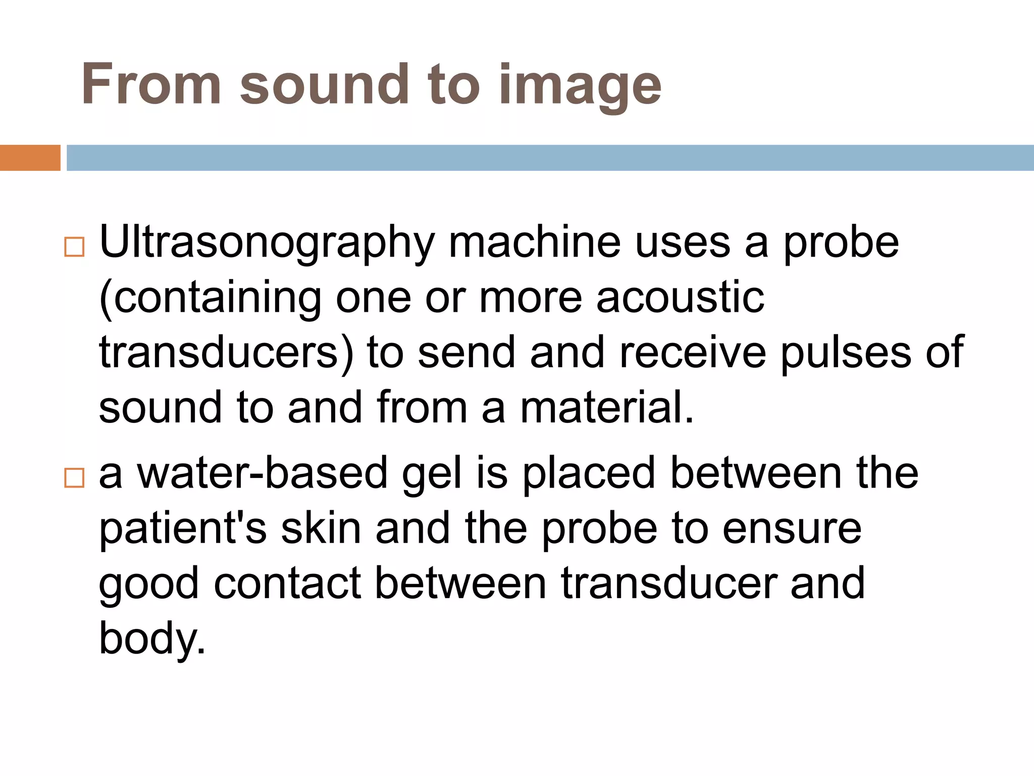
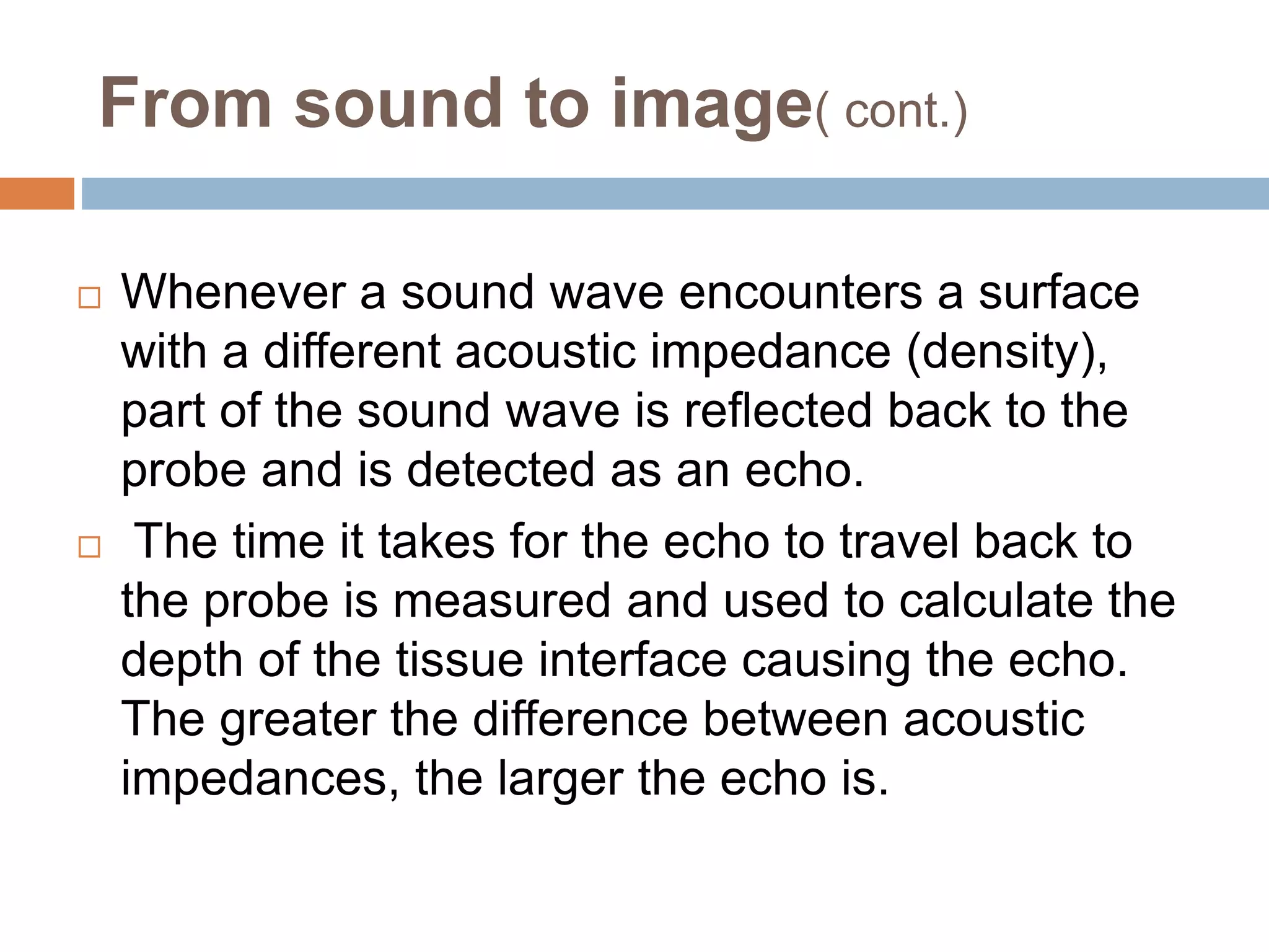
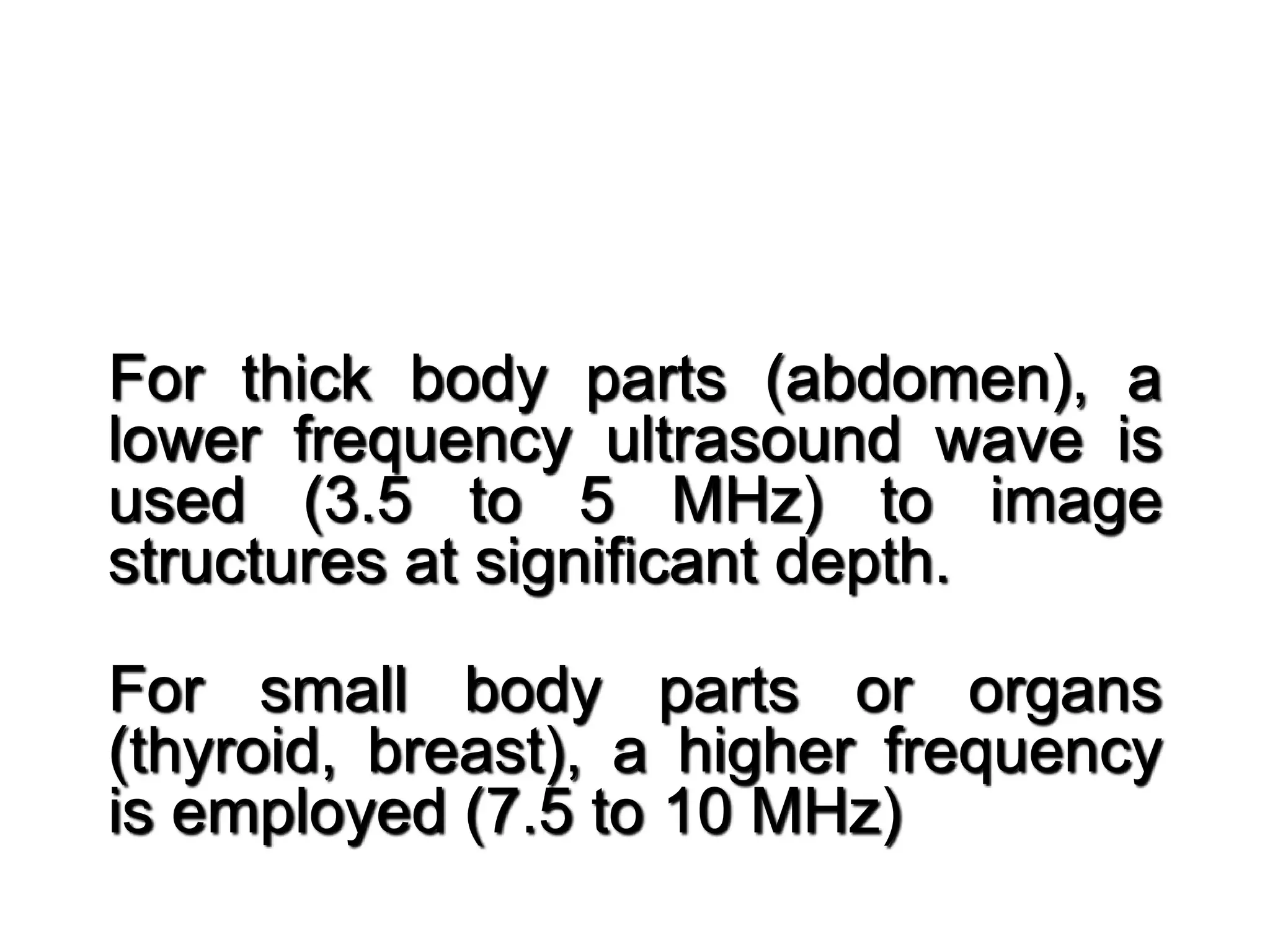
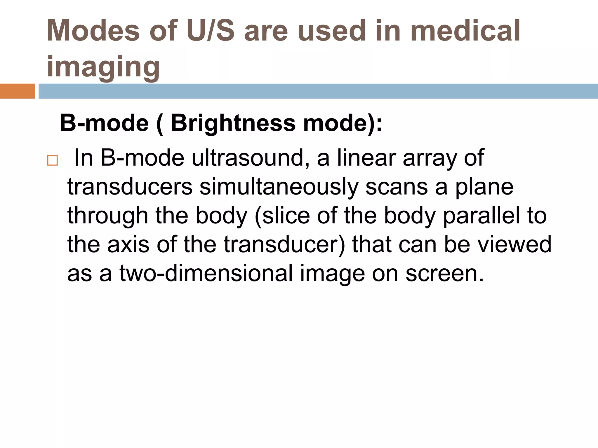
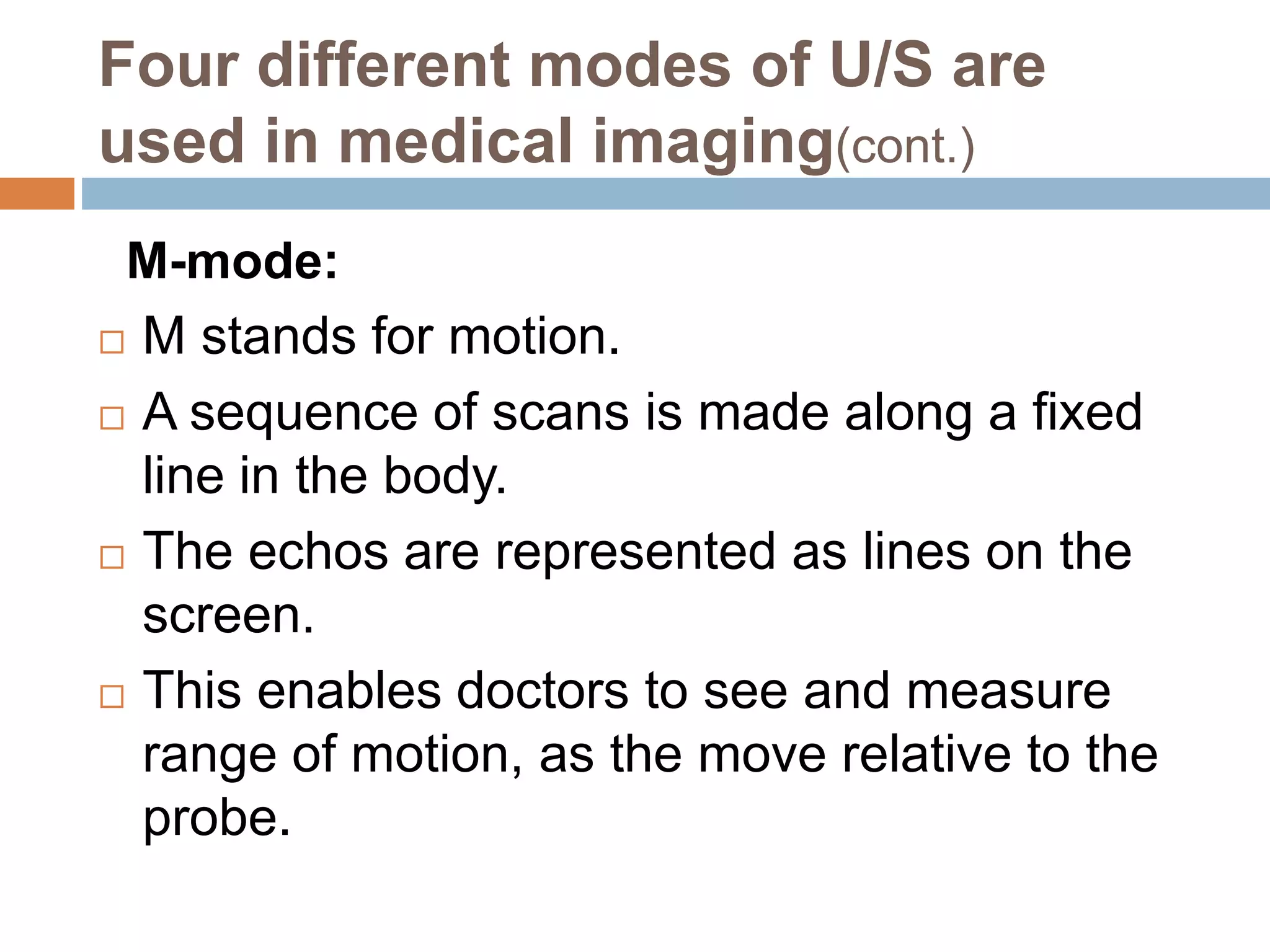
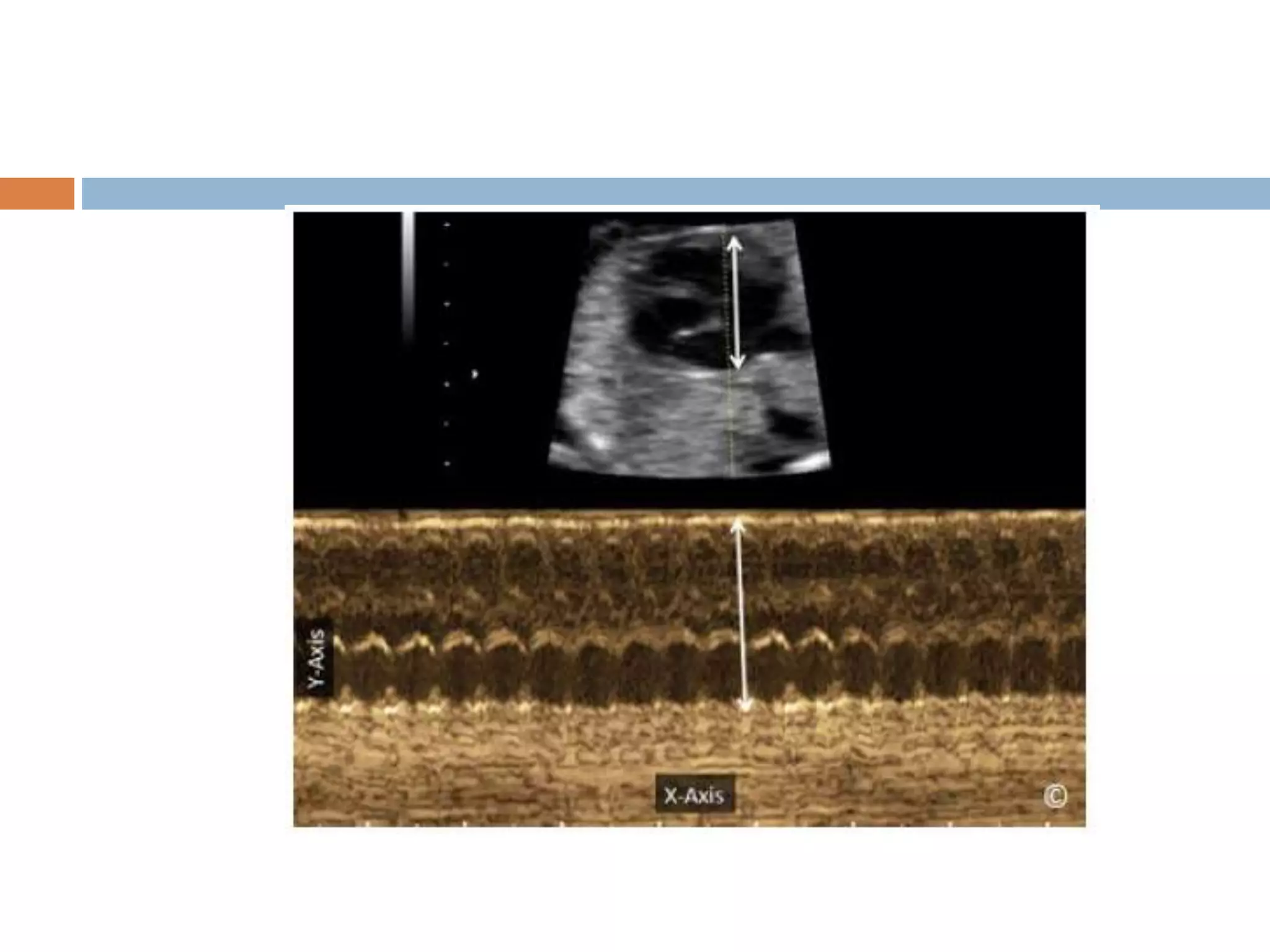
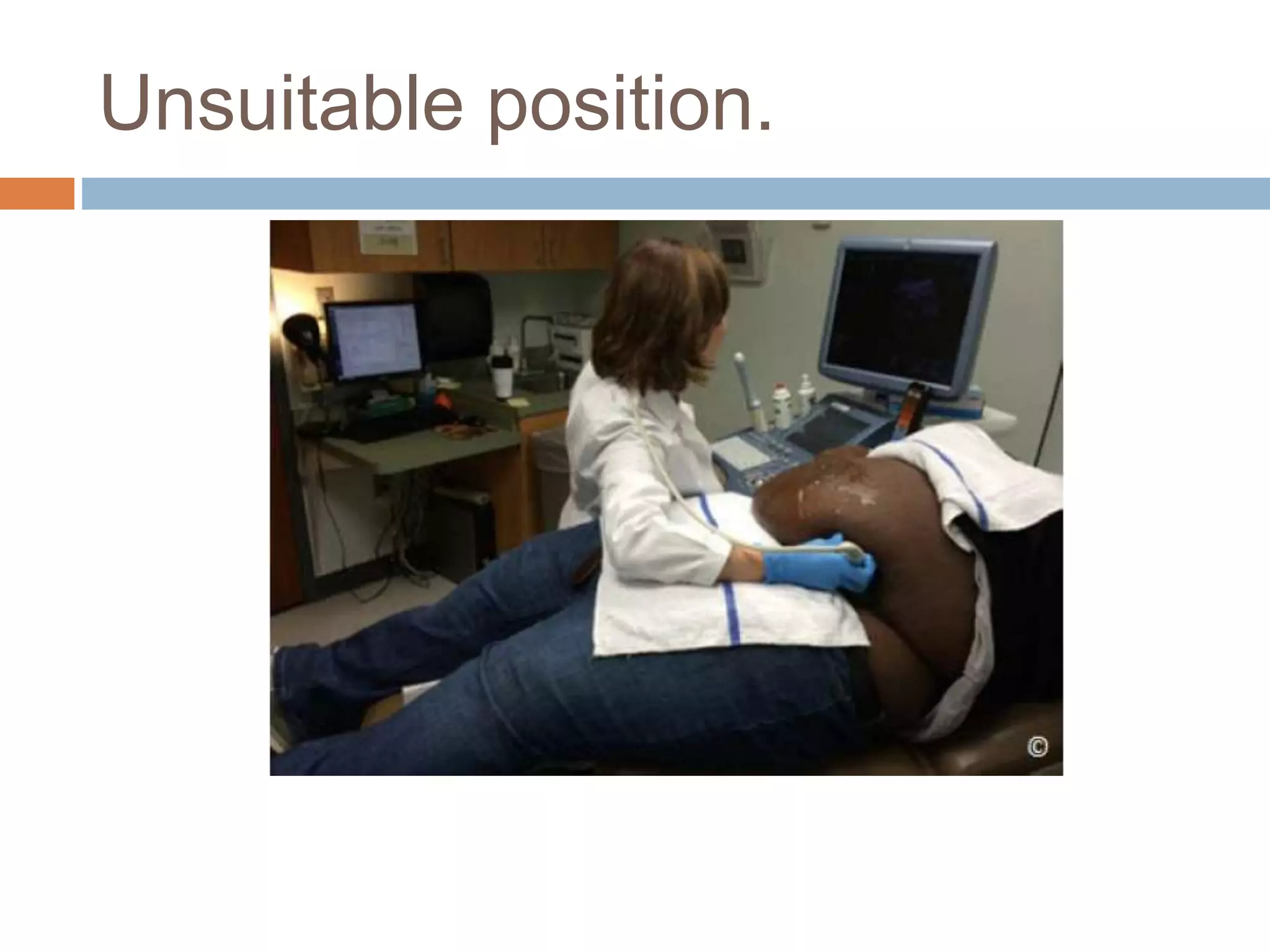
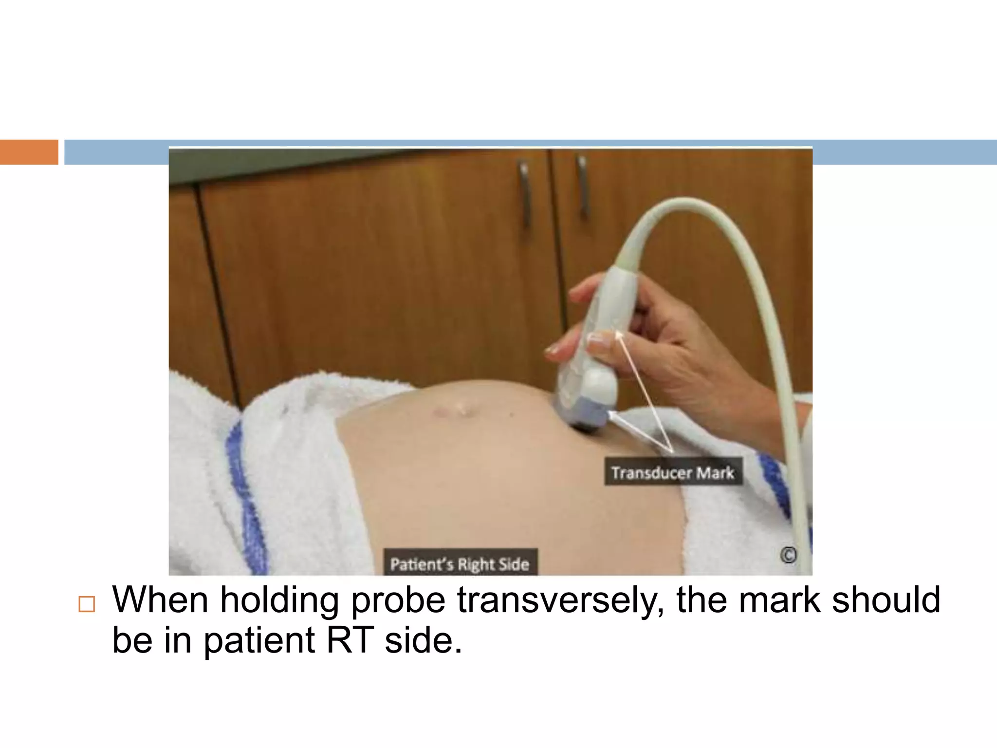
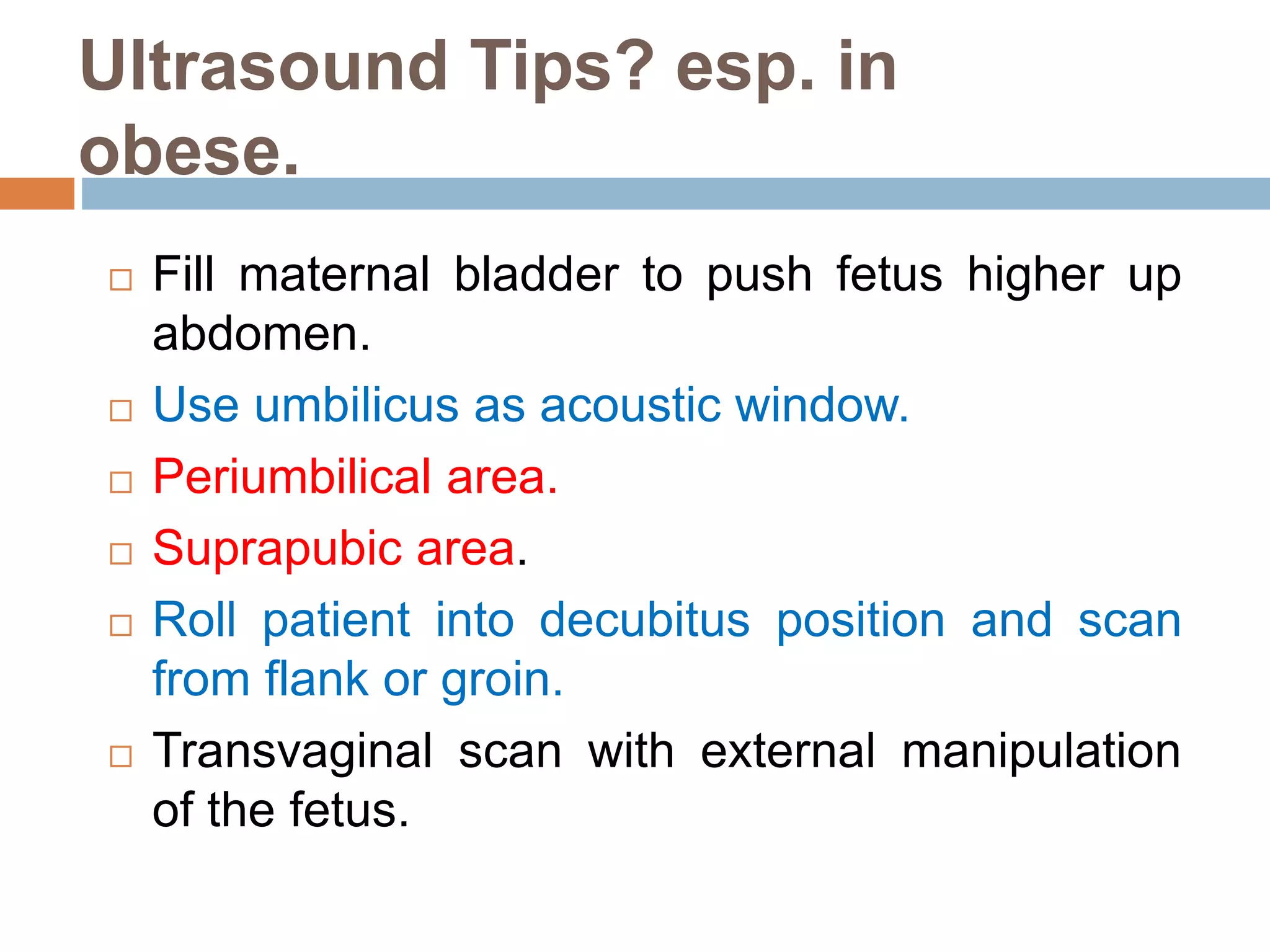
INTRODUCTION
TO
OBSTETRICS
ULTRASOUND
By: Magdy AbdelRahman . 2017
Background on Ultrasound
 Ultrasound waves are sound waves of
frequencies higher than the human ear can hear
(>20 KHz).
 It has been employed in various technologies for
civil and military marine location and navigation
(SONAR = SOund Navigation And Ranging).
Characteristics of Sound
Frequency
Frequency (f) is the number of times the wave oscillates
through a cycle each second (sec) (Hertz: Hz or
cycles/sec)
Infra sound < 15 Hz
Audible sound ~ 15 Hz - 20 kHz
Ultrasound > 20 kHz; for medical usage typically 2-
10 MHz with specialized ultrasound applications up
to 50 MHz
From sound to image
 Ultrasonography machine uses a probe
(containing one or more acoustic
transducers) to send and receive pulses of
sound to and from a material.
 a water-based gel is placed between the
patient's skin and the probe to ensure
good contact between transducer and
body.
From sound to image( cont.)
 Whenever a sound wave encounters a surface
with a different acoustic impedance (density),
part of the sound wave is reflected back to the
probe and is detected as an echo.
 The time it takes for the echo to travel back to
the probe is measured and used to calculate the
depth of the tissue interface causing the echo.
The greater the difference between acoustic
impedances, the larger the echo is.
For thick body parts (abdomen), a
lower frequency ultrasound wave is
used (3.5 to 5 MHz) to image
structures at significant depth.
For small body parts or organs
(thyroid, breast), a higher frequency
is employed (7.5 to 10 MHz)
Modes of U/S are used in medical
imaging
B-mode ( Brightness mode):
 In B-mode ultrasound, a linear array of
transducers simultaneously scans a plane
through the body (slice of the body parallel to
the axis of the transducer) that can be viewed
as a two-dimensional image on screen.
Four different modes of U/S are
used in medical imaging(cont.)
M-mode:
 M stands for motion.
 A sequence of scans is made along a fixed
line in the body.
 The echos are represented as lines on the
screen.
 This enables doctors to see and measure
range of motion, as the move relative to the
probe.
Four different modes of U/S are
used in medical imaging(cont.)
Doppler mode:
 When sound waves strike a moving object
they change their frequency (Doppler effect).
 This mode makes use of the Doppler effect
in measuring and visualizing blood flow.
Strengths ( advantages) of
ultrasound
 It has no known long-term side effects.
 Equipment is widely available and
comparatively flexible.
 Small, easily carried scanners are available;
examination scan be performed at the
bedside.
 Relatively inexpensive compared to other
modes.
 It shows the internal structure of organs.
Risks and side-effects of
ultrasound(cont.)
 World Health Organization’s technical report
series 875 (1998) supports that ultrasound is
harmless: «Diagnostic ultrasound is
recognized as a safe, effective , and highly
flexible imaging modality capable of
providing clinically relevant information about
most parts of the body in a rapid and cost-
effective fashion».
Weaknesses of ultrasound
 The method is operator-dependent. A high level
of skill and experience is needed to acquire
good-quality images and make accurate
diagnoses.
 «The proper, safe, and effective use of
diagnostic ultrasound is highly dependent on the
user, who has a major impact on the
examination's overall benefit.
 Ultrasound device performs very poorly when
there is bone or gas between the transducer
and the organ of interest (lung & intestine).
 Depth penetration of ultrasound waves may is
limited (obese patients).
Ultrasound in Medicine
 There are probes to scan superficially
(through the skin) or intra- cavitatary (inside
the vagina, rectum, esophagus or blood
vessels).
 Ultrasound is used as a diagnostic tool in
many branches in medicine for scanning
solid soft tissue organs and hollow fluid filled
organs or cavities.
How is the procedure performed?
 Trans-abdominal Scan:
For most ultrasound
exams, the patient is
positioned lying face-up
on an examination table
that can be tilted or
moved
How is the procedure performed?
(cont.)
 Trans-vaginal Scan:
For most ultrasound exams, the patient is positioned
lying in lithotomy position on an examination table
that can be tilted or moved. The vaginal probe is
placed in the vagina.
This method usually provides better images of the
pelvic organs (and therefore more information) in
patients who are obese and/or in the early stages of
pregnancy.
This is contraindicated in virgin patients
(Trans-vaginal Scan:Done only by specialist)
Preparing to scan.
Unsuitable position.
 When holding probe transversely, the mark should
be in patient RT side.
Ultrasound Tips? esp. in
obese.
 Fill maternal bladder to push fetus higher up
abdomen.
 Use umbilicus as acoustic window.
 Periumbilical area.
 Suprapubic area.
 Roll patient into decubitus position and scan
from flank or groin.
 Transvaginal scan with external manipulation
of the fetus.
Introduction to obs. ultrasound

Introduction to obs. ultrasound

  • 1.
  • 2.
    Background on Ultrasound Ultrasound waves are sound waves of frequencies higher than the human ear can hear (>20 KHz).  It has been employed in various technologies for civil and military marine location and navigation (SONAR = SOund Navigation And Ranging).
  • 3.
    Characteristics of Sound Frequency Frequency(f) is the number of times the wave oscillates through a cycle each second (sec) (Hertz: Hz or cycles/sec) Infra sound < 15 Hz Audible sound ~ 15 Hz - 20 kHz Ultrasound > 20 kHz; for medical usage typically 2- 10 MHz with specialized ultrasound applications up to 50 MHz
  • 6.
    From sound toimage  Ultrasonography machine uses a probe (containing one or more acoustic transducers) to send and receive pulses of sound to and from a material.  a water-based gel is placed between the patient's skin and the probe to ensure good contact between transducer and body.
  • 7.
    From sound toimage( cont.)  Whenever a sound wave encounters a surface with a different acoustic impedance (density), part of the sound wave is reflected back to the probe and is detected as an echo.  The time it takes for the echo to travel back to the probe is measured and used to calculate the depth of the tissue interface causing the echo. The greater the difference between acoustic impedances, the larger the echo is.
  • 8.
    For thick bodyparts (abdomen), a lower frequency ultrasound wave is used (3.5 to 5 MHz) to image structures at significant depth. For small body parts or organs (thyroid, breast), a higher frequency is employed (7.5 to 10 MHz)
  • 10.
    Modes of U/Sare used in medical imaging B-mode ( Brightness mode):  In B-mode ultrasound, a linear array of transducers simultaneously scans a plane through the body (slice of the body parallel to the axis of the transducer) that can be viewed as a two-dimensional image on screen.
  • 11.
    Four different modesof U/S are used in medical imaging(cont.) M-mode:  M stands for motion.  A sequence of scans is made along a fixed line in the body.  The echos are represented as lines on the screen.  This enables doctors to see and measure range of motion, as the move relative to the probe.
  • 13.
    Four different modesof U/S are used in medical imaging(cont.) Doppler mode:  When sound waves strike a moving object they change their frequency (Doppler effect).  This mode makes use of the Doppler effect in measuring and visualizing blood flow.
  • 14.
    Strengths ( advantages)of ultrasound  It has no known long-term side effects.  Equipment is widely available and comparatively flexible.  Small, easily carried scanners are available; examination scan be performed at the bedside.  Relatively inexpensive compared to other modes.  It shows the internal structure of organs.
  • 15.
    Risks and side-effectsof ultrasound(cont.)  World Health Organization’s technical report series 875 (1998) supports that ultrasound is harmless: «Diagnostic ultrasound is recognized as a safe, effective , and highly flexible imaging modality capable of providing clinically relevant information about most parts of the body in a rapid and cost- effective fashion».
  • 16.
    Weaknesses of ultrasound The method is operator-dependent. A high level of skill and experience is needed to acquire good-quality images and make accurate diagnoses.  «The proper, safe, and effective use of diagnostic ultrasound is highly dependent on the user, who has a major impact on the examination's overall benefit.  Ultrasound device performs very poorly when there is bone or gas between the transducer and the organ of interest (lung & intestine).  Depth penetration of ultrasound waves may is limited (obese patients).
  • 17.
    Ultrasound in Medicine There are probes to scan superficially (through the skin) or intra- cavitatary (inside the vagina, rectum, esophagus or blood vessels).  Ultrasound is used as a diagnostic tool in many branches in medicine for scanning solid soft tissue organs and hollow fluid filled organs or cavities.
  • 18.
    How is theprocedure performed?  Trans-abdominal Scan: For most ultrasound exams, the patient is positioned lying face-up on an examination table that can be tilted or moved
  • 19.
    How is theprocedure performed? (cont.)  Trans-vaginal Scan: For most ultrasound exams, the patient is positioned lying in lithotomy position on an examination table that can be tilted or moved. The vaginal probe is placed in the vagina. This method usually provides better images of the pelvic organs (and therefore more information) in patients who are obese and/or in the early stages of pregnancy. This is contraindicated in virgin patients (Trans-vaginal Scan:Done only by specialist)
  • 20.
  • 21.
  • 22.
     When holdingprobe transversely, the mark should be in patient RT side.
  • 23.
    Ultrasound Tips? esp.in obese.  Fill maternal bladder to push fetus higher up abdomen.  Use umbilicus as acoustic window.  Periumbilical area.  Suprapubic area.  Roll patient into decubitus position and scan from flank or groin.  Transvaginal scan with external manipulation of the fetus.