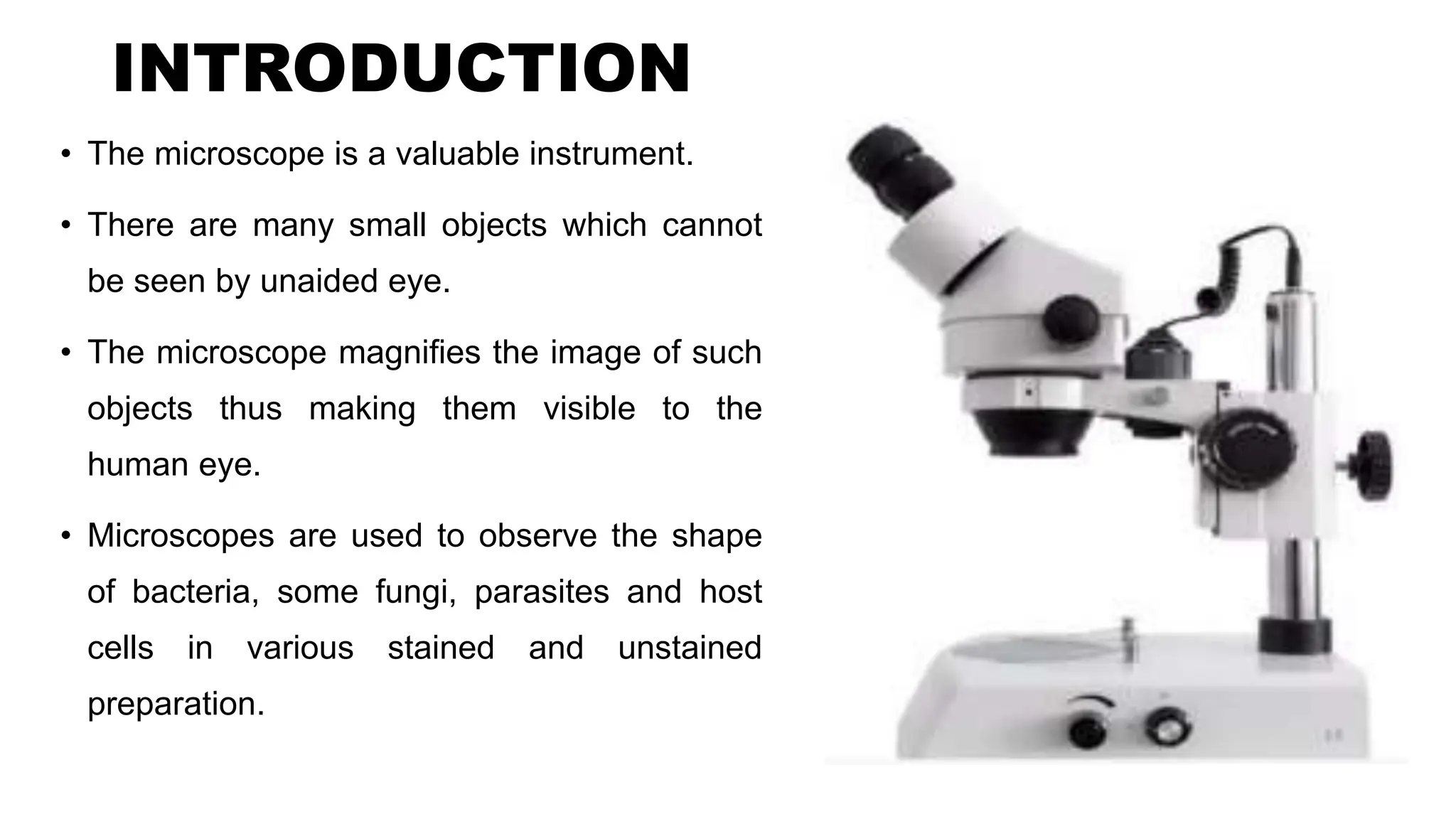
PRESENTATION
BY
ABURA GEOFFREY
DEPT OF MICROBIOLOGY & IMMUNOLOGY
INTRODUCTION
• The microscope is a valuable instrument.
• There are many small objects which cannot
be seen by unaided eye.
• The microscope magnifies the image of such
objects thus making them visible to the
human eye.
• Microscopes are used to observe the shape
of bacteria, some fungi, parasites and host
cells in various stained and unstained
preparation.
Types of a microscope
• Microscope used in clinical practice are light microscopes. They are called light
microscope because they used a beam of light to view specimen.
• A compound light microscope: It’s the most common microscope used in
microbiology. It consists of two lens systems to magnify the image. A
compound light microscope with a single eye-piece is called monocular and
the one with two eye-pieces is called Binocular.
• Electron microscope: microscopes that use a beam of electrons instead of
beam of light and electromagnet instead of glass lenses for focusing are called
electron microscopes. These microscopes provide a higher magnification, used
for observing extremely small microorganism such as viruses.
Parts of a microscope
• The compound microscope is mainly used for studying the structural
details of cells, tissues or sections of organs
• The parts of a compound microscope can be classified into:
1. Non-optical parts
2. Optical parts
Non-optical parts
• Base: Also known as the foot which is either U or horse-shaped. It’s
a metallic structure that supports the entire microscope
• Pillar: The connection between the base and the arm are possible
through the pillar
• Arm: Also known as the limb which is metallic handle forming the
connection between the arm to the inclined joint. The stage and the
body tube is supported by the arm.
Con’t
• Stage: It’s a metallic platform that is fitted to the lower part of the
arm with a hole in the center. The microscopic slides are placed on
the stage.
• Body tube: The main purpose of the body tube is to hold the
objectives and the ocular lenses at the two ends. The end where the
ocular lens is present is known as the head while the end where the
objectives lens is placed is known as the nose piece.
• Adjustment screw: These are two pairs of adjusting screws that
are used either for a coarse adjustment or for fine adjustment.
OPTICAL PARTS
• Diaphragm: The amount of light falling on the object can be controlled
through the diaphragm. Its present below the stage. The disc and Iris are
the two types of diaphragm
• Condenser: Its present below the diaphragm. The focusing of light can
be done by adjusting the condenser by moving it either up or down
• Reflector: A mirror that is attached above the base. On side of the mirror
has a plane mirror while the other side has a concave mirror. When the
light is strong, the plane mirror side is used and when the light is weak,
the concave mirror side is used. The light on the object is directed with
the help of the reflector through the diaphragm and condenser
Con’t
• Objective lenses: These lenses are present over the nose piece.
There are two to three types of objective lenses;
• Low power
• High power
• Oil immersion
• The objective lens is a compound lens that forms a real inverted
image of the image inside the body tube
Con’t
• Ocular lens: The ocular lens is also known as the eye piece. The image of
microscopic objects can be viewed through these lenses.
• There are four types of magnification that can take place in the ocular lens:
• 5x
• 10x
• 15x
• 20x
• The binocular head is the device that uses two eye pieces and has many
mirrors and prisms, which makes the passage of light easier.
Mechanisms of compound microscope
First of all, a specimen is placed between the objective and condenser lens.
Light emitted from the light source is pointed over the specimen with help of a
condenser lens
After that, the light is passed through specimen and comes towards the objective lens
The objective lens captures the light coming from the specimen and creates a
magnified image of the specimen which is called the primary image
Then the objective lens passed this image through the body tube to the ocular lens or
eye piece and again magnifies the image
At last, the viewer can see a clear and magnified image of the specimen through the
eye piece.
Care of the microscope
• Tips for microscope care
Handle with care
Keep lenses clear of slides
Clean after using immersion oil
Cover when not in use
Look after the bulb
Store in a clean dry place
Only use special lens paper or wipes for cleaning the lenses
Keep your user’s Manual in a safe place.
RULES FOR USING MICROSCOPE
Carry the microscope properly
Always begin focusing with the 4X objective
Use the coarse focus only with the 4X objective in place
Use the immersion oil only with the 100X objective (oil immersion) in place
Use only ONE drop of oil
Lower the stage and then remove the slide when you are done
Always clean the microscope when you’re done using a lense paper
Always place the 4X objective over the stage and be sure the stage is at its lowest position before putting
the microscope away
Always turn off the light before putting the microscope away
Always wrap the cord correctly before putting the microscope away
Always return the microscope to the correct cabinet
LECTURE NOTES FOR MICROSCOPE FOR STUDENTS.pptx.

LECTURE NOTES FOR MICROSCOPE FOR STUDENTS.pptx.

  • 1.
    PRESENTATION BY ABURA GEOFFREY DEPT OFMICROBIOLOGY & IMMUNOLOGY
  • 2.
    INTRODUCTION • The microscopeis a valuable instrument. • There are many small objects which cannot be seen by unaided eye. • The microscope magnifies the image of such objects thus making them visible to the human eye. • Microscopes are used to observe the shape of bacteria, some fungi, parasites and host cells in various stained and unstained preparation.
  • 5.
    Types of amicroscope • Microscope used in clinical practice are light microscopes. They are called light microscope because they used a beam of light to view specimen. • A compound light microscope: It’s the most common microscope used in microbiology. It consists of two lens systems to magnify the image. A compound light microscope with a single eye-piece is called monocular and the one with two eye-pieces is called Binocular. • Electron microscope: microscopes that use a beam of electrons instead of beam of light and electromagnet instead of glass lenses for focusing are called electron microscopes. These microscopes provide a higher magnification, used for observing extremely small microorganism such as viruses.
  • 7.
    Parts of amicroscope • The compound microscope is mainly used for studying the structural details of cells, tissues or sections of organs • The parts of a compound microscope can be classified into: 1. Non-optical parts 2. Optical parts
  • 8.
    Non-optical parts • Base:Also known as the foot which is either U or horse-shaped. It’s a metallic structure that supports the entire microscope • Pillar: The connection between the base and the arm are possible through the pillar • Arm: Also known as the limb which is metallic handle forming the connection between the arm to the inclined joint. The stage and the body tube is supported by the arm.
  • 9.
    Con’t • Stage: It’sa metallic platform that is fitted to the lower part of the arm with a hole in the center. The microscopic slides are placed on the stage. • Body tube: The main purpose of the body tube is to hold the objectives and the ocular lenses at the two ends. The end where the ocular lens is present is known as the head while the end where the objectives lens is placed is known as the nose piece. • Adjustment screw: These are two pairs of adjusting screws that are used either for a coarse adjustment or for fine adjustment.
  • 10.
    OPTICAL PARTS • Diaphragm:The amount of light falling on the object can be controlled through the diaphragm. Its present below the stage. The disc and Iris are the two types of diaphragm • Condenser: Its present below the diaphragm. The focusing of light can be done by adjusting the condenser by moving it either up or down • Reflector: A mirror that is attached above the base. On side of the mirror has a plane mirror while the other side has a concave mirror. When the light is strong, the plane mirror side is used and when the light is weak, the concave mirror side is used. The light on the object is directed with the help of the reflector through the diaphragm and condenser
  • 11.
    Con’t • Objective lenses:These lenses are present over the nose piece. There are two to three types of objective lenses; • Low power • High power • Oil immersion • The objective lens is a compound lens that forms a real inverted image of the image inside the body tube
  • 12.
    Con’t • Ocular lens:The ocular lens is also known as the eye piece. The image of microscopic objects can be viewed through these lenses. • There are four types of magnification that can take place in the ocular lens: • 5x • 10x • 15x • 20x • The binocular head is the device that uses two eye pieces and has many mirrors and prisms, which makes the passage of light easier.
  • 13.
    Mechanisms of compoundmicroscope First of all, a specimen is placed between the objective and condenser lens. Light emitted from the light source is pointed over the specimen with help of a condenser lens After that, the light is passed through specimen and comes towards the objective lens The objective lens captures the light coming from the specimen and creates a magnified image of the specimen which is called the primary image Then the objective lens passed this image through the body tube to the ocular lens or eye piece and again magnifies the image At last, the viewer can see a clear and magnified image of the specimen through the eye piece.
  • 14.
    Care of themicroscope • Tips for microscope care Handle with care Keep lenses clear of slides Clean after using immersion oil Cover when not in use Look after the bulb Store in a clean dry place Only use special lens paper or wipes for cleaning the lenses Keep your user’s Manual in a safe place.
  • 15.
    RULES FOR USINGMICROSCOPE Carry the microscope properly Always begin focusing with the 4X objective Use the coarse focus only with the 4X objective in place Use the immersion oil only with the 100X objective (oil immersion) in place Use only ONE drop of oil Lower the stage and then remove the slide when you are done Always clean the microscope when you’re done using a lense paper Always place the 4X objective over the stage and be sure the stage is at its lowest position before putting the microscope away Always turn off the light before putting the microscope away Always wrap the cord correctly before putting the microscope away Always return the microscope to the correct cabinet