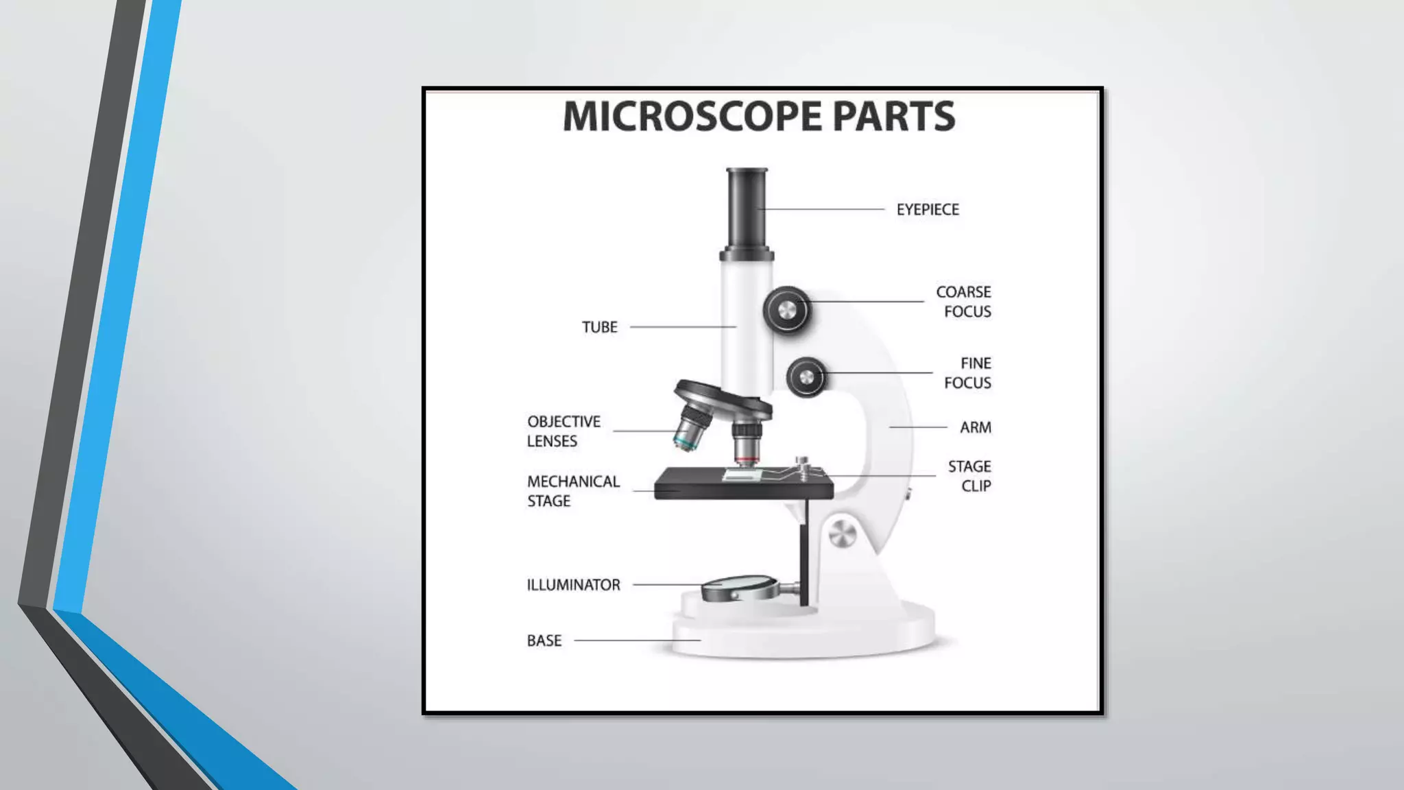
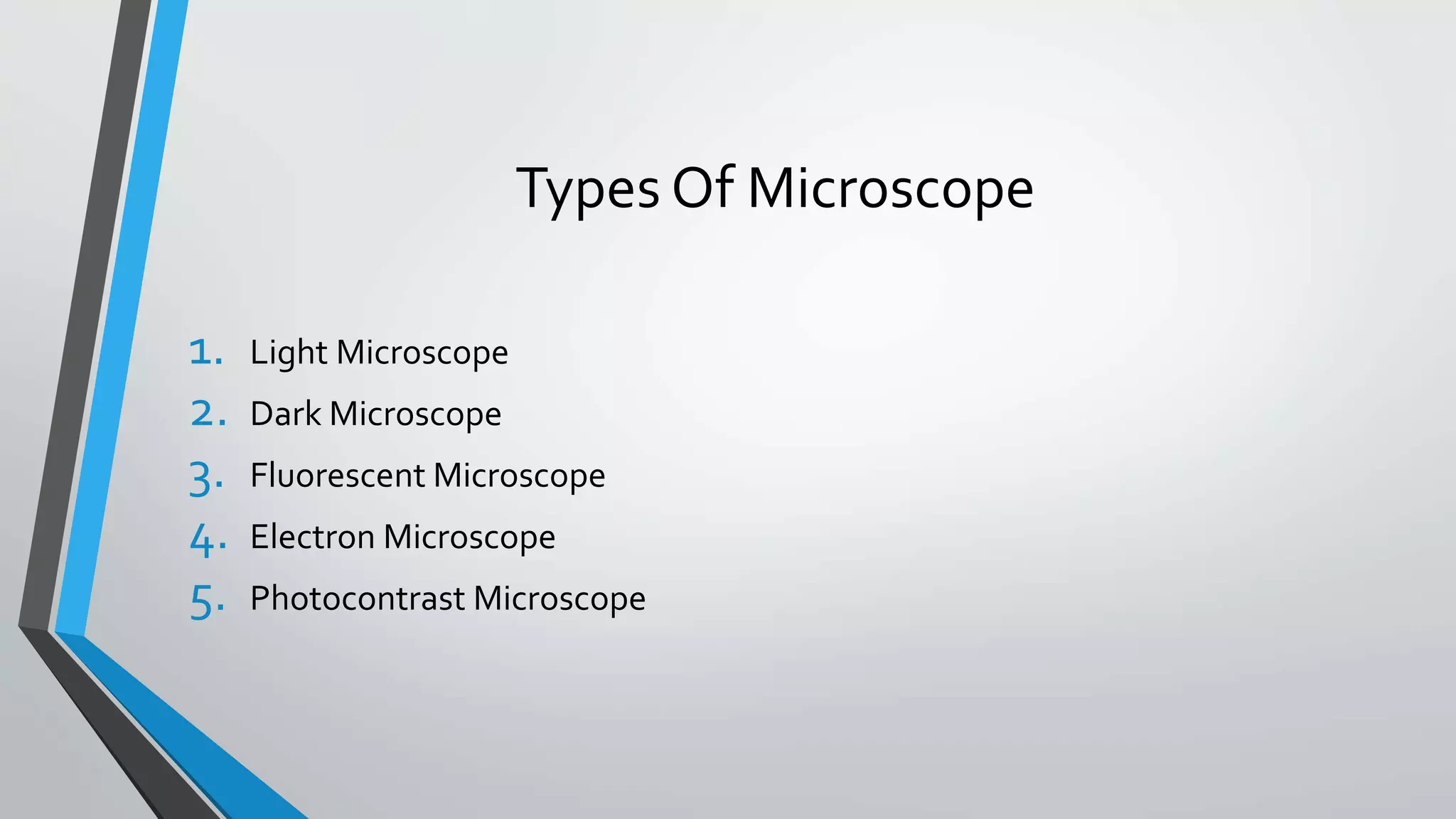
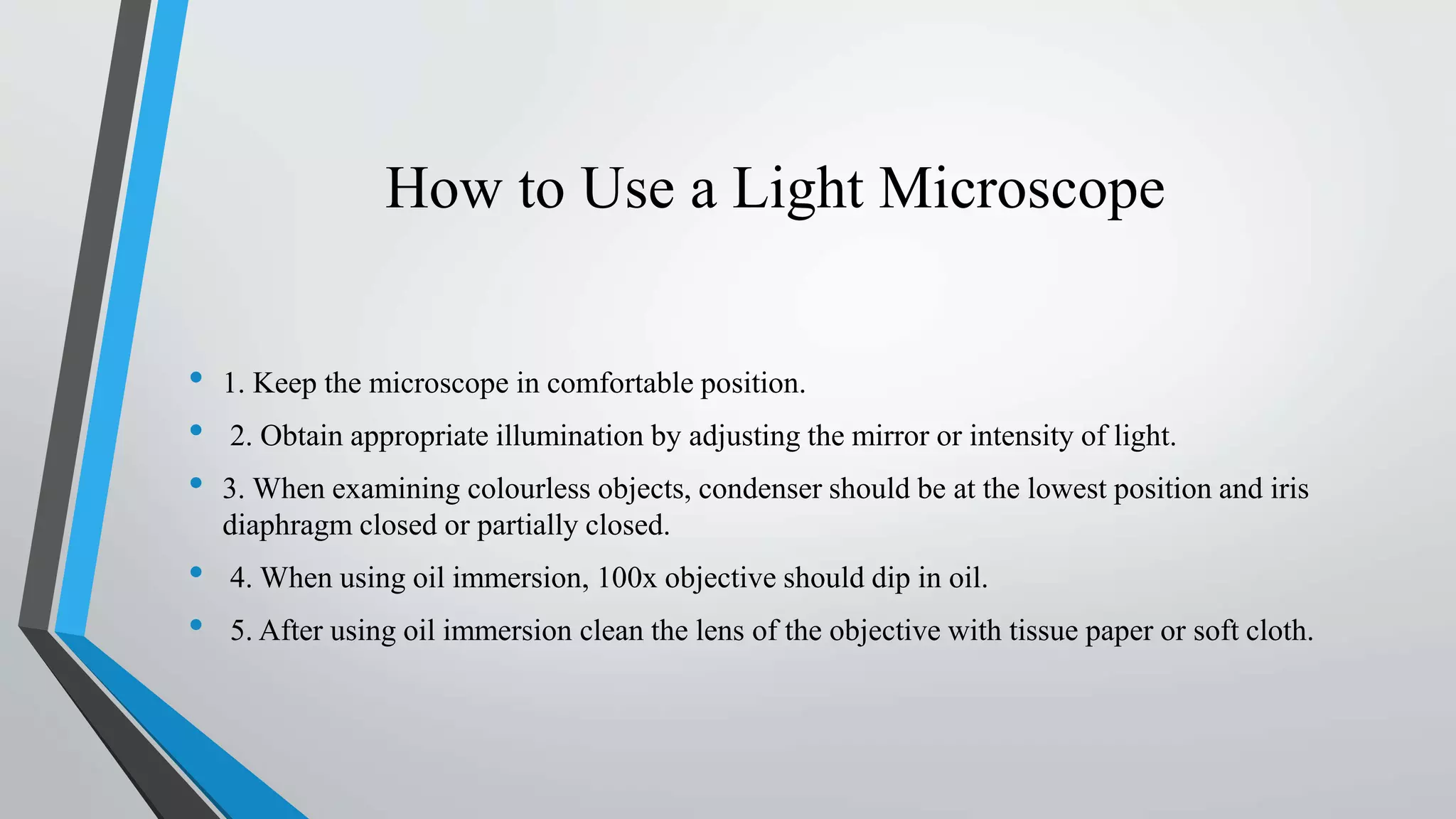
STUDY OF MICROSCOPE
Dr.Utkarsh Singhal
M.B.B.S , M.D (Pathology)
L.L.R.M Medical College, Meerut,U.P
MICROSCOPE
• The word "microscope" is formed by two words: "micros"- small and 'skipein'-to
look.
• Invented by ‘Zacharias Janssen’.
• Microscope is an instrument which makes enlarged images of minute objects, sub
cellular structures, and many more, generally hard to resolved with naked human
eyes.
Types Of Microscope
1. Light Microscope
2. Dark Microscope
3. Fluorescent Microscope
4. Electron Microscope
5. Photocontrast Microscope
Light Microscope
Two type:
1. Simple microscope: This is a simple hand magnifying lens. The magnification power of hand
lens is from 2x to 200x.
2. Compound microscope: This has a battery of lenses which are fitted in a complex instrument.
One type of lens remains near the object (objective lens) and another type of lens near the
observer’s eye (eye piece lens).
Parts of compound microscope :
• Stand: This is horse-shoe shaped in monocular microscope. It gives stability to the microscope.
Binocular microscopes have a variety of ergonomic shapes of stand.
• Body : It consists of a limb which arises from the joint with which microscope can be moved in
comfortable position. The stand and the limb carry the following:
• i. Body tubes
• ii. Stage
• iii. Knobs for coarse and fine adjustment
• Body Tubes There are two tubes: external tube which carries at its lower end a revolving nose piece having
objective lenses of different magnification while internal tube is draw tube which carries at its upper end eye
pieces
• Stage :This is a metallic platform which accommodates glass slide having mounted object over it to be seen.
Stage is attached to the limb just below the level of objectives. It has an aperture in its centre which permits
the light to reach the object. Slide on the stage can be moved horizontally or vertically by two knobs attached
to slide holder.
• Substage: Just below the stage is substage which consists of condenser through which light is focused on the
object. The substage can be moved up and down. The substage has an iris diaphragm, closing and opening of
which controls the amount of light reaching the object.
• Knobs for Coarse and Fine Adjustment For coarse and fine adjustments, knobs are provided on either side
of the body. Coarse adjustment has two bigger knobs, the movement of which moves the body tubes with its
lenses. Fine adjustment has two smaller knobs on either side of the body.
Optical System :Optical system is comprised by different lenses which are fitted into a microscope. It
consists of eye piece, objectives and condensers.
• Eye Piece:In monocular microscope, there is one eye piece while binocular microscope has two. Eye piece
has two planoconvex lenses. Their magnification can be 5x, 10x, or 15x
• Objectives :These are made of a battery of lenses with prisms incorporated in them. Their magnification
power is 4x, 10x, 40x and 100x.
• Condenser: This is made up of two simple lenses and it condenses light on to the object.
Light/Illumination System:
For day light illumination, a mirror is fitted which is plane on one side and concave on the other side. Plane
mirror is used in sunlight while concave in artificial light.
Magnification
• It is the degree of image enlargement. It depends upon the following:
• i. Length of optical tube
• ii. Magnifying power of objective
• iii. Magnifying power of eye piece
= Magnifying power of objective × Magnifying power of eye piece (Mo x Me)
Resolving Power
• Resolving power represents the capacity of the optical system to produce separate images of
objects very close to each other.
• R=
𝟎.𝟔𝟏 𝝀
𝑵𝑨
• Where λ is wavelength of incidental light;
• NA is numerical aperture of lens
• Resolving power of a standard light microscope is around 200 nm.
How to Use a Light Microscope
• 1. Keep the microscope in comfortable position.
• 2. Obtain appropriate illumination by adjusting the mirror or intensity of light.
• 3. When examining colourless objects, condenser should be at the lowest position and iris
diaphragm closed or partially closed.
• 4. When using oil immersion, 100x objective should dip in oil.
• 5. After using oil immersion clean the lens of the objective with tissue paper or soft cloth.
OtherTypes Of Microscope
• BINOCULAR MICROSCOPE : In this the light rays from object is divided in the half angle of 45
degree which permits light.
• Dark Ground Illumination (DGI) : This method is used for examination of unstained living micro-
organisms e.g. Treponema pallidum.
Principle : The micro-organisms are illuminated by an oblique ray of light which does not pass through
the micro-organism. The condenser is blackened in the centre and light passes through its periphery
illuminating the living micro-organism on a glass slide.
• Electron Microscope :This is used for study of ultrastructural details of the tissues and cells. For
electron microscopy, tissue is fixed in 4% glutaraldehyde at 4°C for 4 hours.
Principle: By using an electron beam of light, the resolving power of the microscope is increased to
50,000 to 100,000 times and very small structures can be visualised. In contrast to light microscopy,
resolution of electron microscopy is 0.2 nm or less.
There are two types of electron microscopy:
1. Transmission Electron Microscopy (TEM): TEM helps visualize cell’s cytoplasm and organelles. For
this purpose, ultrathin sections are required. TEM interprets atomic rather than molecular properties of
the tissue and gives two dimensional image of the tissue.
2. Scanning Electron Microscopy (SEM) : SEM helps in the study of cell surface. In this three
dimensional image is produced. The image is produced on cathode ray oscillograph which can also be
amplified. SEM can also be used for fluorescent antibody techniques.
• Fluorescent Microscope: This method is used for demonstration of naturally occurring fluorescent
material and other non-fluorescent substances or micro-organisms after staining with some fluorescent
dyes e.g. Mycobacterium tuberculosis, amyloid, lipids, elastic fibres etc. UV light is used for illumination.
• Principle: Fluorescent microscopy depends upon illumination of a substance with a specific wavelength
(UV region i.e. invisible region) which then emits light at a lower wavelength (visible region).
Precautions:
• For a wet object stage should be horizontal both eye open through a microscope.
• Condensor should be lowered in unstained condition.
• First focus with low power using coarse adjustment for histological/pathological studies.
• In bacteriology and immunology, oil immersion should be used.
MICROSCOPE-Study of Microscope,types.pptx

MICROSCOPE-Study of Microscope,types.pptx

  • 1.
    STUDY OF MICROSCOPE Dr.UtkarshSinghal M.B.B.S , M.D (Pathology) L.L.R.M Medical College, Meerut,U.P
  • 2.
    MICROSCOPE • The word"microscope" is formed by two words: "micros"- small and 'skipein'-to look. • Invented by ‘Zacharias Janssen’. • Microscope is an instrument which makes enlarged images of minute objects, sub cellular structures, and many more, generally hard to resolved with naked human eyes.
  • 4.
    Types Of Microscope 1.Light Microscope 2. Dark Microscope 3. Fluorescent Microscope 4. Electron Microscope 5. Photocontrast Microscope
  • 5.
    Light Microscope Two type: 1.Simple microscope: This is a simple hand magnifying lens. The magnification power of hand lens is from 2x to 200x. 2. Compound microscope: This has a battery of lenses which are fitted in a complex instrument. One type of lens remains near the object (objective lens) and another type of lens near the observer’s eye (eye piece lens). Parts of compound microscope : • Stand: This is horse-shoe shaped in monocular microscope. It gives stability to the microscope. Binocular microscopes have a variety of ergonomic shapes of stand. • Body : It consists of a limb which arises from the joint with which microscope can be moved in comfortable position. The stand and the limb carry the following: • i. Body tubes • ii. Stage • iii. Knobs for coarse and fine adjustment
  • 6.
    • Body TubesThere are two tubes: external tube which carries at its lower end a revolving nose piece having objective lenses of different magnification while internal tube is draw tube which carries at its upper end eye pieces • Stage :This is a metallic platform which accommodates glass slide having mounted object over it to be seen. Stage is attached to the limb just below the level of objectives. It has an aperture in its centre which permits the light to reach the object. Slide on the stage can be moved horizontally or vertically by two knobs attached to slide holder. • Substage: Just below the stage is substage which consists of condenser through which light is focused on the object. The substage can be moved up and down. The substage has an iris diaphragm, closing and opening of which controls the amount of light reaching the object. • Knobs for Coarse and Fine Adjustment For coarse and fine adjustments, knobs are provided on either side of the body. Coarse adjustment has two bigger knobs, the movement of which moves the body tubes with its lenses. Fine adjustment has two smaller knobs on either side of the body. Optical System :Optical system is comprised by different lenses which are fitted into a microscope. It consists of eye piece, objectives and condensers. • Eye Piece:In monocular microscope, there is one eye piece while binocular microscope has two. Eye piece has two planoconvex lenses. Their magnification can be 5x, 10x, or 15x
  • 7.
    • Objectives :Theseare made of a battery of lenses with prisms incorporated in them. Their magnification power is 4x, 10x, 40x and 100x. • Condenser: This is made up of two simple lenses and it condenses light on to the object. Light/Illumination System: For day light illumination, a mirror is fitted which is plane on one side and concave on the other side. Plane mirror is used in sunlight while concave in artificial light.
  • 8.
    Magnification • It isthe degree of image enlargement. It depends upon the following: • i. Length of optical tube • ii. Magnifying power of objective • iii. Magnifying power of eye piece = Magnifying power of objective × Magnifying power of eye piece (Mo x Me)
  • 9.
    Resolving Power • Resolvingpower represents the capacity of the optical system to produce separate images of objects very close to each other. • R= 𝟎.𝟔𝟏 𝝀 𝑵𝑨 • Where λ is wavelength of incidental light; • NA is numerical aperture of lens • Resolving power of a standard light microscope is around 200 nm.
  • 10.
    How to Usea Light Microscope • 1. Keep the microscope in comfortable position. • 2. Obtain appropriate illumination by adjusting the mirror or intensity of light. • 3. When examining colourless objects, condenser should be at the lowest position and iris diaphragm closed or partially closed. • 4. When using oil immersion, 100x objective should dip in oil. • 5. After using oil immersion clean the lens of the objective with tissue paper or soft cloth.
  • 11.
    OtherTypes Of Microscope •BINOCULAR MICROSCOPE : In this the light rays from object is divided in the half angle of 45 degree which permits light. • Dark Ground Illumination (DGI) : This method is used for examination of unstained living micro- organisms e.g. Treponema pallidum. Principle : The micro-organisms are illuminated by an oblique ray of light which does not pass through the micro-organism. The condenser is blackened in the centre and light passes through its periphery illuminating the living micro-organism on a glass slide. • Electron Microscope :This is used for study of ultrastructural details of the tissues and cells. For electron microscopy, tissue is fixed in 4% glutaraldehyde at 4°C for 4 hours. Principle: By using an electron beam of light, the resolving power of the microscope is increased to 50,000 to 100,000 times and very small structures can be visualised. In contrast to light microscopy, resolution of electron microscopy is 0.2 nm or less. There are two types of electron microscopy:
  • 12.
    1. Transmission ElectronMicroscopy (TEM): TEM helps visualize cell’s cytoplasm and organelles. For this purpose, ultrathin sections are required. TEM interprets atomic rather than molecular properties of the tissue and gives two dimensional image of the tissue. 2. Scanning Electron Microscopy (SEM) : SEM helps in the study of cell surface. In this three dimensional image is produced. The image is produced on cathode ray oscillograph which can also be amplified. SEM can also be used for fluorescent antibody techniques. • Fluorescent Microscope: This method is used for demonstration of naturally occurring fluorescent material and other non-fluorescent substances or micro-organisms after staining with some fluorescent dyes e.g. Mycobacterium tuberculosis, amyloid, lipids, elastic fibres etc. UV light is used for illumination. • Principle: Fluorescent microscopy depends upon illumination of a substance with a specific wavelength (UV region i.e. invisible region) which then emits light at a lower wavelength (visible region).
  • 13.
    Precautions: • For awet object stage should be horizontal both eye open through a microscope. • Condensor should be lowered in unstained condition. • First focus with low power using coarse adjustment for histological/pathological studies. • In bacteriology and immunology, oil immersion should be used.