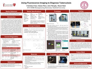
1. Needs Specifications Prototype
Current Diagnostics
Using Fluorescence Imaging to Diagnose Tuberculosis
Christina Chen, Soham More, Jack Murphy, Mansi Shah
2014 SIMR Bioengineering Bootcamp, Stanford University
Needs Statement
Concept Analysis
Introduction
References
Future Work
There are several improvements that we plan to make to our device in
the future.
1. Create a more sensitive light sensor that does not rely on an Arduino
kit.
2. Test the device with CDG-oME instead of fluorescein as a substitute.
3. Use our box to diagnose a variety of other diseases using different
fluorescent dyes.
4. Make the box more robust and user-friendly.
Acknowledgements
A cost effective way to diagnosis Tuberculosis within a day at home in
a non-clinical setting for those with little/no access to physicians.
3.
Materials
• Laser-cut black acrylic plastic (1/8 inch thick) with non-reflective
interior to build the bulk of the box
• 1-inch diameter clear plastic tubing with electrical tape to channel
and concentrate the light
• Arduino kit hooked up with phototransistor and LED light to detect
and indicate the presence of fluorescence
• Electrical Wiring
• LED light with brightness over 50 lumens to optimize fluorescence
• Excitation filter (490 nm) and emission filter (530 nm)
• Fluorescein as a substitute fluorescent dye for CDG-oME
Machinery
• Laser Cutter: LaserCAMM
• Acrylic Glue
• Bandsaw
Testing
Due to limited access to Dr. Rao’s CDG-oME dye, to test the
functionality of our box we used a substitute fluorescent protein called
fluorescein which has similar excitation and emission wavelengths.
We placed a .3125% fluorescein solution into the 2 dram vial, entered
it into the capsule, and turned on our LED and light sensor. We took a
picture through the camera hole and our app was successfully able to
detect the fluorescence. We also filled the vial with water for a
control, and our app did not give a false positive. Our light sensor
worked well and detected a small LED through our emission filter,
but was not sensitive enough to pick up the fluorescent signal.
In 2010, it was believed that one third of the world’s population
was affected by Tuberculosis, especially in developing countries and
areas prone to poverty. TB, caused by the many strains of mycobacteria,
most commonly Mycobacterium tuberculosis (Mtb), is easily spread
through the air, and may potentially infect not only the lungs but also
other vital organs.1 In order to effectively treat this disease, it is
imperative that it is diagnosed in its early stages; nonetheless, with the
current technologies, cost effective, quick, and accurate diagnostic tests
fail to reach the gold standard. Fortunately, recent research from the lab
of Dr. Jianghong Rao at Stanford University has been successful
creating a potential rapid point of care detection method for Mtb using
BlaC (Beta-lactamase, an enzyme specific to and naturally expressed by
Mtb) as a marker and a chemically engineered fluorescent protein
(CDG-OMe) as a detection probe.2 In less than ten minutes, Mtb can be
detected in substances as noninvasive as unprocessed human sputum.
Using the data from this research, the goal of our project is to develop a
fluorescent imaging box that is suitable for not just consumer use but
also for field work in impoverished areas.
Diagnostics Cost Speed Method Need
Doctor?
Skin Test $45 48 - 72
hrs
Tuberculin injected
under skin. Bump
inspection
Yes
Blood Test
(IGRA)
$105 4-24 hrs Proteins centrifuged
with Blood. IGRA
measured.
Yes
Chest X-ray $90 24 hrs X-ray of chest area.
Doctor analyzes images
Yes
Smear $28 24 hrs Stained sputum imaged
under a light
microscope
Yes
Major problems with current diagnostics:
1. Most require two or more
visits to the doctor
2. Clinical visits are too
expensive
3. All require a trained medical
physician
4. Most methods (3/4) are
invasive
Issues one through three pose a problem for they make it difficult
for consumers living in rural and/or impoverished areas to
receive not just basic healthcare but also important diagnostics
such as for TB.
Needs
• Consumer Cost: < $100
• Speed: < 4 hrs
• Portability: < 10 lbs
• Clinical Independence
• Noninvasive
• Safe and Sanitary
Nice to have
• Consumer Cost: <
$50
• Speed: < 2 hrs
• Portability: < 5 lbs
• Aesthetically pleasing
• Reusable
Welfare organizations are also able to purchase this
device and distribute it quickly and efficiently to
hundreds. One device can serve to provide a
diagnosis to large groups.
Target
Consumers:
1. Consumers with
little access to
health care
2. Organizations
working in these
areas
3. The everyday
consumer
1. Breathalyzer – Detect volatile particles created by the bacterium in the
users breath
2. Paper Microscope Analysis - Use Manu Prakash’s paper microscope
to inexpensively analyze a sputum sample
3. Imaging Box - (final concept) explained below
Figure 1: Analysis of current diagnostics1
Figure 2: Systems level diagram
We greatly appreciate the help from our BioE Bootcamp TAs: Beatriz
Collazo, Paul Hichwa, Elaine Ng, and Heather Waters; advice from our
mentors: Vander Harris and Farah Memon; and guidance from Dr. Michael
Lin, MD, PhD, Dr. Joseph Shih, PhD, and Dr. Jim Cybulski, PhD.
A
B
C
D/E
Figure 3: Final Prototype
(A) LED Light (>50 lumen) provides the light energy required for the
fluorescence of the dye mixture.
(B) The excitation filter (490 nm) allows only blue light from the LED to
excite the fluorescent dye and the emission filter (530 nm) ensures that
only the green light emitted from the dye reaches the signal processor.
(C) The user sputum will be placed in a 2 dram vial and mixed with the
CDG-oME fluorescent dye.
(D) In order to pick up light, we set up a light sensor consisting of two LED
lights and an Arduino kit. Arduinos provide an open source, cross-
platform, and inexpensive way to build circuits. In addition to emitting
light, LEDs can serve as tools to pick up light if inserted, within the
circuit, similarly as a photodiode
(E) The iPhone application is built with Objective C and Swift through
Xcode’s dynamic interface. When users open the application they are
prompted to take a picture through the default camera application. Once
the picture is taken, an image-processing algorithm analyzes the picture
for a spot, of 10 pixels and higher, that fits within a certain RGB range.
If the application receives a match, it releases appropriate output.
The goal of this project was to make an at-home diagnostic for
Tuberculosis. To do so, we created a light-tight box that uses
fluorescence to detect the presence of Mycobacterium tuberculosis. We
were able to successfully detect the fluorescence with the iPhone app and
are currently working to modify and improve both our light sensor and
the fluorescent signal amplification components within our box. This
model serves as a proof of concept for a non-invasive, non-clinical
method for diagnosing Tuberculosis.
1. Tuberculosis (TB). Centers for Disease Control and Prevention.
http://www.cdc.gov/tb/. Accessed July 20, 2014.
2. Rao, Jianghong et al. Rapid point-of-care detection of the tuberculosis
pathogen using a BlaC-specific fluorogenic probe.
Figure 6: Code for
the Arduino
Figure 5: Positive
result from iPhone
app
Figure 4: Light sensor
Conclusions
Prototype Progression
Figure 7: Original Prototype Figure 8: Final Prototype
In our second prototype, we limited the distance between the vial, the
LED light source, and light sensor to strengthen the fluorescent signal
and to facilitate signal detection.
Figure 10: Fluorescein spectraFigure 9: Initial and sensor prototype,
laying side by side
.
User Sputum In TBox
Fluorescein
Vial
490 nm
Excitation
Filter
LED Light
Battery A
490 nm
530 nm
(Fluorescence)
Emission
Filter
Sensor
530 nm
(Fluorescence)
Battery A
Energy
Volts/Watts
Indicator/
Processor
Battery A
User
Interface
Blinking Color change Manual
User
Figure 11: Light Passage during diagnostic
490 nm
530 nm
490 nm
530 nm
White light