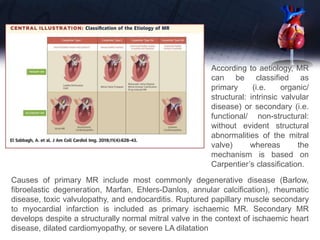
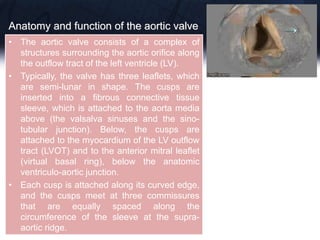
This document discusses the echocardiographic assessment of valvular regurgitation, focusing on mitral and tricuspid regurgitation. It describes the anatomy and causes of mitral and tricuspid regurgitation. Quantitative methods for assessing regurgitation severity include vena contracta width, proximal isovelocity surface area, and regurgitant volume. Consequences of significant regurgitation include left ventricular and atrial enlargement. Echocardiography is important for evaluating regurgitation severity and mechanisms to guide treatment decisions.
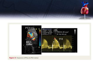
Mitral regurgitation (MR) is a common form of valvular heart disease that is associated with significant morbidity and
mortality. Treatment decisions are completely dependent on accurate diagnosis of both mechanism and severity of MR,
which can be challenging and is often done incorrectly. Transthoracic echocardiography is the most commonly used
imaging test for MR; transesophageal echocardiography is often needed to better define morphology and MR severity,
and is essential for guiding transcatheter therapies for MR. Multidetector computed tomography has become the standard to assess whether transcatheter valve replacement is an option because of its ability to assess valve sizing, access,
and potential left ventricular outflow tract obstruction. Finally, cine cardiac magnetic resonance has been recommended
by recent guidelines to quantify MR severity when the distinction between moderate and severe MR is indeterminate by
echocardiography. This paper focuses on the main questions to be answered by imaging techniques and illustrates some
common tips, tricks, and pitfalls in the assessment of MR.
The main hemodynamic interactions that may impact on the diagnosis of multiple and mixed Multiple and Mixed Valvular Heart Diseases:HOW TO USE IMAGINGThe interplay of multiple valve pathology.The clinical challenge of concomitant aortic and mitral valve stenosis
.
.
Knee anatomy and clinical tests 2024.pdfvimalpl1234
This includes all relevant anatomy and clinical tests compiled from standard textbooks, Campbell,netter etc..It is comprehensive and best suited for orthopaedicians and orthopaedic residents.
Muktapishti is a traditional Ayurvedic preparation made from Shoditha Mukta (Purified Pearl), is believed to help regulate thyroid function and reduce symptoms of hyperthyroidism due to its cooling and balancing properties. Clinical evidence on its efficacy remains limited, necessitating further research to validate its therapeutic benefits.
Basavarajeeyam is a Sreshta Sangraha grantha (Compiled book ), written by Neelkanta kotturu Basavaraja Virachita. It contains 25 Prakaranas, First 24 Chapters related to Rogas& 25th to Rasadravyas.
Ozempic: Preoperative Management of Patients on GLP-1 Receptor Agonists Saeid Safari
Preoperative Management of Patients on GLP-1 Receptor Agonists like Ozempic and Semiglutide
ASA GUIDELINE
NYSORA Guideline
2 Case Reports of Gastric Ultrasound
NVBDCP.pptx Nation vector borne disease control programSapna Thakur
NVBDCP was launched in 2003-2004 . Vector-Borne Disease: Disease that results from an infection transmitted to humans and other animals by blood-feeding arthropods, such as mosquitoes, ticks, and fleas. Examples of vector-borne diseases include Dengue fever, West Nile Virus, Lyme disease, and malaria.
Antimicrobial stewardship to prevent antimicrobial resistanceGovindRankawat1
India is among the nations with the highest burden of bacterial infections.
India is one of the largest consumers of antibiotics worldwide.
India carries one of the largest burdens of drug‑resistant pathogens worldwide.
Highest burden of multidrug‑resistant tuberculosis,
Alarmingly high resistance among Gram‑negative and Gram‑positive bacteria even to newer antimicrobials such as carbapenems.
NDM‑1 ( New Delhi Metallo Beta lactamase 1, an enzyme which inactivates majority of Beta lactam antibiotics including carbapenems) was reported in 2008
These simplified slides by Dr. Sidra Arshad present an overview of the non-respiratory functions of the respiratory tract.
Learning objectives:
1. Enlist the non-respiratory functions of the respiratory tract
2. Briefly explain how these functions are carried out
3. Discuss the significance of dead space
4. Differentiate between minute ventilation and alveolar ventilation
5. Describe the cough and sneeze reflexes
Study Resources:
1. Chapter 39, Guyton and Hall Textbook of Medical Physiology, 14th edition
2. Chapter 34, Ganong’s Review of Medical Physiology, 26th edition
3. Chapter 17, Human Physiology by Lauralee Sherwood, 9th edition
4. Non-respiratory functions of the lungs https://academic.oup.com/bjaed/article/13/3/98/278874
Here is the updated list of Top Best Ayurvedic medicine for Gas and Indigestion and those are Gas-O-Go Syp for Dyspepsia | Lavizyme Syrup for Acidity | Yumzyme Hepatoprotective Capsules etc
DISSERTATION on NEW DRUG DISCOVERY AND DEVELOPMENT STAGES OF DRUG DISCOVERYNEHA GUPTA
The process of drug discovery and development is a complex and multi-step endeavor aimed at bringing new pharmaceutical drugs to market. It begins with identifying and validating a biological target, such as a protein, gene, or RNA, that is associated with a disease. This step involves understanding the target's role in the disease and confirming that modulating it can have therapeutic effects. The next stage, hit identification, employs high-throughput screening (HTS) and other methods to find compounds that interact with the target. Computational techniques may also be used to identify potential hits from large compound libraries.
Following hit identification, the hits are optimized to improve their efficacy, selectivity, and pharmacokinetic properties, resulting in lead compounds. These leads undergo further refinement to enhance their potency, reduce toxicity, and improve drug-like characteristics, creating drug candidates suitable for preclinical testing. In the preclinical development phase, drug candidates are tested in vitro (in cell cultures) and in vivo (in animal models) to evaluate their safety, efficacy, pharmacokinetics, and pharmacodynamics. Toxicology studies are conducted to assess potential risks.
Before clinical trials can begin, an Investigational New Drug (IND) application must be submitted to regulatory authorities. This application includes data from preclinical studies and plans for clinical trials. Clinical development involves human trials in three phases: Phase I tests the drug's safety and dosage in a small group of healthy volunteers, Phase II assesses the drug's efficacy and side effects in a larger group of patients with the target disease, and Phase III confirms the drug's efficacy and monitors adverse reactions in a large population, often compared to existing treatments.
After successful clinical trials, a New Drug Application (NDA) is submitted to regulatory authorities for approval, including all data from preclinical and clinical studies, as well as proposed labeling and manufacturing information. Regulatory authorities then review the NDA to ensure the drug is safe, effective, and of high quality, potentially requiring additional studies. Finally, after a drug is approved and marketed, it undergoes post-marketing surveillance, which includes continuous monitoring for long-term safety and effectiveness, pharmacovigilance, and reporting of any adverse effects.
ABDOMINAL TRAUMA in pediatrics part one.drhasanrajab
Abdominal trauma in pediatrics refers to injuries or damage to the abdominal organs in children. It can occur due to various causes such as falls, motor vehicle accidents, sports-related injuries, and physical abuse. Children are more vulnerable to abdominal trauma due to their unique anatomical and physiological characteristics. Signs and symptoms include abdominal pain, tenderness, distension, vomiting, and signs of shock. Diagnosis involves physical examination, imaging studies, and laboratory tests. Management depends on the severity and may involve conservative treatment or surgical intervention. Prevention is crucial in reducing the incidence of abdominal trauma in children.
share - Lions, tigers, AI and health misinformation, oh my!.pptxTina Purnat
• Pitfalls and pivots needed to use AI effectively in public health
• Evidence-based strategies to address health misinformation effectively
• Building trust with communities online and offline
• Equipping health professionals to address questions, concerns and health misinformation
• Assessing risk and mitigating harm from adverse health narratives in communities, health workforce and health system
Adv. biopharm. APPLICATION OF PHARMACOKINETICS : TARGETED DRUG DELIVERY SYSTEMSAkankshaAshtankar
MIP 201T & MPH 202T
ADVANCED BIOPHARMACEUTICS & PHARMACOKINETICS : UNIT 5
APPLICATION OF PHARMACOKINETICS : TARGETED DRUG DELIVERY SYSTEMS By - AKANKSHA ASHTANKAR
263778731218 Abortion Clinic /Pills In Harare ,sisternakatoto
263778731218 Abortion Clinic /Pills In Harare ,ABORTION WOMEN’S CLINIC +27730423979 IN women clinic we believe that every woman should be able to make choices in her pregnancy. Our job is to provide compassionate care, safety,affordable and confidential services. That’s why we have won the trust from all generations of women all over the world. we use non surgical method(Abortion pills) to terminate…Dr.LISA +27730423979women Clinic is committed to providing the highest quality of obstetrical and gynecological care to women of all ages. Our dedicated staff aim to treat each patient and her health concerns with compassion and respect.Our dedicated group ABORTION WOMEN’S CLINIC +27730423979 IN women clinic we believe that every woman should be able to make choices in her pregnancy. Our job is to provide compassionate care, safety,affordable and confidential services. That’s why we have won the trust from all generations of women all over the world. we use non surgical method(Abortion pills) to terminate…Dr.LISA +27730423979women Clinic is committed to providing the highest quality of obstetrical and gynecological care to women of all ages. Our dedicated staff aim to treat each patient and her health concerns with compassion and respect.Our dedicated group of receptionists, nurses, and physicians have worked together as a teamof receptionists, nurses, and physicians have worked together as a team wwww.lisywomensclinic.co.za/
3. INTRODUCTION
• Doppler echocardiography is critical to initial and
longitudinal assessment of patients with valvular
regurgitation.
• Doppler echocardiography not only detects the
presence of regurgitation, but also permits to
understand mechanisms of regurgitation,
quantification of its severity, and repercussions.
4. • In practice, the evaluation of valvular regurgitation starts
with 2D TTE, which can orient readily to a severe
regurgitation in the presence of a major valvular defect or
to a minor leak when the valve anatomy and leaflet motion
are normal.
• The aetiology (cause of the valve disease) and mechanism
of the regurgitation (lesion/deformation resulting in valve
dysfunction) including the dysfunction type (cusp motion
abnormality) are described according to the Carpentier’s
classification of leaflet motion.
5. The Carpentier’s classification of leaflet motion :
Type I: normal leaflet motion
Type II: excessive motion
Type III: restrictive motion
6. • A careful assessment of the regurgitant jet by
colour Doppler, using multiple views, can rapidly
diagnose minimal regurgitation, which requires a
priori no further quantification.
• In the other cases, the use of a more quantitative
method is advised when feasible [vena contracta
(VC); proximal isovelocity surface area (PISA)]
7.
8.
9.
10. • Normal mitral valve function depends on perfect function of the
complex interaction between the mitral leaflets, the subvalvular
apparatus (chordae tendineae, and papillary muscles), the mitral
annulus, and the LV.
• An imperfection in any one of these components can cause the valve
to leak.
• The normal mitral valve has two leaflets; the posterior attached to the
two-thirds of the annular plane and the anterior, to the remaining one-
third.
• They are each divided into three scallops: A1, A2, A3 and P1, P2, P3.
A1 and P1 correspond to the external, anterolateral portion of their
respective leaflet, close to the anterolateral commissure, and the left
atrial appendage. In opposite, A3 and P3 are internal, close the
posteromedial commissure, and the tricuspid annulus
11. Causes of primary MR include most commonly degenerative disease (Barlow,
fibroelastic degeneration, Marfan, Ehlers-Danlos, annular calcification), rheumatic
disease, toxic valvulopathy, and endocarditis. Ruptured papillary muscle secondary
to myocardial infarction is included as primary ischaemic MR. Secondary MR
develops despite a structurally normal mitral valve in the context of ischaemic heart
disease, dilated cardiomyopathy, or severe LA dilatation
According to aetiology, MR
can be classified as
primary (i.e. organic/
structural: intrinsic valvular
disease) or secondary (i.e.
functional/ non-structural:
without evident structural
abnormalities of the mitral
valve) whereas the
mechanism is based on
Carpentier’s classification.
12.
13.
14. • The TTE parasternal short-axis view and the TOE transgastric view at
08 permit the assessment of the six scallops and, with colour Doppler
imaging, the localization of the origin of the regurgitant jet may identify
prolapsing segments. The TTE parasternal long-axis and the TOE
sagittal view at 1208 classically show A2 and P2.
• In apical four-chamber view, appreciation of A3, A2, and P1 (internal to
external) is possible as the TTE two-chamber view or the TOE view at
408 to 608 with TOE displays P3, A2, and P1 (left to right, bi-
commissural view). The precise location of the involved leaflets/
scallops, the description of the presence and extent of calcifications
and the extent of anatomic changes are fundamental parameters to be
provided. A flail leaflet, a ruptured papillary muscle or a large
coaptation defect is specific for severe MR.
15. Assessment of MR severity
Colour flow Doppler (Colour flow imaging)
• Colour flow imaging is the most common way to assess MR
severity.
• The general assumption is that as the severity of the MR
increases, the size and the extent of the jet into the LA also
increase. Theoretically, larger colour jets that extend deep
into the LA represent more MR than smaller thin jets that
appear just beyond the mitral leaflets. However, the
relationship between jet size and MR severity presents a
large range of variability because, in addition to
regurgitation severity, the colour flow display depends on
many technical and haemodynamic factors
16. VC width
• The VC is the area of the jet as it leaves the regurgitant
orifice; it reflects thus the regurgitant orifice area.
• The VC is typically imaged in a view perpendicular to the
commissural line.
Assessment of MR severity
17. Averaging measurements over at least two to three beats
and using two orthogonal planes, whenever possible is
recommended. A VC ,3 mm indicates mild MR, whereas a
width ≥7 mm defines severe MR.
Assessment of MR severity
18.
19.
20. The flow convergence method.
• The flow convergence method is the most recommended
quantitative approach whenever feasible. The apical four-
chamber view is classically recommended for optimal
visualization of the PISA. However, the parasternal long or
short-axis view is often useful for visualization of the PISA
in case of anterior mitral valve prolapse.
• The area of interest is optimized by lowering imaging depth
and reducing the Nyquist limit to 15– 40 cm/s. The radius of
the PISA is measured at mid-systole using the first aliasing.
R Vol and EROA are obtained using the standard formulas.
21.
22.
23. Consequences of MR
• The presence of severe primary MR has significant haemodynamic effects,
primarily on the LV and LA.
• In acute primary MR, the LV is classically not enlarged while the LV ejection
fraction usually increases I
• The regurgitant jet area (RJA) as well as the mitral E wave velocity increase
with the severity of MR. In severe MR, the continuous wave Doppler signal of
the regurgitant jet is truncated, triangular, and intense. Notching of the
continuous wave envelope (cut-off sign) can occur in severe MR. TVI, time-
velocity integral. Mild MR, normal pulmonary vein flow pattern; moderate MR,
blunt forward systolic pulmonary vein flow in a patient with moderate MR;
severe MR, reversed systolic pulmonary flow in a patient with severe MR. S,
systolic wave; (D) diastolic wave.
• Secondary MR has a different physiology, as it is the consequence of an initial
ventricular disease. The LV and LA dilatation are in excess to the degree of
MR. The LA pressure is often elevated despite lower regurgitant volume than
in primary MR
24.
25. INTRODUCTION
• Tricuspid regurgitation (TR) is a common condition and its prevalence is
only expected to increase with the aging population, rising rates of atrial
fibrillation, and increased use of intracardiac devices (e.g.,
pacemakers).
• The prevalence and clinical impact of TR was largely overlooked until
recently. A contemporary cohort showed 4.5% of the community and
12% of hospitalized patients had significant TR (equal to moderate or
greater severity), which increased to 23% among patients with heart
failure with reduced ejection fraction (HFrEF). Over 90% of TR is due to
functional etiologies, including left-sided valvular disease, pulmonary
hypertension, left ventricular (LV) dysfunction, or dilation of the tricuspid
apparatus resulting in an incompetent valve.5,6 TR is associated with
heart failure hospitalizations (RR 1.73) and all-cause mortality (RR 1.61
and 3.44 for moderate and severe TR, respectively).
• Echocardiography is an essential tool to assess the suitability of and
support percutaneous interventions.
26. Anatomy of the Tricuspid Valve
• The TV typically has three
leaflets: anterior (A), septal (S),
and posterior (P). The relative
circumferential length ratios of
the A:S:P leaflets in healthy
patients is 1:1:0.75; the
anterior leaflet is the longest
radially and most mobile while
the septal leaflet is the shortest
and least mobile.
• Additionally, anatomical
variants may generate complex
regurgitant jets making
intervention more challenging.
27. Transthoracic Echocardiographic
Assessment of the Tricuspid Valve
• Assessing right ventricle (RV) size and function, RV systolic
pressure, right atrial (RA) size and estimated pressure, and left-
sided heart disease are all important.
• Evaluation of the tricuspid annulus size, leaflet coaptation, and
leaflet tethering and motion can help determine the
etiology. The annulus is normally a saddle shape, but with
dilation becomes flatter and more circular.
• Alternatively, leaflet malcoaptation may occur secondary to
distortion of the papillary muscles and chordae in an elongated
and dilated RV.
28. The Carpentier’s classification remains the most
common used functional classification in TR:
• Type I: leaflet perforation (infective endocarditis)
or more frequently by annular dilatation;
• Type II:prolapse of one or more leaflets (tricuspid
valve prolapse); and
• Type III: restricted motion as the consequence of
rheumatic disease, significant calcifications, toxic
valvulopathy, functional TR
29. Tricuspid valve morphology
• The three main TTE views allowing the tricuspid valve
visualization are the parasternal (long-axis view of RV
inflow, short-axis view at the level of the aortic valve), the
apical 4-chamber and the subcostal views.
• It should be stressed that TR is most often seen in patients
with multiple valvular disease especially aortic or mitral
valve disease. In secondary TR, increased tethering
(apical displacement of the tricuspid leaflets) can be
evaluated by the measurement of the systolic tenting area
(area between the tricuspid annulus and the tricuspid
leaflets body) and the coaptation distance (distance
between the tricuspid annular plane and the point of
coaptation) in mid-systole from the apical 4-chamber view
30. Assessment of TR severity
Colour flow Doppler
• Grading the severity of TR is in principle similar to
MR. However, because standards for determining
the TR severity are less robust than for MR, the
algorithms for relating colour flow-derived
parameters to TR severity are less well developed.
31. VC width
• The VC width of the TR is typically imaged in the apical four
chamber view using the same settings as for MR.
• Averaging measurements over at least two to three beats is
recommended. A VC ≥7 mm is in favour of severe, whereas
a diameter ,6 mm may be either mild or moderate TR. The
geometry of the regurgitant orifice of secondary TR is
usually more complex than the one of MR and not
necessarily circular. A poor correlation has been suggested
between the 2D VC width and the 3D assessment of the
EROA. This could underline the poor accuracy of the 2D VC
width in eccentric jets. With 3D echo, an EROA .75 mm2
seems to indicate severe TR. However, these data need to
be confirmed in further studies.
32. Key point :
When feasible, the
measurement of the VC is
recommended to quantify TR.
A VC width ≥7 mm defines
severe TR. Lower values are
difficult to interpret. In case of
multiple jets, the respective
values of the VC width are not
additive.
The assessment of the VC by
3D echo is still reserved to
research purposes.
33.
34. Continuous wave Doppler
• Continuous wave Doppler of TR jet As for MR, the CW envelope of the TR jet
can be a guide to TR severity. A dense TR signal with a full envelope indicates
more severe TR than a faint signal.
• The CW Doppler envelope may be truncated (notched) with a triangular contour
and an early peak velocity (blunt), which is indicative of elevated right atrial
pressure or a prominent regurgitant pressure wave in the RA due to severe TR.
Marked respiratory variation (decreased TR velocity with inspiration) suggests
an elevated RA pressure (Kussmaul’s sign on physical examination).
• The TR jet can be used to determine RV or pulmonary artery systolic pressure.
This is done by calculating the RV to RA pressure gradient using the modified
Bernoullie quation and then adding an assumed RA pressure.
• To note, the velocity of the TR jet by itself does not provide useful information
about the severity of TR. For instance, massive TR is often associated with a
low jet velocity (,2 m/s) as there is near equalization of RV and RA pressures.66
In some case, a high-velocity jet that represents only mild TR maybe present
when severe pulmonary hypertension is present.
35.
36. Consequences of TR
• Signs of severe TR include RA and RV dilatation, a dilatedand pulsatile inferior vena cava
and hepatic vein, a dilated coronary sinus, and systolic bowingof the interatrialseptum
towardsthe LA. An end-systolic RV eccentricity index .2—obtained by dividing the longest
right lateral distance by the distance connecting the ventricular septum and the RV free
wall—is in favour of severe TR (sensitivity of 79%).55 Conversely, a poor correlation
between the RV area, the fractional areachangeand the degree of TR hasbeen reported.
Moreover, evidence of right heart dilatation is not specific for TR but can be noted in other
conditions (pulmonary valve regurgitation left-to-right atrial shunt, anomalous venous
return). However, its absence suggests milder degree of TR. RV dysfunction may be seen
inbothleft-sidedandright-sided valvularheartdisease.Inunoperated patients with mitral
valve disease, subnormal RV ejection fraction at rest is associated with decreased
exercise tolerance and mortality. In operated patients, an RV ejection fraction ,20%
predicted postoperative death.67 Progressive RV dysfunction may also result from
chronic TR. In a recent study, it has been shown that flail tricuspid valve is associated
with decreased survival and increasing risk of heart failure.68 This indicates that
sustained severe TR for a long time could lead to significant RV dysfunction. As for the
LV, the RV ejection fraction is a crude estimate of RV function. It is dependent on loading
conditions, ventricular interaction as well as myocardial structure. Other parameters
(tricuspid annular plane systolic excursion or TAPSE, systolic myocardial velocities) are
currently available for a better assessment of RV function but they still need to be
validated in both left- and right-sided valve diseases. Both the TAPSE and the systolic
38. Augustine, Daniel X., et al. "Echocardiographic assessment of pulmonary hypertension: a guideline protocol from the British Society of
Echocardiography." Echo Research & Practice 5.3 (2018): G11-G24.
39. Augustine, Daniel X., et al. "Echocardiographic assessment of pulmonary hypertension: a guideline protocol from the British Society of
Echocardiography." Echo Research & Practice 5.3 (2018): G11-G24.
40.
41. Anatomy and function of the pulmonary valve
The pulmonary valve is a three leaflet structure,
anatomically similar to the aortic valve.
The pulmonic valve structure is however thinner
because of the lower pressures in the right than
in the left heart system.
42. Aetiology and mechanisms
Pulmonary regurgitation may be caused by :
• Congenital anomalies (quadricuspid or bicuspid valves)
• Hypoplasia
• Post-repair of Tetralogy of Fallot
• Prolapse of the pulmonary valve.
• Other causes include infective endocarditis, carcinoid
syndrome, and rheumatic heart disease.
44. Assessment of PR severity
Colour flow imaging
• Detection of PR relies almost exclusively on colour flow
imaging. PR is diagnosed by documenting a diastolic jet in the
right ventricular (RV) outflow tract directed towards the RV.
• Functional PR jets are usually very small, central, and spindle
shaped.
45. • In severe PR, where equalization of the diastolic pulmonary artery and RV
pressures occurs earlier in diastole, the colour jet area can be brief and
inaccurate (dependency on the driving pressure).
• The maximum colour jet diameter (width) is measured in diastole
immediately below the pulmonic valve (at the junction of the RV outflow
tract and pulmonary annulus) in the parasternal short-axis view or from the
subcostal view.
46. VC width
Although the VC width is probably a more accurate method than the
jet width to evaluate PR severity by colour Doppler, it lacks
validation studies. The shape of the VC is complex in most cases.
47. Continuous wave doppler
• In mild PR, there is a slow deceleration of the jet velocity.
• A rapid deceleration rate with termination of flow in mid-to-
late diastole is not specific but compatible with severe
regurgitation.
52. INTRO
Aortic regurgitation (AR) is a common valvular disease.
Echocardiography plays a valuable role in the assessment
and management of patients with AR.
53. Anatomy and function of the aortic valve
• The aortic valve consists of a complex of
structures surrounding the aortic orifice along
the outflow tract of the left ventricle (LV).
• Typically, the valve has three leaflets, which
are semi-lunar in shape. The cusps are
inserted into a fibrous connective tissue
sleeve, which is attached to the aorta media
above (the valsalva sinuses and the sino-
tubular junction). Below, the cusps are
attached to the myocardium of the LV outflow
tract (LVOT) and to the anterior mitral leaflet
(virtual basal ring), below the anatomic
ventriculo-aortic junction.
• Each cusp is attached along its curved edge,
and the cusps meet at three commissures
that are equally spaced along the
circumference of the sleeve at the supra-
aortic ridge.
54. Aetiology and mechanisms of AR
• AR results from disease of either the aortic leaflets or the
aortic root that distorts the leaflets and prevents their correct
apposition.
• Common causes of leaflet abnormalities that result in AR
include senile leaflet calcifications, bicuspid aortic valve,
infective endocarditis, and rheumatic fever.
• Aortic causes of AR include annulo-aortic ectasia (idiopathic
root dilatation), Marfan’s syndrome, aortic dissection,
collagen vascular disease, and syphilis.
• The Carpentier’s classification is also commonly used to
describe the mechanism of A
55. Colour flow imaging
• Using colour flow Doppler, the regurgitant jet into the LV
during diastole can be visualized from multiple views.
• The colour jet area and length are weakly correlated to the
degree of AR, are affected by the aortic to LV diastolic
pressure gradient and LV compliance, and are often
overestimated in the apical views.
• Central jets are suggestive of rheumatic disease
• Eccentric jets are often associated with aortic valve
prolapse or perforation.
57. Eccentric jets are often associated with aortic valve
prolapse or perforation (PLAX)
58.
59. Consequences of AR on LV size and function
• The presence of severe AR has
significant haemodynamic effects,
primarily on the LV.
• AR imposes additional volume load
on the LV. In acute AR, the LV is
classically not enlarged, while in the
chronic situation, the LV
progressively dilates and
irreversible LV damage may
occur.
62. Imaging of the flow convergence zone is obtained from the apical three or
five-chamber or parasternal long-axis or upper right parasternal
views.
The radius of the PISA is measured at diastole using the first aliasing.
R Vol and EROA are obtained using the standard formulas.