Downloaded 106 times


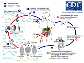
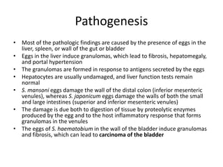


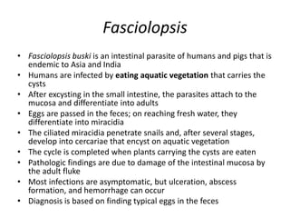


The document discusses several species of trematodes (parasitic flatworms) that infect humans, including their life cycles, transmission, pathogenesis, clinical findings, and diagnosis. It covers the important trematodes Schistosoma (blood flukes), Clonorchis sinensis (liver fluke), Paragonimus westermani (lung fluke), Fasciola hepatica, and Fasciolopsis buski. It provides details on the life cycles, symptoms, and laboratory diagnosis of Schistosoma and Fasciola infections.


