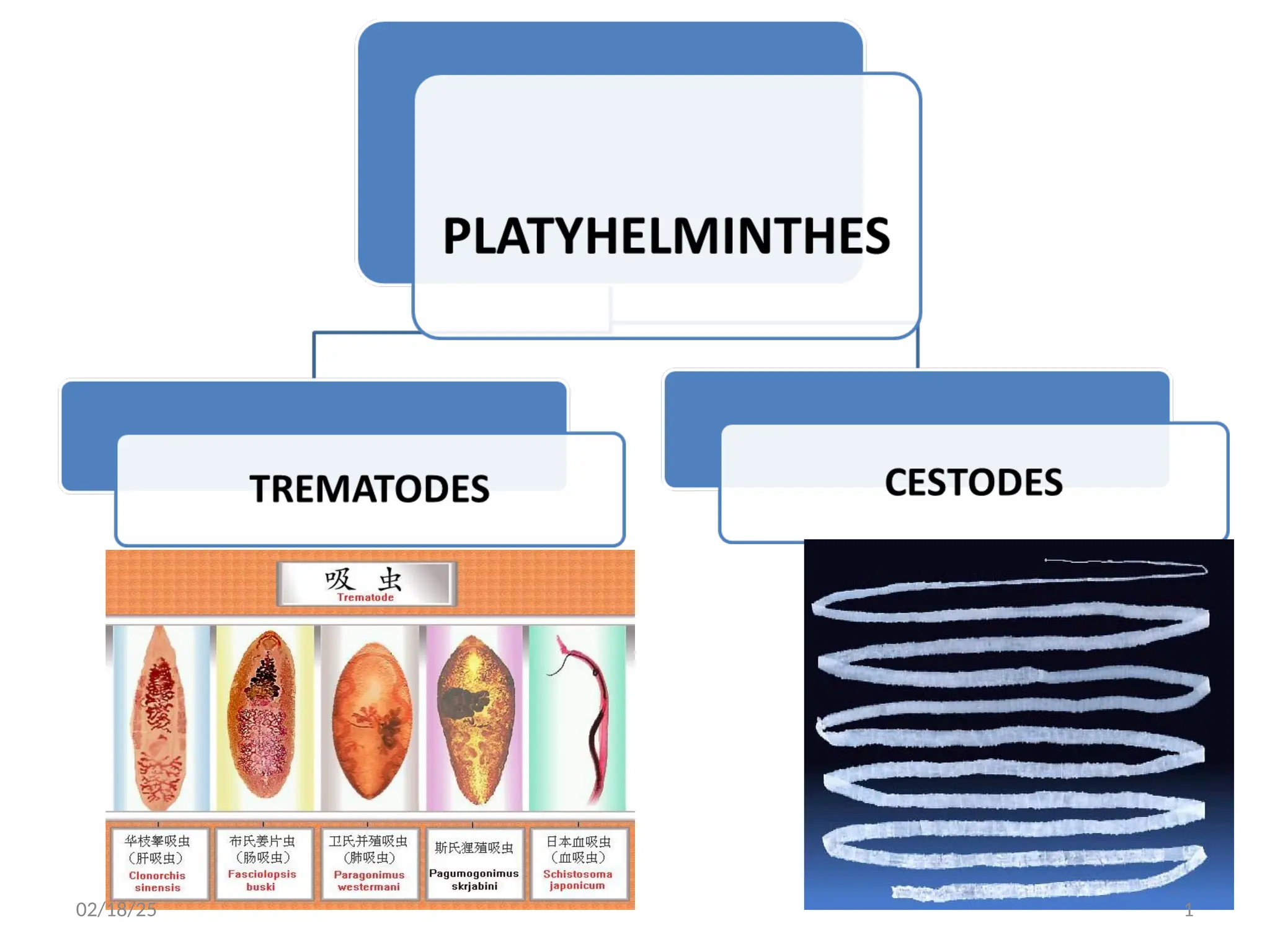
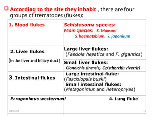
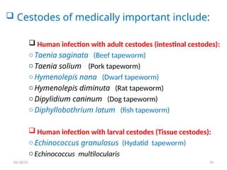
According tothe site they inhabit , there are four
groups of trematodes (flukes):
3
1. Blood flukes Schistosoma species:
Main species: S. Mansoni
S. haematobium, S. japonicum
2. Liver flukes
(in the liver and biliary duct)
Large liver flukes:
(Fasciola hepatica and F. gigantica)
Small liver flukes:
Clonorchis sinensis, Opisthorchis viverrini
3. Intestinal flukes
Large intestinal fluke:
(Fasciolopsis buski)
Small intestinal flukes:
(Metagonimus and Heterophyes)
Paragonimus westermani 4. Lung fluke
02/18/25
4.
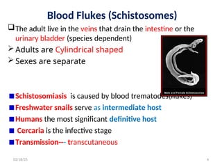
Blood Flukes (Schistosomes)
Theadult live in the veins that drain the intestine or the
urinary bladder (species dependent)
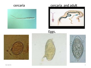
Adults are Cylindrical shaped
Sexes are separate
Schistosomiasis is caused by blood trematodes(flukes)
Freshwater snails serve as intermediate host
Humans the most significant definitive host
Cercaria is the infective stage
Transmission--- transcutaneous
4
02/18/25
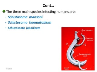
5.
Cont…
The three mainspecies infecting humans are:
- Schistosoma mansoni
- Schistosoma haematobium
- Schistosoma japonicum
5
02/18/25
6.
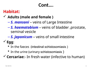
Cont….
Habitat:
Adults (maleand female )
– S. mansoni – veins of Large Intestine
– S. haematobium – veins of bladder ,prostate,
seminal vesicle
– S. japonicum – veins of small intestine
Egg:
In the faeces (intestinal schistosomiasis )
In the urine (urinary schistosomiasis )
Cercariae:- In fresh water (infective to human)
6
02/18/25
7.
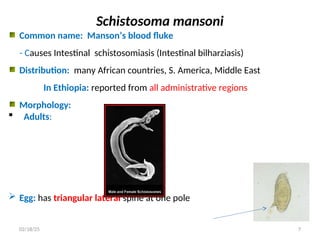
Schistosoma mansoni
Common name:Manson’s blood fluke
- Causes Intestinal schistosomiasis (Intestinal bilharziasis)
Distribution: many African countries, S. America, Middle East
In Ethiopia: reported from all administrative regions
Morphology:
Adults:
Egg: has triangular lateral spine at one pole
7
02/18/25
8.
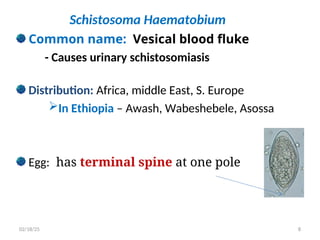
Schistosoma Haematobium
Common name:Vesical blood fluke
- Causes urinary schistosomiasis
Distribution: Africa, middle East, S. Europe
In Ethiopia – Awash, Wabeshebele, Asossa
Egg: has terminal spine at one pole
8
02/18/25
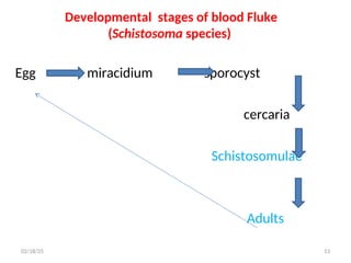
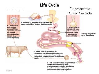
Life cycle ofSchistosoma species
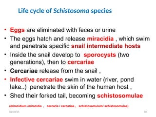
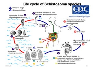
• Eggs are eliminated with feces or urine
• The eggs hatch and release miracidia , which swim
and penetrate specific snail intermediate hosts
• Inside the snail develop to sporocysts (two
generations), then to cercariae
• Cercariae release from the snail ,
• Infective cercariae swim in water (river, pond
lake..) penetrate the skin of the human host ,
• Shed their forked tail, becoming schistosomulae
(miracidium /miracidia , cercaria / cercariae , schistosomulum/ schistosomulae)
10
02/18/25
11.
Cont…
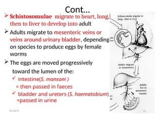
Schistosomulae migrateto heart, lung,
then to liver to develop into adult
Adults migrate to mesenteric veins or
veins around urinary bladder, depending
on species to produce eggs by female
worms
The eggs are moved progressively
toward the lumen of the:
intestine(S. mansoni )
= then passed in faeces
bladder and ureters (S. haematobium)
=passed in urine
11
02/18/25
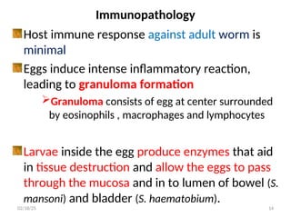
Immunopathology
Host immune responseagainst adult worm is
minimal
Eggs induce intense inflammatory reaction,
leading to granuloma formation
Granuloma consists of egg at center surrounded
by eosinophils , macrophages and lymphocytes
Larvae inside the egg produce enzymes that aid
in tissue destruction and allow the eggs to pass
through the mucosa and in to lumen of bowel (S.
mansoni) and bladder (S. haematobium).
14
02/18/25
15.
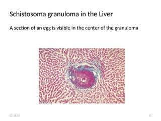
Schistosoma granuloma inthe Liver
A section of an egg is visible in the center of the granuloma
15
02/18/25
16.
Clinical Features
Syndromes include:
1.Cercarial dermatitis :- Penetration of skin by
cercariae causes transient dermatitis (swimmers' itch)
2. Acute schistosomiasis (Katayama's fever) may
occur weeks after the initial infection, especially by
S. mansoni and S. japonicum.
Associated with heavy primary infection and the
initiation of egg production
Manifestations include fever, abdominal pain,
diarrhea, hepatosplenomegaly, lymphadenopathy
and eosinophilia.
16
02/18/25
17.
3. Chronic schistosomiasis
May have mild, chronic bloody stools due to passage of
egg through intestinal wall
Host reaction to eggs lodged in the intestinal/bladder
mucosa leads to the formation of
- Granuloma
- Ulceration and
- Thickening of the wall
..
17
02/18/25
18.
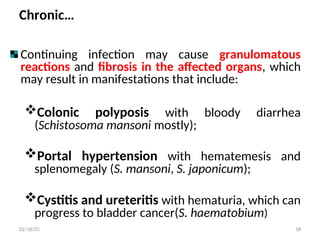
Chronic…
Continuing infection maycause granulomatous
reactions and fibrosis in the affected organs, which
may result in manifestations that include:
Colonic polyposis with bloody diarrhea
(Schistosoma mansoni mostly);
Portal hypertension with hematemesis and
splenomegaly (S. mansoni, S. japonicum);
Cystitis and ureteritis with hematuria, which can
progress to bladder cancer(S. haematobium)
18
02/18/25
19.
Chronic…
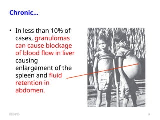
• In lessthan 10% of
cases, granulomas
can cause blockage
of blood flow in liver
causing
enlargement of the
spleen and fluid
retention in
abdomen.
19
02/18/25
20.
Laboratory Diagnosis
Microscopic identificationof eggs in stool or urine is the
most practical method for diagnosis.
Stool examination should be performed when infection
with S. mansoni or S. japonicum is suspected
urine examination should be performed if
S. haematobium is suspected
Treatment
Praziquantel: effective against all species
20
02/18/25
21.
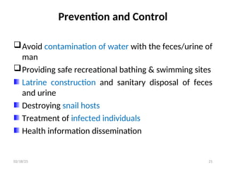
Prevention and Control
Avoidcontamination of water with the feces/urine of
man
Providing safe recreational bathing & swimming sites
Latrine construction and sanitary disposal of feces
and urine
Destroying snail hosts
Treatment of infected individuals
Health information dissemination
21
02/18/25
22.
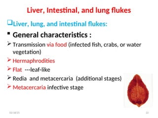
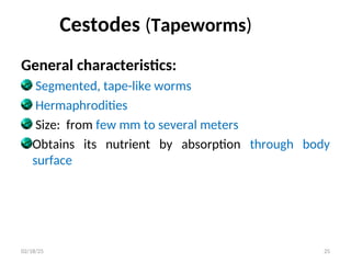
Liver, Intestinal, andlung flukes
Liver, lung, and intestinal flukes:
General characteristics :
Transmission via food (infected fish, crabs, or water
vegetation)
Hermaphrodities
Flat ---leaf-like
Redia and metacercaria (additional stages)
Metacercaria infective stage
22
02/18/25
23.
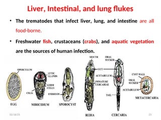
Liver, Intestinal, andlung flukes
• The trematodes that infect liver, lung, and intestine are all
food-borne.
• Freshwater fish, crustaceans (crabs), and aquatic vegetation
are the sources of human infection.
Developmental stages :
23
02/18/25
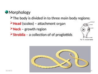
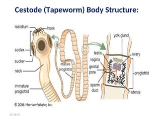
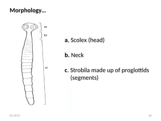
Morphology
The body isdivided in to three main body regions:
Head (scolex) – attachment organ
Neck – growth region
Strobila – a collection of of proglottids
26
02/18/25
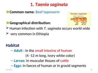
1. Taenia saginata
Commonname: Beef tapeworm
Geographical distribution:
Human infection with T. saginata occurs world wide
very common in Ethiopia
Habitat
– Adult : in the small intesine of human
(4 -12 m long, ivory white color)
– Larvae: in muscular tissues of cattle
– Eggs: in faeces of human or in gravid segments
30
02/18/25
31.
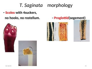
T. Saginata morphology
-Scolex with 4suckers,
no hooks, no rostellum. - Proglottid(segement)
31
02/18/25
32.
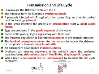
Transmission and LifeCycle
Humans are the DH while cattle are the IH
The infective form for humans is cysticercus bovis
A person is infected with T. saginata after consuming raw or undercooked
beef containing cysticerci
In the small intestine the process of strobilization start & adult worm
forms.
Eggs are produced in the gravid segment of the worm
Cattle while grazing, ingest eggs along with their food
The ingested eggs hatch to liberate oncospheres in the animals intestine
The hooked oncospheres penetrate the mucosa to invade bloodstream
or lymphatic's to reach muscles of cattle
An oncosphere develop into cysticercus bovis
Cysticerci can develop anywhere in the animal’s body; the preferred
locations being striated muscles of hind limbs, diaphragm & tongue
When beef is consumed raw or undercooked by humans the life cycle
completes.
32
02/18/25
33.
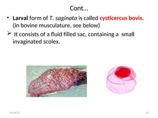
Cont…
• Larval formof T. saginata is called cysticercus bovis.
(in bovine musculature, see below)
It consists of a fluid filled sac, containing a small
invaginated scolex.
33
02/18/25
Clinical features andpathology
T. saginata infections are usually asymptomatic
When the infection is symptomatic; vague abdominal
discomfort, nausea, weakness and weight loss may be
present.
Very rarely migrating segments may cause appendicitis
or cholangitis
Entanglement of worms may lead to intestinal
obstruction
35
02/18/25
36.
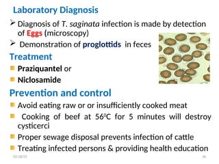
Laboratory Diagnosis
Diagnosisof T. saginata infection is made by detection
of Eggs (microscopy)
Demonstration of proglottids in feces
Treatment
Praziquantel or
Niclosamide
Prevention and control
Avoid eating raw or or insufficiently cooked meat
Cooking of beef at 560
C for 5 minutes will destroy
cysticerci
Proper sewage disposal prevents infection of cattle
Treating infected persons & providing health education
36
02/18/25
37.
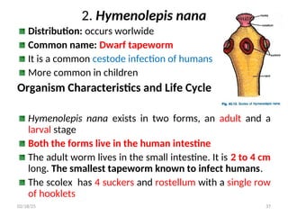
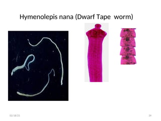
2. Hymenolepis nana
Distribution:occurs worlwide
Common name: Dwarf tapeworm
It is a common cestode infection of humans
More common in children
Organism Characteristics and Life Cycle
Hymenolepis nana exists in two forms, an adult and a
larval stage
Both the forms live in the human intestine
The adult worm lives in the small intestine. It is 2 to 4 cm
long. The smallest tapeworm known to infect humans.
The scolex has 4 suckers and rostellum with a single row
of hooklets
37
02/18/25
38.
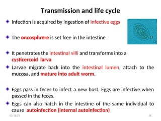
Transmission and lifecycle
Infection is acquired by ingestion of infective eggs
The oncosphrere is set free in the intestine
It penetrates the intestinal villi and transforms into a
cysticercoid larva
Larvae migrate back into the intestinal lumen, attach to the
mucosa, and mature into adult worm.
Eggs pass in feces to infect a new host. Eggs are infective when
passed in the feces.
Eggs can also hatch in the intestine of the same individual to
cause autoinfection (internal autoinfection)
38
02/18/25
Clinical features andpathology
Even with a large number of intestinal worms the
infection is usually asymptomatic
When symptomatic, patients have( especially children)
- anorexia, abdominal pain and
- diarrhoea
40
02/18/25
41.
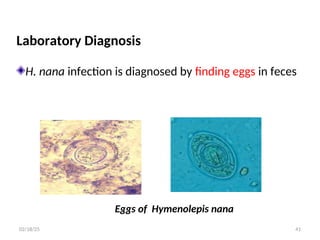
Laboratory Diagnosis
H. nanainfection is diagnosed by finding eggs in feces
Eggs of Hymenolepis nana
41
02/18/25
42.
Treatment
Praziquantel: the treatmentof choice (acts against
both adult and larval forms of the parasite)
Niclosamide
Prevention and control
• Good personal hygiene and improved sanitation can
prevent the disease
• Health education
• Treatment of infected persons
42
02/18/25
43.
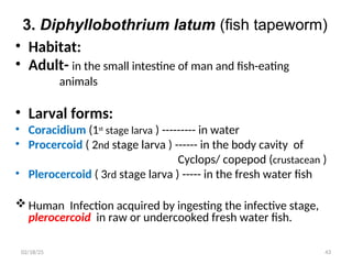
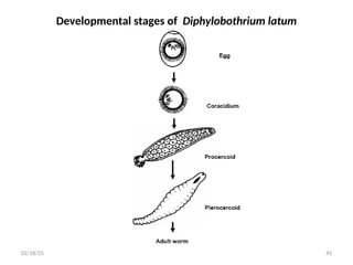
3. Diphyllobothrium latum(fish tapeworm)
• Habitat:
• Adult- in the small intestine of man and fish-eating
animals
• Larval forms:
• Coracidium (1st
stage larva ) --------- in water
• Procercoid ( 2nd stage larva ) ------ in the body cavity of
Cyclops/ copepod (crustacean )
• Plerocercoid ( 3rd stage larva ) ----- in the fresh water fish
Human Infection acquired by ingesting the infective stage,
plerocercoid in raw or undercooked fresh water fish.
43
02/18/25
Life cycle
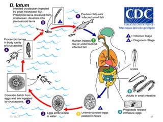
• Immatureeggs passed with in the feces of human.
• Eggs mature and yield oncospheres which develop into a coracidia.
• Coracidia ingested by a suitable freshwater crustacean (copepod 1st
IH),
develop into procercoid larvae .
• Ingestion of the copepod by 2nd
IH (small freshwater fish), the procercoid
larvae are released from the crustacean and migrate into the fish flesh,
develop into a plerocercoid larvae.
• After ingestion of the infected fish by the human host, the plerocercoid
develop into mature adult tapeworms which will reside in the small
intestine of human.
• The adults of D. latum attach to the intestinal mucosa by means of the
two bilateral groves (bothria) of their scolex .
46
02/18/25
47.
Clinical features andpathology
Clinical symptoms may be mild, depending on the number
of worms
Major symptoms:
Abdominal pain
Diarrhoea, constipation
Weight loss, intestinal obstruction
Eosinophilia
As many as 40% of D. latum carriers may have low serum
levels of vitamin B12, presumably because of the
competition between the host and the worm for dietary
vitamin B12.
47
02/18/25
48.
Laboratory diagnosis
• Diagnosisof D. latum infection is made by the
recovery of eggs in human feces. ( microscopy)
Treatment
• praziquantel
Prevention and control
• Avoid eating raw, poorly cooked,or pickled fish
• Proper disposal of faeces
• Treatment of individuals and health education
// 48
02/18/25
49.
Human infection withlarval cestodes
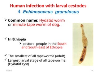
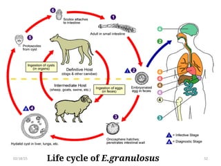
4. Echinococcus granulosus
Common name: Hydatid worm
or minute tape worm of dog.
In Ethiopia
pastoral people in the South
and South-East of Ethiopia
The smallest of all tapeworms (adult)
Largest larval stage of all tapeworms
(Hydatid cyst)
49
02/18/25
50.
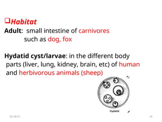
Habitat
Adult: small intestineof carnivores
such as dog, fox
Hydatid cyst/larvae: in the different body
parts (liver, lung, kidney, brain, etc) of human
and herbivorous animals (sheep)
50
02/18/25
51.
Cont…
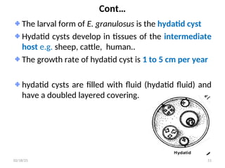
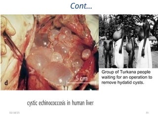
The larval formof E. granulosus is the hydatid cyst
Hydatid cysts develop in tissues of the intermediate
host e.g. sheep, cattle, human..
The growth rate of hydatid cyst is 1 to 5 cm per year
hydatid cysts are filled with fluid (hydatid fluid) and
have a doubled layered covering.
51
02/18/25
Clinical features andPathology
The symptoms depend upon the location and
size of the hydatid cyst.
• Causes obstruction and pressure on vital
organs
The cyst grows slowly but continuously
The majority of hydatid cyst occur in the liver
Liver cysts cause obstructive jaundice
53
02/18/25
54.
Clinical …
• Cystsin the lungs may produce cough, dyspnea
and chest pain. Cyst rupture can lead to pulmonary
abscesses
• Large abdominal cysts ----- increasing discomfort
• Kidney cysts cause renal dysfunction
• Brain cysts produce intracranial pressure and
epilepsy
• In bones, the growing hydatid cyst leads to bone
erosion and pathological fractures.
//
54
02/18/25
Laboratory diagnosis
Serological diagnosis– ELISA for Antibody detection
Examination of cystic fluid (for brood capsules and protoscolices) following
surgical removal of a cyst or fine needle aspiration
In symptomatic hydatid cyst: X-ray, CT scan, ultrasound studies are useful
56
02/18/25
57.
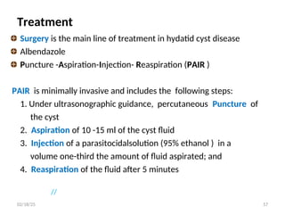
Treatment
Surgery is themain line of treatment in hydatid cyst disease
Albendazole
Puncture -Aspiration-Injection- Reaspiration (PAIR )
PAIR is minimally invasive and includes the following steps:
1. Under ultrasonographic guidance, percutaneous Puncture of
the cyst
2. Aspiration of 10 -15 ml of the cyst fluid
3. Injection of a parasitocidalsolution (95% ethanol ) in a
volume one-third the amount of fluid aspirated; and
4. Reaspiration of the fluid after 5 minutes
//
57
02/18/25
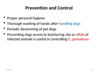
58.
Prevention and Control
Proper personal hygiene
Thorough washing of hands after handling dogs
Periodic deworming of pet dogs
Preventing dogs access to butchering site or offals of
infected animals is useful in controlling E. granulosus
58
02/18/25
Editor's Notes
#11 Adults can live 20 to 30 years b/s as the worm develop in the portal circulation ,they elaborate remarkable defense against host resistance.they coat themselves with substance the host recognize as self ( they absorb the host serum or RBC)
#17 Eggs swept by the blood flow become trapped in the liver and induce granuloma formation and fibrosis around the portal vessel and branches in the liver (periportal fibrosis)
Large scale peri porat fibrsis will result in reduced blood flow and increased portal blood pressure (portal hypertension)
Ectopic lesion: The damage to the central nervous system ( brain, spinal ) may cause paralysis (monoplegia, hemiplegia ), pulmonary hypertension , left ventricle hypertrophy
Colaterla veins formed near the lower part of the oesphagus may brust due to mechanical stress and leading to massive bleeding and vomiting of blood
#55 Cyst of E. granulosus histological section through through cyst wall


![CASE_PRESENTATION_ON_subdural_hematoma(SDH)[1 FINAL PPT]-1.pptx](https://cdn.slidesharecdn.com/ss_thumbnails/casepresentationonsubduralhematomasdh1finalppt-1-260129172522-d405d375-thumbnail.jpg?width=640&height=640&fit=bounds)
