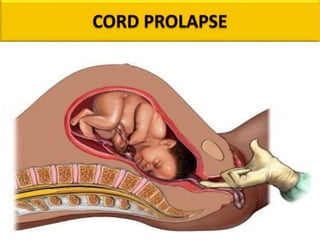
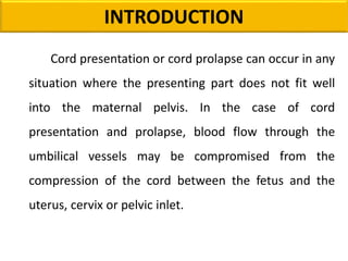
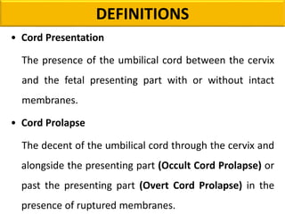
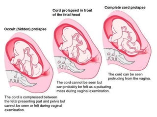
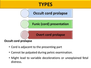
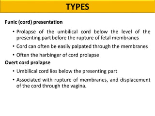
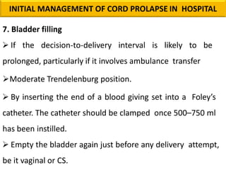
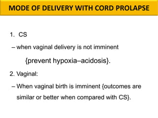
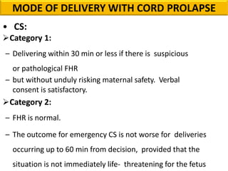
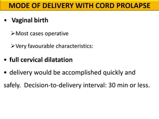
MRS. SONY SARA P.J
ASSO. PROFESSOR
MSC (N),OBSTETRICS AND GYNECOLOGICAL NURSING
GANGA COLLEGE OF NURSING
COIMBATORE
CONTENT OVERVIEW
Introduction
Definition
Types
Incidence and Epidemiology
Risk Factors
Consequences
Diagnosis
Prevention
Management
Management in Community Setting
Prognosis
Conclusion
INTRODUCTION
Cord presentation or cord prolapse can occur in any
situation where the presenting part does not fit well
into the maternal pelvis. In the case of cord
presentation and prolapse, blood flow through the
umbilical vessels may be compromised from the
compression of the cord between the fetus and the
uterus, cervix or pelvic inlet.
Where cord prolapse has occurred the cord is
vulnerable to compression, umbilical vein occlusion, and
umbilical artery vasospasm, which can compromise fetal
oxygenation.
Cord prolapse is a life threatening obstetric
emergency that may result in fetal asphyxia or death.
Caesarean section is the safest birth option for the viable
fetus, especially in the first and early second stage of
labour.
INTRODUCTION
DEFINITIONS
• Cord Presentation
The presence of the umbilical cord between the cervix
and the fetal presenting part with or without intact
membranes.
• Cord Prolapse
The decent of the umbilical cord through the cervix and
alongside the presenting part (Occult Cord Prolapse) or
past the presenting part (Overt Cord Prolapse) in the
presence of ruptured membranes.
TYPES
Occult cord prolapse
• Cord is adjacent to the presenting part
• Cannot be palpated during pelvic examination.
• Might lead to variable decelerations or unexplained fetal
distress.
Occult cord prolapse
Funic (cord) presentation
Overt cord prolapse
Funic (cord) presentation
• Prolapse of the umbilical cord below the level of the
presenting partbefore the rupture of fetal membranes
• Cord can often be easily palpated through the membranes
• Often the harbinger of cord prolapse
Overt cord prolapse
• Umbilical cord lies below the presenting part
• Associated with rupture of membranes, and displacement
of the cord through the vagina.
TYPES
INCIDENCE
• Overall Incidence of Cord prolapse: 0.1% - 0.6%.
• Cephalic presentation: 0.5%
• Breech presentation: 1%.
 0.5% frank breech
 complete breech 5%
 footling breech 15%, and
 transverse lie 20%
RISK FACTORS (Maternal)
Multiparity Polyhydramnios Abnormal placentation
Low birth weight Pelvic deformities Second twin
Breech/Malpresentations Transverse, Oblique and
unstable lie
High head at onset of labour
Fetal Congenital
anomalies
Long umbilical cord
RISK FACTORS (Fetal)
OBSTETRIC
PROCEDURES
AMNIOTOMY
INTERNAL PODALIC
VERSION
INDUCTION
OFLABOUR
EXTERNAL CEPHALIC
VERSION
RISK FACTORS
CONSEQUENCES
Cord
compression
Umbilical artery
vasospasm
Birth asphyxia
Hypoxic
Ischemic
Encephalopathy
Perinatal death
S.NO EXAMINATION RATIONALIZATION
1.
Vaginal examination 1. Visualization of the cord at the introitus
2. Feeling the cord within the vagina
2.
Obstetric abdominal
examination:
Reduction in the fetal
heart rate detected by
Pinard examination.
1. Compression of the cord by the presenting
part and vasospasm of the umbilical vessels will
lead to fetal hypoxia.
2. Fetal heart rate can also be assesses by CTG
or doppler.
3. Cardiotocography
(CTG)
Not necessary for the diagnosis but will indicate
fetal hypoxia.
DIAGNOSTIC TEST
CLINICAL FINDINGS
Occult prolapse:
• It is rarely palpated during
pelvic examination.
• This condition can be inferred
only if fetal heart rate changes
• Bradycardia :
a heart rate of less than 120 beats/ mins.
Overt cord prolapse:
It can be diagnosed simply
by visualizing the cord
protruding from the introitus or
by palpating loops of cord in the
vaginal canal by fingers.
CLINICAL FINDINGS
Funic presentation
It is made by pelvic
examination if loops of cord are
palpated through the
membranes.
CLINICAL FINDINGS
PREVENTION
1. Admission if
a.Transverse, oblique or unstable lie after 37+6 weeks
b.Noncephalic presentations and preterm
prelabour rupture of the membranes
2. Vaginal examination & monitoring FHR for
abnormalities.
– In the presence of risk factors cord presentation
or prolapse should be excluded at every vaginal
examination.
3. Avoid ARM if
– Presenting part is mobile
– Cord is felt below the presenting part
IF ARM necessary:
Performed with arrangements for immediate CS.
Upward pressure on the presenting part should be kept
to a minimum once membranes have been ruptured.
PREVENTION
Where artificial rupture of membranes (ARM) is
indicated it should only be performed by senior medical or
midwifery staff in the following circumstances (consider the
need to exclude cord presentation on ultrasound before
ARM for:
– High, ill-fitting presenting part & Unstable lie
– Polyhydramnios
4. Caesarean section:
– When cord presentation in established labour
PREVENTION
PRINCIPLES OF MANAGEMENT
• Call for help
• Continuous fetal monitoring
• O2 by face mask
• Immediate delivery
– Essentially by CS – allow instrumental/vaginal delivery if
deemed quicker
– Inform anesthetist, pediatrician, and OR staff
– Patient consent
• Funic reduction (rarely used)
– Replacement of the umbilical cord into the uterus
• Funic decompression/elevation of the presenting part
– Two fingers/hand in the vagina + elevation of the presenting
part
– Steep Trendelenburg or knee-chest position
– Insertion of Foley’s catheter and filling the urinary bladder
(500–750 mL)
• Tocolysis
– Not a first line management – can be considered if prolonged
interval to delivery is expected
• Keep the cord moist
– With cord extruding in the vagina and delivery is not
imminent
PRINCIPLES OF MANAGEMENT
MANAGEMENT
Speed is of the essence and perinatal outcome is
largely dictated by the diagnosis-delivery interval.
The three components of management are:
1. Prevent or relieve cord compression and vasospasm
2. Fetal assessment
3. Prompt delivery of the infant
MANAGEMENT
Overt Cord Prolapse
• The diagnosis of overt cord prolapse demands
immediate action to preserve the life of the fetus.
• An immediate pelvic examination should be
performed to determine cervical effacement and
dilatation, station of the presenting part, and strength
and frequency of pulsations within the cord vessels
If the fetus is viable, the patient should be placed
in the knee– chest position, and the examiner should
apply continuous upward pressure against the
presenting part to lift and maintain the fetus away from
the prolapsed cord until preparations for cesarean
delivery are complete.
MANAGEMENT
MANAGEMENT
• Alternatively, 400–700 mL of saline can be instilled into
the bladder in order to elevate the presenting part.
• Oxygen should be given to the mother until the
anesthesiologist is prepared to administer a rapid-acting
inhalation anesthetic for delivery.
• If heart rate changes, should not delay preparation for
cesarean delivery and a pediatric team should be on standby
in the event immediate resuscitation of the newborn is
necessary.
MANAGEMENT
1. Prevent/relieve cord compression and vasospasm
• Manual replacement & Bladder filling
• Adjust maternal position
Manual replacement
• Manual elevation
• Funic reduction
MANAGEMENT
BLADDER FILLING
• If the decision to delivery interval is likely to be prolonged,
elevation through bladder filling may be more practical.
• Introduced by Vagoin 1970
• It is essential to empty the bladder again just before
any delivery attempt, be it vaginal or CS.
• Physiologically inhibits uterine contraction.
MANAGEMENT
MATERNAL POSITION ADJUSTMENT
• Knee-chest position (Genu-pectoral)
• Gives maximum elevation of the presenting part.
• Provides good initial evaluation of the presenting part.
• A tiring posture to maintain.
• If any length of time is involved, move to the Sim’s
lateral position
• Sim’s lateral position
• More relaxed and dignified for the patient.
• Elevate buttocks with pillow
• Tredelenburg position
• A head-down tilt.
• Very tiring
Sim’s lateral position
Knee-chest position
(Genu-pectoral)
• Tredelenburg position
MATERNAL POSITION ADJUSTMENT
MANAGEMENT
2. FETAL ASSESSMENT
IS THE BABY VIABLE?
Interventions for fetal reasons are not necessary for:
• Already dead baby
• Too immature to survive (e.g. before age of fetal viability)
• Lethal fetal anomaly (e.g. anencephaly)
In these cases, allow labour to progress and deliver
vaginally unless there’s a contraindication to vaginal
delivery.
MANAGEMENT
IF BABY IS ALIVE
• Quickest way to tell is by palpating the presence or
absence of pulsations in the cord.
• Beware of mistaking folds of membranes or tips of
fetal fingers and toes for the cord. Or clinician’s finger
pulsation.
• Absent pulsations should be confirmed between
contractions in case cord compression is released and
pulsations return.
• Fetal heart auscultation best determines whether or not
the fetus is alive. Electronic fetal heart monitoring using
fetal scalp electrode may be useful. Real-time USS if
available.
MANAGEMENT
MANAGEMENT
3. Prompt delivery
CERVIX FULLY DILATED
• Vaginal birth can be attempted at full dilatation if it is
anticipated that delivery would be accomplished within
20 minutes from diagnosis.
• Depending on the circumstances, this may involve delivery
by forceps, vacuum or breech extraction.
• Breech extraction e.g after IPV for 2nd twin, or for singleton
breechbabies with presenting part distending the perineum
MANAGEMENT
CERVIX NOT FULLY DILATED
An immediate Caesarean Section (usually within 30
minutes) is the recommended mode of delivery in cases of cord
prolapse when vaginal delivery is not imminent, in order to
prevent hypoxia-acidosis.
• The 30-minute decision-to-delivery interval is the target for CS.
Some investigators have noted that the interval to
delivery had little effect on Apgar scores if they delivered
within 30 minutes.
MANAGEMENT
• The presenting part should be kept elevated during
induction of anaesthesia and placement of
sterile sheets.
• Remember to drain bladder before incision.
• Recheck fetal heart before incision.
• Regional anaesthesia may be considered in consultation
with an experienced anaesthetist
• A practitioner competent in the resuscitation of the
newborn, usually a neonatologist, should attend all
deliveries with cord prolapse.
• Neonates born after cord prolapse are at significant risk
of needing neonatal resuscitation, as evidenced by a
high rate of low APGAR scores (<7)
MANAGEMENT
MANAGEMENT
• OCCULT CORD PROLAPSE
The various modalities of management aim at raising the
pelvis, and therefore bring the cervix to a higher level
than the fundus of the uterus.
• Depends on the type of cord prolapse.
• OCCULT PROLAPSE
Immediate VE to rule out cord prolapse
Left lateral position & O2 to mother
Discontinue oxytocin infusion if in place
Allow labor to progress if FH returns to normal and no
further insult.
Continuous fetal heart rate monitoring
Amnioinfusion
CS if cord compression pattern continues
MANAGEMENT
• If occult cord prolapse is suspected, the patient should be
placed in the lateral Sims or trendelenburg position.
• Oxygen should be administered to the mother, and the
fetal heart rate should be continuously monitored
electronically.
• If the cord compression pattern persists or recurs to the
point of fetal jeopardy (moderate to severe variable
decelerations or bradycardia), a rapid cesarean section
should be accomplished.
MANAGEMENT
MANAGEMENT
OR
Lateral sims Trendelenburg position
MANAGEMENT
Funic Presentation
• The patient at term with funic presentation should be
delivered by cesarean section prior to membrane rupture.
• The most conservative approach is to hospitalize the
patient on bed rest in the Sims or Trendelenburg position in
an attempt to reposition the cord within the uterine cavity.
• Serial ultrasonographic examinations should be performed to
ascertain cord position, presentation, and gestational age.
INITIAL MANAGEMENT OF CORD PROLAPSE IN
HOSPITAL
1. Assistance should be immediately called
2. Preparations made for immediate delivery Manual
replacement of the prolapsed cord above the presenting
part to allow continuation of labour is not recommended.
3. Prevent vasospasm:
– Minimal handling of loops of cord lying outside the
vagina.
4. Prevent cord compression: Presenting part be elevated
either
– manually or by
– filling the urinary bladder. & knee–chest position or
– head-down tilt (preferably in left-lateral position).
5. Tocolysis
– while preparing for CS if there are
– persistent FHR abnormalities after attempts to prevent
compression mechanically and when the delivery is
likely to be delayed.
INITIAL MANAGEMENT OF CORD PROLAPSE IN HOSPITAL
– Swabs soaked in warm saline are wrapped around
the cord: unproven benefit.
6. Manual elevation:
– By inserting a gloved hand or two fingers in the vagina
and pushing the presenting part upwards. A variation is
to remove the hand from the vagina once the presenting
part is above the pelvic brim and apply continuous
suprapubic pressure upwards.
– Excessive displacement may encourage more cord
to prolapse.
INITIAL MANAGEMENT OF CORD PROLAPSE IN HOSPITAL
7. Bladder filling
 If the decision-to-delivery interval is likely to be
prolonged, particularly if it involves ambulance transfer
Moderate Trendelenburg position.
 By inserting the end of a blood giving set into a Foley’s
catheter. The catheter should be clamped once 500–750 ml
has been instilled.
 Empty the bladder again just before any delivery attempt,
be it vaginal or CS.
INITIAL MANAGEMENT OF CORD PROLAPSE IN HOSPITAL
MODE OF DELIVERY WITH CORD PROLAPSE
1. CS
– when vaginal delivery is not imminent
{prevent hypoxia–acidosis}.
2. Vaginal:
– When vaginal birth is imminent {outcomes are
similar or better when compared with CS}.
• CS:
Category 1:
– Delivering within 30 min or less if there is suspicious
or pathological FHR
– but without unduly risking maternal safety. Verbal
consent is satisfactory.
Category 2:
– FHR is normal.
– The outcome for emergency CS is not worse for deliveries
occurring up to 60 min from decision, provided that the
situation is not immediately life- threatening for the fetus
MODE OF DELIVERY WITH CORD PROLAPSE
• Vaginal birth
Most cases operative
Very favourable characteristics:
• full cervical dilatation
• delivery would be accomplished quickly and
safely. Decision-to-delivery interval: 30 min or less.
MODE OF DELIVERY WITH CORD PROLAPSE
Breech extraction:
– Performed after internal podalic version for the
second twin.
Forceps or ventouse:
– Depend on clinical circumstances and level of skill. No
difference in neonatal outcomes for fetal distress
MODE OF DELIVERY WITH CORD PROLAPSE
MANAGEMENT
• Neonatal care
– Neonatologist should attend
– Paired cord blood samples for pH and base
excess measurement
{strong predictive value of a normal paired cord blood
gas for the exclusion of intrapartum related hypoxic–
ischemic brain damage}
Management in community settings
There’s an increase in perinatal mortality in cases of
cord prolapse occurring outside the hospital, even
compared with an unmonitored fetus whose cord
prolapsed while in the hospital.
Women should be advised, over the telephone if
necessary, to assume the knee-chest or steep
Trendelenburg position while waiting for hospital transfer.
•During emergency ambulance transfer, the knee–chest is
potentially unsafe and the left-lateral position should be
used.
lateral position
Elevate presenting part: manual or bladder filling
 Prevent vasospasm: minimal handling of loops of cord
lying outside the vagina.
Management in community settings
• All women with cord prolapse should be advised to be
transferred to the nearest consultant unit for delivery,
unless an immediate vaginal examination by a
competent professional reveals that a spontaneous
vaginal delivery is imminent.
• Preparations for transfer should still be made.
• The presenting part should be elevated during
transfer by either manual or bladder filling methods.
Management in community settings
PROGNOSIS
Maternal
Maternal complications include those related to
anesthesia, blood loss, and infection following cesarean
section or operative vaginal delivery. Maternal recovery is
generally complete
Neonatal
– If the diagnosis is made early and the duration of
complete cord occlusion is less than 5 minutes, the
prognosis is good.
– If complete cord occlusion has occurred for longer
than 5 minutes or if intermittent partial cord
occlusion has occurred over a prolonged period of
time, fetal damage or death may occur
PROGNOSIS
DOCUMENTATION ITEMS
• Time of occurrence of cord prolapse
• Fetal status at cord prolapse and till delivery
• Condition of the cord (color, pulsations)
• Time of notification/arrival of obstetrician and other
health care personnel
• Maneuvers carried out to relieve cord compression
• Fetal response to interventions/maneuvers
• Time of entering the theatre and start of CS –
if carried out
• Type of anesthesia
• Diagnosis-to-delivery interval
• The condition of the baby at delivery including cord pH
• Neonatal outcome
DOCUMENTATION ITEMS
SUMMARY OF PRACTICE RECOMMENDATIONS
• Risk of cord presentation/prolapse is increased after
artificial rupture of the membranes (ARM) or sudden
spontaneous rupture of the forewaters with
malpresentation or high presenting part.
• Perform a vaginal examination to exclude or confirm the
presence of cord presentation/prolapse if sudden
appearance of persistent, deep, fetal variable
decelerations or prolonged fetal bradycardia.
• Once cord prolapse is diagnosed, treat as an obstetric
emergency.
• If the woman’s cervix is fully dilated, expedite vaginal birth.
• If the woman’s cervix is not fully dilated, the priority is to
relieve pressure on the cord by elevating the presenting part
while preparations are made for an emergency caesarean
section.
• If the cord is not pulsating, confirm fetal death with
ultrasound and plan for vaginal birth.
SUMMARY OF PRACTICE RECOMMENDATIONS
CONCLUSION
Umbilical cord prolapse is a well known obstetric
emergency that requires prompt delivery to avoid
potentially devastating fetal outcomes. There is no
evidence that umbilical cord prolapse can be prevented,
but rapid diagnosis and delivery has been shown to be
advantageous. If managed improperly, it can lead to
significant fetal morbidity and mortality. Prompt,
appropriate management of this condition, however, has
been shown to have favourable overall outcomes.
REFERENCES
1. Royal College of Obstetricians and Gynaecologists. Green-top Guideline
No. 50,Umbilical Cord Prolapse. UK, 2014.
2. The Royal Australian and New Zealand College of Obstetricians and
Gynaecologists. PROMPT Practical Obstetric Multi Professional Training™
Course Manual Australian and New Zealand Edition. Melbourne, Victoria;
2012.
3. Bush M, Eddleman K, Belogolovkin V. Umbilical cord prolapse [Internet].
UptoDate. [cited 2019 Oct 10] Available from: www.uptodate.com; 2019.
4. South Australian Pregnancy Outcome Unit. Pregnancy Outcome in South
Australia 2016. Pregnancy Outcome Unit, Prevention and Population
Health Branch, Adelaide: Government of South Australia; 2018
REFERENCES
• D.C.Dutta,”Textbook of Obstetrics including Perinatology and
Contraception". Seventh Edition.
• J.B. Sharma, “Midwifery & Gynaecological Nursing” Avichal Publishing
company:1st edition
• Jacob, Annamma (2009). A Comprehensive Textbook of Midwifery.Second
Edition. New Delhi: Jaypee Brothers Medical Publishers.
CORD PROLAPSE
CORD PROLAPSE

CORD PROLAPSE

  • 1.
    MRS. SONY SARAP.J ASSO. PROFESSOR MSC (N),OBSTETRICS AND GYNECOLOGICAL NURSING GANGA COLLEGE OF NURSING COIMBATORE
  • 3.
    CONTENT OVERVIEW Introduction Definition Types Incidence andEpidemiology Risk Factors Consequences Diagnosis Prevention Management Management in Community Setting Prognosis Conclusion
  • 4.
    INTRODUCTION Cord presentation orcord prolapse can occur in any situation where the presenting part does not fit well into the maternal pelvis. In the case of cord presentation and prolapse, blood flow through the umbilical vessels may be compromised from the compression of the cord between the fetus and the uterus, cervix or pelvic inlet.
  • 5.
    Where cord prolapsehas occurred the cord is vulnerable to compression, umbilical vein occlusion, and umbilical artery vasospasm, which can compromise fetal oxygenation. Cord prolapse is a life threatening obstetric emergency that may result in fetal asphyxia or death. Caesarean section is the safest birth option for the viable fetus, especially in the first and early second stage of labour. INTRODUCTION
  • 6.
    DEFINITIONS • Cord Presentation Thepresence of the umbilical cord between the cervix and the fetal presenting part with or without intact membranes. • Cord Prolapse The decent of the umbilical cord through the cervix and alongside the presenting part (Occult Cord Prolapse) or past the presenting part (Overt Cord Prolapse) in the presence of ruptured membranes.
  • 8.
    TYPES Occult cord prolapse •Cord is adjacent to the presenting part • Cannot be palpated during pelvic examination. • Might lead to variable decelerations or unexplained fetal distress. Occult cord prolapse Funic (cord) presentation Overt cord prolapse
  • 9.
    Funic (cord) presentation •Prolapse of the umbilical cord below the level of the presenting partbefore the rupture of fetal membranes • Cord can often be easily palpated through the membranes • Often the harbinger of cord prolapse Overt cord prolapse • Umbilical cord lies below the presenting part • Associated with rupture of membranes, and displacement of the cord through the vagina. TYPES
  • 10.
    INCIDENCE • Overall Incidenceof Cord prolapse: 0.1% - 0.6%. • Cephalic presentation: 0.5% • Breech presentation: 1%.  0.5% frank breech  complete breech 5%  footling breech 15%, and  transverse lie 20%
  • 11.
    RISK FACTORS (Maternal) MultiparityPolyhydramnios Abnormal placentation Low birth weight Pelvic deformities Second twin
  • 12.
    Breech/Malpresentations Transverse, Obliqueand unstable lie High head at onset of labour Fetal Congenital anomalies Long umbilical cord RISK FACTORS (Fetal)
  • 13.
  • 14.
  • 15.
    S.NO EXAMINATION RATIONALIZATION 1. Vaginalexamination 1. Visualization of the cord at the introitus 2. Feeling the cord within the vagina 2. Obstetric abdominal examination: Reduction in the fetal heart rate detected by Pinard examination. 1. Compression of the cord by the presenting part and vasospasm of the umbilical vessels will lead to fetal hypoxia. 2. Fetal heart rate can also be assesses by CTG or doppler. 3. Cardiotocography (CTG) Not necessary for the diagnosis but will indicate fetal hypoxia. DIAGNOSTIC TEST
  • 16.
    CLINICAL FINDINGS Occult prolapse: •It is rarely palpated during pelvic examination. • This condition can be inferred only if fetal heart rate changes • Bradycardia : a heart rate of less than 120 beats/ mins.
  • 17.
    Overt cord prolapse: Itcan be diagnosed simply by visualizing the cord protruding from the introitus or by palpating loops of cord in the vaginal canal by fingers. CLINICAL FINDINGS
  • 18.
    Funic presentation It ismade by pelvic examination if loops of cord are palpated through the membranes. CLINICAL FINDINGS
  • 19.
    PREVENTION 1. Admission if a.Transverse,oblique or unstable lie after 37+6 weeks b.Noncephalic presentations and preterm prelabour rupture of the membranes 2. Vaginal examination & monitoring FHR for abnormalities. – In the presence of risk factors cord presentation or prolapse should be excluded at every vaginal examination.
  • 20.
    3. Avoid ARMif – Presenting part is mobile – Cord is felt below the presenting part IF ARM necessary: Performed with arrangements for immediate CS. Upward pressure on the presenting part should be kept to a minimum once membranes have been ruptured. PREVENTION
  • 21.
    Where artificial ruptureof membranes (ARM) is indicated it should only be performed by senior medical or midwifery staff in the following circumstances (consider the need to exclude cord presentation on ultrasound before ARM for: – High, ill-fitting presenting part & Unstable lie – Polyhydramnios 4. Caesarean section: – When cord presentation in established labour PREVENTION
  • 22.
    PRINCIPLES OF MANAGEMENT •Call for help • Continuous fetal monitoring • O2 by face mask • Immediate delivery – Essentially by CS – allow instrumental/vaginal delivery if deemed quicker – Inform anesthetist, pediatrician, and OR staff – Patient consent • Funic reduction (rarely used) – Replacement of the umbilical cord into the uterus
  • 23.
    • Funic decompression/elevationof the presenting part – Two fingers/hand in the vagina + elevation of the presenting part – Steep Trendelenburg or knee-chest position – Insertion of Foley’s catheter and filling the urinary bladder (500–750 mL) • Tocolysis – Not a first line management – can be considered if prolonged interval to delivery is expected • Keep the cord moist – With cord extruding in the vagina and delivery is not imminent PRINCIPLES OF MANAGEMENT
  • 24.
    MANAGEMENT Speed is ofthe essence and perinatal outcome is largely dictated by the diagnosis-delivery interval. The three components of management are: 1. Prevent or relieve cord compression and vasospasm 2. Fetal assessment 3. Prompt delivery of the infant
  • 25.
    MANAGEMENT Overt Cord Prolapse •The diagnosis of overt cord prolapse demands immediate action to preserve the life of the fetus. • An immediate pelvic examination should be performed to determine cervical effacement and dilatation, station of the presenting part, and strength and frequency of pulsations within the cord vessels
  • 26.
    If the fetusis viable, the patient should be placed in the knee– chest position, and the examiner should apply continuous upward pressure against the presenting part to lift and maintain the fetus away from the prolapsed cord until preparations for cesarean delivery are complete. MANAGEMENT
  • 27.
    MANAGEMENT • Alternatively, 400–700mL of saline can be instilled into the bladder in order to elevate the presenting part. • Oxygen should be given to the mother until the anesthesiologist is prepared to administer a rapid-acting inhalation anesthetic for delivery. • If heart rate changes, should not delay preparation for cesarean delivery and a pediatric team should be on standby in the event immediate resuscitation of the newborn is necessary.
  • 28.
    MANAGEMENT 1. Prevent/relieve cordcompression and vasospasm • Manual replacement & Bladder filling • Adjust maternal position Manual replacement • Manual elevation • Funic reduction
  • 29.
    MANAGEMENT BLADDER FILLING • Ifthe decision to delivery interval is likely to be prolonged, elevation through bladder filling may be more practical. • Introduced by Vagoin 1970 • It is essential to empty the bladder again just before any delivery attempt, be it vaginal or CS. • Physiologically inhibits uterine contraction.
  • 30.
    MANAGEMENT MATERNAL POSITION ADJUSTMENT •Knee-chest position (Genu-pectoral) • Gives maximum elevation of the presenting part. • Provides good initial evaluation of the presenting part. • A tiring posture to maintain. • If any length of time is involved, move to the Sim’s lateral position • Sim’s lateral position • More relaxed and dignified for the patient. • Elevate buttocks with pillow • Tredelenburg position • A head-down tilt. • Very tiring
  • 31.
    Sim’s lateral position Knee-chestposition (Genu-pectoral) • Tredelenburg position MATERNAL POSITION ADJUSTMENT
  • 32.
    MANAGEMENT 2. FETAL ASSESSMENT ISTHE BABY VIABLE? Interventions for fetal reasons are not necessary for: • Already dead baby • Too immature to survive (e.g. before age of fetal viability) • Lethal fetal anomaly (e.g. anencephaly) In these cases, allow labour to progress and deliver vaginally unless there’s a contraindication to vaginal delivery.
  • 33.
    MANAGEMENT IF BABY ISALIVE • Quickest way to tell is by palpating the presence or absence of pulsations in the cord. • Beware of mistaking folds of membranes or tips of fetal fingers and toes for the cord. Or clinician’s finger pulsation.
  • 34.
    • Absent pulsationsshould be confirmed between contractions in case cord compression is released and pulsations return. • Fetal heart auscultation best determines whether or not the fetus is alive. Electronic fetal heart monitoring using fetal scalp electrode may be useful. Real-time USS if available. MANAGEMENT
  • 35.
    MANAGEMENT 3. Prompt delivery CERVIXFULLY DILATED • Vaginal birth can be attempted at full dilatation if it is anticipated that delivery would be accomplished within 20 minutes from diagnosis. • Depending on the circumstances, this may involve delivery by forceps, vacuum or breech extraction. • Breech extraction e.g after IPV for 2nd twin, or for singleton breechbabies with presenting part distending the perineum
  • 36.
    MANAGEMENT CERVIX NOT FULLYDILATED An immediate Caesarean Section (usually within 30 minutes) is the recommended mode of delivery in cases of cord prolapse when vaginal delivery is not imminent, in order to prevent hypoxia-acidosis. • The 30-minute decision-to-delivery interval is the target for CS. Some investigators have noted that the interval to delivery had little effect on Apgar scores if they delivered within 30 minutes.
  • 37.
    MANAGEMENT • The presentingpart should be kept elevated during induction of anaesthesia and placement of sterile sheets. • Remember to drain bladder before incision. • Recheck fetal heart before incision. • Regional anaesthesia may be considered in consultation with an experienced anaesthetist
  • 38.
    • A practitionercompetent in the resuscitation of the newborn, usually a neonatologist, should attend all deliveries with cord prolapse. • Neonates born after cord prolapse are at significant risk of needing neonatal resuscitation, as evidenced by a high rate of low APGAR scores (<7) MANAGEMENT
  • 39.
    MANAGEMENT • OCCULT CORDPROLAPSE The various modalities of management aim at raising the pelvis, and therefore bring the cervix to a higher level than the fundus of the uterus. • Depends on the type of cord prolapse. • OCCULT PROLAPSE Immediate VE to rule out cord prolapse Left lateral position & O2 to mother
  • 40.
    Discontinue oxytocin infusionif in place Allow labor to progress if FH returns to normal and no further insult. Continuous fetal heart rate monitoring Amnioinfusion CS if cord compression pattern continues MANAGEMENT
  • 41.
    • If occultcord prolapse is suspected, the patient should be placed in the lateral Sims or trendelenburg position. • Oxygen should be administered to the mother, and the fetal heart rate should be continuously monitored electronically. • If the cord compression pattern persists or recurs to the point of fetal jeopardy (moderate to severe variable decelerations or bradycardia), a rapid cesarean section should be accomplished. MANAGEMENT
  • 42.
  • 43.
    MANAGEMENT Funic Presentation • Thepatient at term with funic presentation should be delivered by cesarean section prior to membrane rupture. • The most conservative approach is to hospitalize the patient on bed rest in the Sims or Trendelenburg position in an attempt to reposition the cord within the uterine cavity. • Serial ultrasonographic examinations should be performed to ascertain cord position, presentation, and gestational age.
  • 44.
    INITIAL MANAGEMENT OFCORD PROLAPSE IN HOSPITAL 1. Assistance should be immediately called 2. Preparations made for immediate delivery Manual replacement of the prolapsed cord above the presenting part to allow continuation of labour is not recommended. 3. Prevent vasospasm: – Minimal handling of loops of cord lying outside the vagina.
  • 45.
    4. Prevent cordcompression: Presenting part be elevated either – manually or by – filling the urinary bladder. & knee–chest position or – head-down tilt (preferably in left-lateral position). 5. Tocolysis – while preparing for CS if there are – persistent FHR abnormalities after attempts to prevent compression mechanically and when the delivery is likely to be delayed. INITIAL MANAGEMENT OF CORD PROLAPSE IN HOSPITAL
  • 46.
    – Swabs soakedin warm saline are wrapped around the cord: unproven benefit. 6. Manual elevation: – By inserting a gloved hand or two fingers in the vagina and pushing the presenting part upwards. A variation is to remove the hand from the vagina once the presenting part is above the pelvic brim and apply continuous suprapubic pressure upwards. – Excessive displacement may encourage more cord to prolapse. INITIAL MANAGEMENT OF CORD PROLAPSE IN HOSPITAL
  • 47.
    7. Bladder filling If the decision-to-delivery interval is likely to be prolonged, particularly if it involves ambulance transfer Moderate Trendelenburg position.  By inserting the end of a blood giving set into a Foley’s catheter. The catheter should be clamped once 500–750 ml has been instilled.  Empty the bladder again just before any delivery attempt, be it vaginal or CS. INITIAL MANAGEMENT OF CORD PROLAPSE IN HOSPITAL
  • 48.
    MODE OF DELIVERYWITH CORD PROLAPSE 1. CS – when vaginal delivery is not imminent {prevent hypoxia–acidosis}. 2. Vaginal: – When vaginal birth is imminent {outcomes are similar or better when compared with CS}.
  • 49.
    • CS: Category 1: –Delivering within 30 min or less if there is suspicious or pathological FHR – but without unduly risking maternal safety. Verbal consent is satisfactory. Category 2: – FHR is normal. – The outcome for emergency CS is not worse for deliveries occurring up to 60 min from decision, provided that the situation is not immediately life- threatening for the fetus MODE OF DELIVERY WITH CORD PROLAPSE
  • 50.
    • Vaginal birth Mostcases operative Very favourable characteristics: • full cervical dilatation • delivery would be accomplished quickly and safely. Decision-to-delivery interval: 30 min or less. MODE OF DELIVERY WITH CORD PROLAPSE
  • 51.
    Breech extraction: – Performedafter internal podalic version for the second twin. Forceps or ventouse: – Depend on clinical circumstances and level of skill. No difference in neonatal outcomes for fetal distress MODE OF DELIVERY WITH CORD PROLAPSE
  • 52.
    MANAGEMENT • Neonatal care –Neonatologist should attend – Paired cord blood samples for pH and base excess measurement {strong predictive value of a normal paired cord blood gas for the exclusion of intrapartum related hypoxic– ischemic brain damage}
  • 53.
    Management in communitysettings There’s an increase in perinatal mortality in cases of cord prolapse occurring outside the hospital, even compared with an unmonitored fetus whose cord prolapsed while in the hospital. Women should be advised, over the telephone if necessary, to assume the knee-chest or steep Trendelenburg position while waiting for hospital transfer.
  • 54.
    •During emergency ambulancetransfer, the knee–chest is potentially unsafe and the left-lateral position should be used. lateral position Elevate presenting part: manual or bladder filling  Prevent vasospasm: minimal handling of loops of cord lying outside the vagina. Management in community settings
  • 55.
    • All womenwith cord prolapse should be advised to be transferred to the nearest consultant unit for delivery, unless an immediate vaginal examination by a competent professional reveals that a spontaneous vaginal delivery is imminent. • Preparations for transfer should still be made. • The presenting part should be elevated during transfer by either manual or bladder filling methods. Management in community settings
  • 56.
    PROGNOSIS Maternal Maternal complications includethose related to anesthesia, blood loss, and infection following cesarean section or operative vaginal delivery. Maternal recovery is generally complete
  • 57.
    Neonatal – If thediagnosis is made early and the duration of complete cord occlusion is less than 5 minutes, the prognosis is good. – If complete cord occlusion has occurred for longer than 5 minutes or if intermittent partial cord occlusion has occurred over a prolonged period of time, fetal damage or death may occur PROGNOSIS
  • 58.
    DOCUMENTATION ITEMS • Timeof occurrence of cord prolapse • Fetal status at cord prolapse and till delivery • Condition of the cord (color, pulsations) • Time of notification/arrival of obstetrician and other health care personnel • Maneuvers carried out to relieve cord compression • Fetal response to interventions/maneuvers
  • 59.
    • Time ofentering the theatre and start of CS – if carried out • Type of anesthesia • Diagnosis-to-delivery interval • The condition of the baby at delivery including cord pH • Neonatal outcome DOCUMENTATION ITEMS
  • 60.
    SUMMARY OF PRACTICERECOMMENDATIONS • Risk of cord presentation/prolapse is increased after artificial rupture of the membranes (ARM) or sudden spontaneous rupture of the forewaters with malpresentation or high presenting part. • Perform a vaginal examination to exclude or confirm the presence of cord presentation/prolapse if sudden appearance of persistent, deep, fetal variable decelerations or prolonged fetal bradycardia.
  • 61.
    • Once cordprolapse is diagnosed, treat as an obstetric emergency. • If the woman’s cervix is fully dilated, expedite vaginal birth. • If the woman’s cervix is not fully dilated, the priority is to relieve pressure on the cord by elevating the presenting part while preparations are made for an emergency caesarean section. • If the cord is not pulsating, confirm fetal death with ultrasound and plan for vaginal birth. SUMMARY OF PRACTICE RECOMMENDATIONS
  • 62.
    CONCLUSION Umbilical cord prolapseis a well known obstetric emergency that requires prompt delivery to avoid potentially devastating fetal outcomes. There is no evidence that umbilical cord prolapse can be prevented, but rapid diagnosis and delivery has been shown to be advantageous. If managed improperly, it can lead to significant fetal morbidity and mortality. Prompt, appropriate management of this condition, however, has been shown to have favourable overall outcomes.
  • 63.
    REFERENCES 1. Royal Collegeof Obstetricians and Gynaecologists. Green-top Guideline No. 50,Umbilical Cord Prolapse. UK, 2014. 2. The Royal Australian and New Zealand College of Obstetricians and Gynaecologists. PROMPT Practical Obstetric Multi Professional Training™ Course Manual Australian and New Zealand Edition. Melbourne, Victoria; 2012. 3. Bush M, Eddleman K, Belogolovkin V. Umbilical cord prolapse [Internet]. UptoDate. [cited 2019 Oct 10] Available from: www.uptodate.com; 2019. 4. South Australian Pregnancy Outcome Unit. Pregnancy Outcome in South Australia 2016. Pregnancy Outcome Unit, Prevention and Population Health Branch, Adelaide: Government of South Australia; 2018
  • 64.
    REFERENCES • D.C.Dutta,”Textbook ofObstetrics including Perinatology and Contraception". Seventh Edition. • J.B. Sharma, “Midwifery & Gynaecological Nursing” Avichal Publishing company:1st edition • Jacob, Annamma (2009). A Comprehensive Textbook of Midwifery.Second Edition. New Delhi: Jaypee Brothers Medical Publishers.