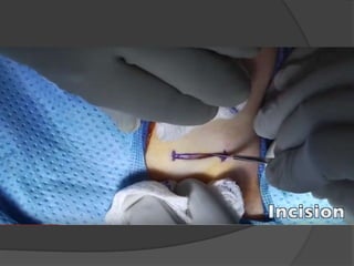
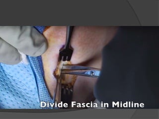
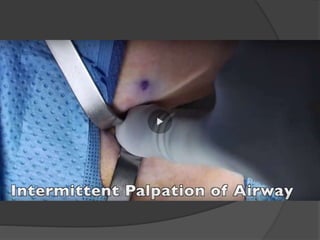
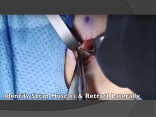
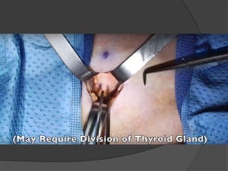
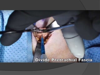
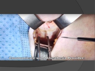
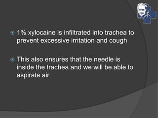
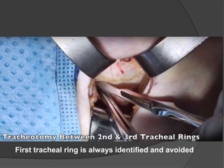
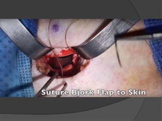
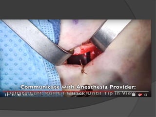
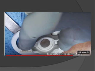
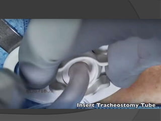
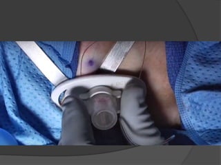
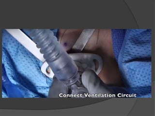
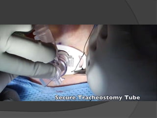
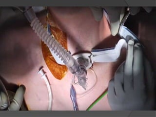
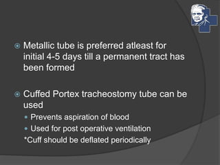
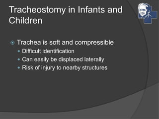
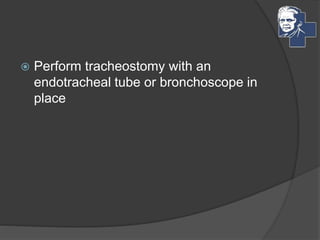
Dr Venkatesh Karthikeyan
CRRI
VMCH&RI
Mail : 4852012@gmail.com
 It is a procedure where
 an opening is made in the anterior wall of
the trachea
 And a channel is constructed between the
trachea and skin surface of neck by inserting
a tracheostomy tube
Indications
 Upper Airway obstructions
 Pulmonary toilet and airway protection
 Assisted Ventilation
Upper airway obstruction
 Causes may be intra – laryngeal or extra
– laryngeal
 Congenital
 Traumatic
 Infections
 Tumors
 Neurogenic
Congenital
 Pierre Robin Syndrome
 Subglottic stenosis
 Laryngeal web
 Laryngomalacia
Traumatic
 External trauma – maxillofacial injuries,
neck contusion, laryngeal fractures
 Foreign bodies
 Burns
 Instrumentation
 Prolonged intubation
Infections
 Acute epiglottitis
 ALTB
 Retropharygeal abscess
 Laryngeal diptheria
Tumors
 Laryngeal papillomatosis
 Hemangiomas
 Lymphangiomas
 Carcinoma
 Others
Neurogenic
 Bilateral vocal cord paralysis
Pulmonary toilet and airway
protection
 Head injury
 Drug intoxication
 Encephalitis
 Burns
 Shock lung
 Chest injury
 Cervical cord degeneration
Assisted Ventilation
 Aspiration
 Maxillofacial injuries
 Laryngeal cleft
 Neurogenic
 Tracheo – Esophageal fistula
 Flail chest
 Pneumothorax
 Pneumomediastinum
 Respiratory Distress syndrome
 Poliomyelitis
 Polyneuritis
Functions of Tracheostomy
 Relieves upper airway obstruction
 Decreases dead space
 Facilitates cleaning of Tracheobronchial
tree
 Protects against aspiration
 Reduces resistance of airflow
 Increases total compliance and alveolar
ventilation
 Helps in Intermittent positive pressure
ventilation
 Helps in induction of anaesthesia
Types of Tracheostomy
 According to Purpose
 Temporary
 Permanent
 According to timing of surgery
 Elective
 Emergency
 According to site of Stoma
 High (1st ring)
 Mid (2-4th rings)
 Low (below 4th ring)
Procedure
 Anesthesia
 General
 Local
Position
 Supine
 Pillow under the shoulders to allow
maximum extension of neck
 Head and neck must be held in midline
○ Symphysis menti and suprasternal notch must
be in one line
Incision
 5 cm long
 Two finger breath above the sternal notch
Horizontal incision
 In Elective procedures
 Cosmetically better scar
Vertical Incision
 In emergencies
 Easy access
 Minimal bleeding
Incision is deepened to expose
the strap muscles
 1% xylocaine is infiltrated into trachea to
prevent excessive irritation and cough
 This also ensures that the needle is
inside the trachea and we will be able to
aspirate air
First tracheal ring is always identified and avoided
An inferiorly based flap of tracheal ring
cartilage is made and sutured to the stomal
skin
 Metallic tube is preferred atleast for
initial 4-5 days till a permanent tract has
been formed
 Cuffed Portex tracheostomy tube can be
used
 Prevents aspiration of blood
 Used for post operative ventilation
*Cuff should be deflated periodically
Tracheostomy in Infants and
Children
 Trachea is soft and compressible
 Difficult identification
 Can easily be displaced laterally
 Risk of injury to nearby structures
 Perform tracheostomy with an
endotracheal tube or bronchoscope in
place
 Rose position Mediastinal structures
like apical pleura protrude into the neck
Injury  Pneumothorax
 Tracheotomy itself is suffice
 Suture placed on either side of the
incision on the trachea
 Tracheal lumen is narrow
 Deep incision
 Tracheo – esophageal fistula
Selection of Tracheostomy tube
 Large tube – Necrosis of tracheal rings
 Long tube – Impinges upon carina or
enter right bronchus
 Post of X ray
 Position of tube
 Rule out pneumothorax
Post op Management
 Cleaning and changing of inner tube
 Tracheobronchial toilet
 Humidification and prevention of crust
formation
 Nursing care to avoid infection
 Nasogastric feeding maybe needed
initially
 Physiotherapy
Decanulation
 By reduction of size of tube
 Closing the tube with a spigot
Complications
 Immediate
 Intermediate
 Late
Immediate Complications
 Primary hemorrhage
 Injury to blood vessels, jugular veins,
carotid artery
 Injury to RLN
 Pneumothorax / Pneumomediastinum
 Apnea due to sudden release of CO2
 Damage to cricoid and trachea
 Cardiac arrest
 Collapse of lung
Intermediate Complications
 False passage – Displacement of tube
 Crusting and Blocking of Inner tube
 Atelectasis and lung abscess
 Surgical emphysema
 Tracheo-oesophageal fistula
 Dysphagia
 Secondary infection
Late complications
 Hemorrhage
 Laryngo-tracheal stenosis
 Tracheo – cutaneous fistula
 Difficult decannulation
 Corrosion of tracheostomy tube
 Keloid formation over scar
Reference
 A Short Practice of Otorhinolaryngology
by Prof.K.K. Ramalingam
 Diseases of Ear, Nose and Throat by PL
Dhingra
 https://www.youtube.com/watch?v=77Wi
5Z3FOGk (Mayo Clinic)
Thank you

Tracheostomy

  • 2.
  • 3.
     It isa procedure where  an opening is made in the anterior wall of the trachea  And a channel is constructed between the trachea and skin surface of neck by inserting a tracheostomy tube
  • 4.
    Indications  Upper Airwayobstructions  Pulmonary toilet and airway protection  Assisted Ventilation
  • 5.
    Upper airway obstruction Causes may be intra – laryngeal or extra – laryngeal  Congenital  Traumatic  Infections  Tumors  Neurogenic
  • 6.
    Congenital  Pierre RobinSyndrome  Subglottic stenosis  Laryngeal web  Laryngomalacia
  • 7.
    Traumatic  External trauma– maxillofacial injuries, neck contusion, laryngeal fractures  Foreign bodies  Burns  Instrumentation  Prolonged intubation
  • 8.
    Infections  Acute epiglottitis ALTB  Retropharygeal abscess  Laryngeal diptheria
  • 9.
    Tumors  Laryngeal papillomatosis Hemangiomas  Lymphangiomas  Carcinoma  Others
  • 10.
  • 11.
    Pulmonary toilet andairway protection  Head injury  Drug intoxication  Encephalitis  Burns  Shock lung  Chest injury  Cervical cord degeneration
  • 12.
    Assisted Ventilation  Aspiration Maxillofacial injuries  Laryngeal cleft  Neurogenic  Tracheo – Esophageal fistula
  • 13.
     Flail chest Pneumothorax  Pneumomediastinum  Respiratory Distress syndrome  Poliomyelitis  Polyneuritis
  • 14.
    Functions of Tracheostomy Relieves upper airway obstruction  Decreases dead space  Facilitates cleaning of Tracheobronchial tree  Protects against aspiration  Reduces resistance of airflow  Increases total compliance and alveolar ventilation  Helps in Intermittent positive pressure ventilation  Helps in induction of anaesthesia
  • 15.
    Types of Tracheostomy According to Purpose  Temporary  Permanent  According to timing of surgery  Elective  Emergency  According to site of Stoma  High (1st ring)  Mid (2-4th rings)  Low (below 4th ring)
  • 16.
  • 17.
  • 18.
    Position  Supine  Pillowunder the shoulders to allow maximum extension of neck  Head and neck must be held in midline ○ Symphysis menti and suprasternal notch must be in one line
  • 19.
    Incision  5 cmlong  Two finger breath above the sternal notch
  • 20.
    Horizontal incision  InElective procedures  Cosmetically better scar
  • 21.
    Vertical Incision  Inemergencies  Easy access  Minimal bleeding
  • 23.
    Incision is deepenedto expose the strap muscles
  • 30.
     1% xylocaineis infiltrated into trachea to prevent excessive irritation and cough  This also ensures that the needle is inside the trachea and we will be able to aspirate air
  • 31.
    First tracheal ringis always identified and avoided
  • 32.
    An inferiorly basedflap of tracheal ring cartilage is made and sutured to the stomal skin
  • 41.
     Metallic tubeis preferred atleast for initial 4-5 days till a permanent tract has been formed  Cuffed Portex tracheostomy tube can be used  Prevents aspiration of blood  Used for post operative ventilation *Cuff should be deflated periodically
  • 42.
    Tracheostomy in Infantsand Children  Trachea is soft and compressible  Difficult identification  Can easily be displaced laterally  Risk of injury to nearby structures
  • 43.
     Perform tracheostomywith an endotracheal tube or bronchoscope in place
  • 44.
     Rose positionMediastinal structures like apical pleura protrude into the neck Injury  Pneumothorax
  • 45.
     Tracheotomy itselfis suffice  Suture placed on either side of the incision on the trachea  Tracheal lumen is narrow  Deep incision  Tracheo – esophageal fistula
  • 46.
    Selection of Tracheostomytube  Large tube – Necrosis of tracheal rings  Long tube – Impinges upon carina or enter right bronchus  Post of X ray  Position of tube  Rule out pneumothorax
  • 47.
    Post op Management Cleaning and changing of inner tube  Tracheobronchial toilet  Humidification and prevention of crust formation  Nursing care to avoid infection  Nasogastric feeding maybe needed initially  Physiotherapy
  • 48.
    Decanulation  By reductionof size of tube  Closing the tube with a spigot
  • 49.
  • 50.
    Immediate Complications  Primaryhemorrhage  Injury to blood vessels, jugular veins, carotid artery  Injury to RLN  Pneumothorax / Pneumomediastinum  Apnea due to sudden release of CO2  Damage to cricoid and trachea  Cardiac arrest  Collapse of lung
  • 51.
    Intermediate Complications  Falsepassage – Displacement of tube  Crusting and Blocking of Inner tube  Atelectasis and lung abscess  Surgical emphysema  Tracheo-oesophageal fistula  Dysphagia  Secondary infection
  • 52.
    Late complications  Hemorrhage Laryngo-tracheal stenosis  Tracheo – cutaneous fistula  Difficult decannulation  Corrosion of tracheostomy tube  Keloid formation over scar
  • 53.
    Reference  A ShortPractice of Otorhinolaryngology by Prof.K.K. Ramalingam  Diseases of Ear, Nose and Throat by PL Dhingra  https://www.youtube.com/watch?v=77Wi 5Z3FOGk (Mayo Clinic)
  • 54.

Editor's Notes