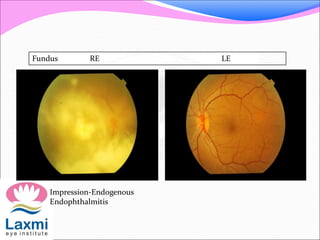
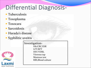
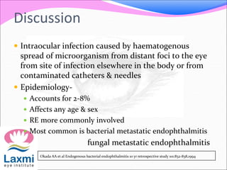
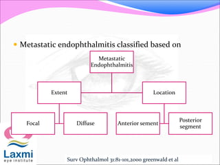
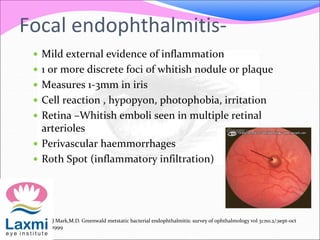
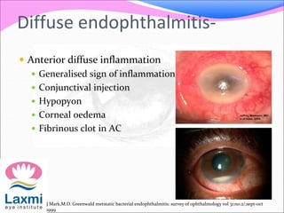
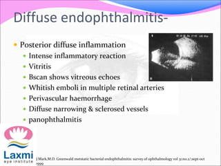
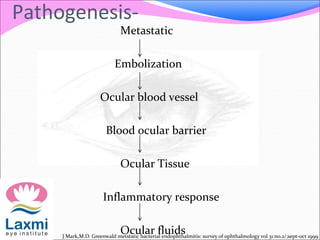
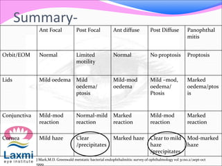
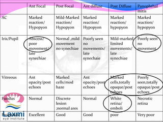
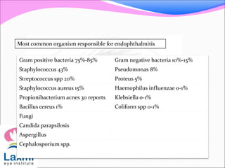
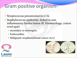
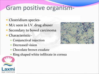
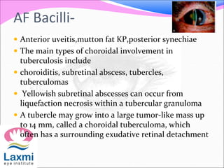
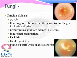
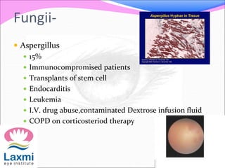
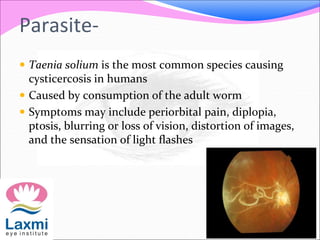
Metastatic Endophthalmitis
Presenter-Dr Shubhangini J
Moderator-Dr Sachin Fegde
๏‚— Definition
๏‚— Classification
๏‚— Predisposing factors
๏‚— Histo-pathological description
๏‚— Organism causing metastatic endophthalmitis
๏‚— Investigation
๏‚— Management
Definition-
๏‚— Inflammation within the anterior segment or
posterior segment or both, concurrent with partial
thickness involvement of an adjacent ocular wall
๏‚— Associated with-
๏‚— Decreased vision
๏‚— Pain
๏‚— Conjunctival hyperemia
๏‚— Lid oedema
๏‚— Anterior chamber & vitreous cellular reaction
๏‚— Hypopyon
Classification of
Endophthalmitis
Infectious
Exogenous
Post surgical
1. Acute Onset
2. Delayed Onset
3. Bleb associated
Non surgical
1. Post-traumatic
Endogenous
Hematogenous Spread
Sterile
Lens Induced
Toxic
S Michael Kresloff, Endophthalmitis survey of ophthalmology vol
43:no.3:nov-dec 1998
Case
๏‚— Mrs K P 45 yrs old female housewife from panvel
presented to us on 3-9-2013
๏‚— Chief complaint โ€“
๏‚— RE Diminution of vision since 1 month
RE Pain since 1 month
History
๏‚— Diminution of vision:
๏‚— gradual
๏‚— No h/o flashes of light
๏‚— Pain
๏‚— Dull aching
History
๏‚— Past Ocular history:
๏‚— H/o spectacle use since 7yrs
๏‚— No h/o ocular surgery
๏‚— No h/o ocular trauma
๏‚— No h/o similar complaints in the past
๏‚— h/o of hospitalisation 3 months back for fever&
generalised bodyache
๏‚— Personal history insignificant
๏‚— Family history insignificant
Examination-
๏‚— Head posture normal
๏‚— Orthophoria
๏‚— Extraocular movements: Full & free
๏‚— Visual acuity
RE LE
Visual Acuity CF FC, <N36 6/9, N6
Refraction Plano +1.00 DS
Intra ocular Pressure 10 18
Ocular Examination-
๏‚— On anterior segment examination
RE LE
Lids N N
Conjunctiva N N
Cornea Clear Clear
AC Cells ++++ ND Quiet
Pupil TAPD NSRL
Lens Clear Clear
Fundus RE LE
Impression-Endogenous
Endophthalmitis
Differential Diagnosis-
๏‚— Tuberculosis
๏‚— Toxoplasma
๏‚— Toxocara
๏‚— Sarcoidosis
๏‚— Haradaโ€™s disease
๏‚— Syphilitic uveitis
Investigation-
Hb/CBC/ESR
LFT/RFT
HIV/VDRL
Vitreous tap
Mantoux test
RBS,Blood culture
Treatment-
๏‚— Predforte eye drop 6t/day
๏‚— Atropine eye drop 3t/day
๏‚— Vitrectomy+BB + EL+silicon oil implantation NVP
Discussion
๏‚— Intraocular infection caused by haematogenous
spread of microorganism from distant foci to the eye
from site of infection elsewhere in the body or from
contaminated catheters & needles
๏‚— Epidemiology-
๏‚— Accounts for 2-8%
๏‚— Affects any age & sex
๏‚— RE more commonly involved
๏‚— Most common is bacterial metastatic endophthalmitis
fungal metastatic endophthalmitis
Okada AA et al Endogenous bacterial endophthalmitis 10 yr retrospective study 101:832-838,1994
Discussion-
๏‚— Most common site- uveal tract,choroid which are
most vascular tissues
๏‚— Study on ocular oncology service of wills eye hospital,
philadelphia(1996) , in a sample size of 420 patients
had observed breast cancer(47%) most common
tumor forming ocular metastatis followed by
lung(21%)
๏‚— Predisposing factors-
๏‚— Immunocompromised patients
๏‚— Intravenous drug abuse
๏‚— Diabetes milletus
๏‚— Chronic renal failure
๏‚— Malignancy
๏‚— Dental surgery
๏‚— Contaminated intravenous fluid
J Mark,M.D. Greenwald metstatic bacterial endophthalmitis: survey of ophthalmology vol 31:no.2/;sept-oct
1999
๏‚— Metastatic endophthalmitis classified based on
Metastatic
Endophthalmitis
Extent
Focal Diffuse
Location
Anterior sement
Posterior
segment
Surv Ophthalmol 31:81-101,2000 greenwald et al
Focal endophthalmitis-
๏‚— Mild external evidence of inflammation
๏‚— 1 or more discrete foci of whitish nodule or plaque
๏‚— Measures 1-3mm in iris
๏‚— Cell reaction , hypopyon, photophobia, irritation
๏‚— Retina โ€“Whitish emboli seen in multiple retinal
arterioles
๏‚— Perivascular haemmorrhages
๏‚— Roth Spot (inflammatory infiltration)
J Mark,M.D. Greenwald metstatic bacterial endophthalmitis: survey of ophthalmology vol 31:no.2/;sept-oct
1999
Diffuse endophthalmitis-
๏‚— Anterior diffuse inflammation
๏‚— Generalised sign of inflammation
๏‚— Conjunctival injection
๏‚— Hypopyon
๏‚— Corneal oedema
๏‚— Fibrinous clot in AC
J Mark,M.D. Greenwald metstatic bacterial endophthalmitis: survey of ophthalmology vol 31:no.2/;sept-oct
1999
Diffuse endophthalmitis-
๏‚— Posterior diffuse inflammation
๏‚— Intense inflammatory reaction
๏‚— Vitritis
๏‚— Bscan shows vitreous echoes
๏‚— Whitish emboli in multiple retinal arteries
๏‚— Perivascular haemorrhage
๏‚— Diffuse narrowing & sclerosed vessels
๏‚— panophthalmitis
J Mark,M.D. Greenwald metstatic bacterial endophthalmitis: survey of ophthalmology vol 31:no.2/;sept-oct
1999
Histopathological features-
๏‚— Focal inflammation
๏‚— Inflammatory cells occluding
the lumen of vessels
surrounding tissue
๏‚— Diffuse inflammation
๏‚— Inflammatory cells
๏‚— Necrosis
๏‚— Haemmorhage of involved
tissue
Infitration of all the intraocular structure by inflammatory
cells
Pathogenesis-
Metastatic
Embolization
Ocular blood vessel
Blood ocular barrier
Ocular Tissue
Inflammatory response
Ocular fluidsJ Mark,M.D. Greenwald metstatic bacterial endophthalmitis: survey of ophthalmology vol 31:no.2/;sept-oct 1999
Summary-
Ant Focal Post Focal Ant diffuse Post Diffuse Panophthal
mitis
Orbit/EOM Normal Limited
motility
Normal No proptosis Proptosis
Lids Mild oedema Mild
oedema/
ptosis
Mild-mod
oedema
Mild โ€“mod,
oedema/
Ptosis
Marked
oedema/ptos
is
Conjunctiva Mild-mod
reaction
Normal-mild
reaction
Marked
reaction
Mild-mod
reaction
Marked
reaction
Cornea Mild haze Clear
/precipitates
Marked haze Clear to mild
haze
/precipitates
Mod-marked
haze
J Mark,M.D. Greenwald metstatic bacterial endophthalmitis: survey of ophthalmology vol 31:no.2/;sept-oct
1999
Ant Focal Post Focal Ant diffuse Post Diffuse Panophthal
mitis
AC Marked
reaction/
Hypopyon
Mild-Marked
reaction/
Hypopyon
Marked
reaction/
Hypopyon
Marked
reaction/
hypopyon
Marked
reaction/
hypopyon
Iris/Pupil Abscess/
poor
movement/
late
synechiae
Normal ,mild
movement
no synechiae
Poorly seen
no
movements/
late
synechaie
Mild-marked
limited
movements/
late
synechiae
Poorly seen
no
movement
Vitreous Ant
opacity/post
echoes
Marked
cells/mod
haze
Ant
opacity/post
echoes
Marked
cells,totally
opaque/post
echoes
Poorly
seen,totally
opaque/post
echoes
Fundus Normal Discrete
lesion
;normal ares
Normal White
retina/
emboli
Necrotic
retina
Prognosis Excellent Good Good poor Very poor
Most common organism responsible for endophthalmitis
Gram positive bacteria 75%-85% Gram negative bacteria 10%-15%
Staphylococcus 43% Pseudomonas 8%
Streptococcus spp 20% Proteus 5%
Staphylococcus aureus 15% Haemophilus influenzae 0-1%
Propionibacterium acnes 30 reports Klebsiella 0-1%
Bacillus cereus 1% Coliform spp 0-1%
Fungi
Candida parapsilosis
Aspergillus
Cephalosporium spp.
Gram positive organism-
๏‚— Streptococcus pneumoniae(m/c) &
๏‚— Staphylococcus epidermis- linked to non
inflammatory fundus lesion (R. Haemorrhage, cotton
wool spot)
๏‚— secondary to meningitis
๏‚— Endocarditis
๏‚— Malignant neoplasm(breast cancer m/c)
Gram positive organism-
๏‚— Clostridium species-
๏‚— M/c seen in I.V. drug abuser
๏‚— Secondary to bowel carcinoma
๏‚— Characteristic-
๏‚— Conjunctival injection
๏‚— Decreased vision
๏‚— Chocolate brown exudate
๏‚— Ring shaped white infiltrate in cornea
Gram positive bacilli-
๏‚— Listeria monocytogenes
๏‚— Mild external inflammation
๏‚— Absent systemic signs of infection
๏‚— Indolent infection
๏‚— Brown hypopyon
๏‚— Without corneal involvement
Gram negative organism-
๏‚— Haemophilus influenzae
๏‚— Present in a manner similar to meningococcus with
bacteremia
๏‚— Meningitis
๏‚— Anterior diffuse inflammation of eye
๏‚— Post subretinal abscess confusing it with disciform scar
or a choroidal tumor
AF Bacilli-
๏‚— Nocardia asteroides(AF bacilli)
๏‚— Lead to BEE secondary to dissemination from
pulmonary foci
๏‚— Infecting choroid
๏‚— Proliferating to produce chorioretinitis& vitritis
๏‚— Posterior subretinal abscess
๏‚— Seen in immunocompromised patients
AF Bacilli-
๏‚— Mycobacterium Tuberculosis(AF bacilli)
๏‚— Disseminates from pulmonary focus
๏‚— Infecting & proliferating chorioretinitis
๏‚— Vitritis
๏‚— Acute endophthalmitis
AF Bacilli-
๏‚— Anterior uveitis,mutton fat KP,posterior synechiae
๏‚— The main types of choroidal involvement in
tuberculosis include
๏‚— choroiditis, subretinal abscess, tubercles,
tuberculomas
๏‚— Yellowish subretinal abscesses can occur from
liquefaction necrosis within a tubercular granuloma
๏‚— A tubercle may grow into a large tumor-like mass up
to 14 mm, called a choroidal tuberculoma, which
often has a surrounding exudative retinal detachment
Fungii-
๏‚— Candida albicans
๏‚— 75-80%
๏‚— It forms germ tube in serum that embolize and lodges
in choriocapillaries
๏‚— Creamy retinal infiltrate extends in vitreous
๏‚— Intraretinal haemmorhage
๏‚— Papillitis
๏‚— Focal choroiditis
๏‚— String of pearls(white opacities/snowball)
Fungii-
๏‚— Aspergillus
๏‚— 15%
๏‚— Immunocompromised patients
๏‚— Transplants of stem cell
๏‚— Endocarditis
๏‚— Leukemia
๏‚— I.V. drug abuse,contaminated Dextrose infusion fluid
๏‚— COPD on corticosteriod therapy
Parasite-
๏‚— Taenia solium is the most common species causing
cysticercosis in humans
๏‚— Caused by consumption of the adult worm
๏‚— Symptoms may include periorbital pain, diplopia,
ptosis, blurring or loss of vision, distortion of images,
and the sensation of light flashes
Parasite-
๏‚— On fundus examination living form of cysticercosis
has the features of an undulating, expanding and
contracting โ€œpearlโ€ with intermittent evagination and
invagination of the protoscolex
๏‚— This may result in an inflammatory chorioretinitis
Ocular ultrasonography- subretinal cyst
๏‚— Assays for eosinophilia in anterior chamber fluid
sample
Evaluation-
๏‚— Complete history
๏‚— Physical examination
๏‚— Specific evaluation
๏‚— E.C.G. for endo carditis,
๏‚— CXR PA View,CT Scan,
๏‚— Sputum ,urine for culture sensitivity
๏‚— ESR,BUN,Creatinine
๏‚— CT/MRI Orbit
๏‚— PCR
๏‚— Culture of CSF,throat swab, stool,indwelling catheter
When to cultureโ€ฆ..??
๏‚— Presence of systemic infection
๏‚— Signs of acute or chronic intraocular inflammation in
absence of extraocular culture
๏‚— In presence of culture positive-systemic infection with
sign of inflammation, unresponsive to antibiotic
therapy
๏‚— To rule out suspected malignancy after a negative
systemic work up
What to cultureโ€ฆ??
๏‚— Culture of blood, urine, aqueous, vitreous, CSF &
wound culture & smear indicated
๏‚— To locate site of original infection
๏‚— Document systemic involvement
How to cultureโ€ฆ.???
๏‚— Aqueous material can be obtained by
๏‚— 30 gauge needle in tuberculin syringe
๏‚— Limbal stab incision required
๏‚— 0.1-0.2ml of fluid to be aspirated
๏‚— Vitreous biopsy performed via-
๏‚— Pars plana,1,2,3,port vitrectomy probe OR
๏‚— By 25-27gauge needle in tuberculin syringe
๏‚— 0.1-0.2ml aspirated.
Treatment-
๏‚— Managed similar to acute post operative infectious
endophthalmitis
๏‚— Non ocular culture sensitivity data to guide initial
therapy
๏‚— Specific therapy to begun after ocular culture
Treatment-
๏‚— Mild endogenous endophthalmitis- focal metastatic
abscess in anterior & posterior segment
๏‚— Topical & systemic therapy
๏‚— Vitrectomy done for removal of infecting organism
,endotoxin, exotoxin & vitreous membrane , vitreous
opacities , better distribution of intravitreal antibiotic
Treatment for gram positive organism-
๏‚— Because most cases are caused by gram positive
organisms, vancomycin- (broad-spectrum activity
against most gram positive species) has become
an agent of choice
๏‚— Non toxic in recommended clinical dosage.
๏‚— Thus vancomycin 1 mg in (0.1 ml) BD is given
intra vitreally after blood culture & vitreous tap
Arch Ophth 1999; 117: 1023-1027
Treatment for gram negative
organism-
๏‚— Ceftazidine has emerged as on alternative
๏‚— More effective than aminoglycosides
๏‚— Retinal toxicity studies in primates reveal concentration of
2.25 mg/0.1 ml to be safe after vitreous tap
๏‚— Excellent ocular penentration
๏‚— After 2 weeks to shift on oral tab cefuroxime 500mg BD
Arch Ophthalmol 1994; 112: 48-53
Br. J. Ophth 97; 81: 1006-15
Treatment for fungal endophalmitis-
๏‚— If vitreous is minimally involved culture/smear is
positive for fungus- oral Fluconazole /vitrectomy to
be considered
๏‚— In metastatic aspergillus endophthalmitis
๏‚— IntraVitreal. amphotericin B (5-10ยตg) with IntraVitreal.
Dexamethasone(400ยตg)
๏‚— Repeated for persistance disease after5-7 days in
nonvitrectomised eye & after 2 days in vitrectomised
eye
๏‚— Systemically I.V. amphotericin(0.005mg/0.1ml)/
itraconazole is advocated
๏‚— Forvitreous seeding- vitrectomy
Treatment for TB -
๏‚— For metastatic ocular TB approach to neuro physician
is mandatory to start empirical therapy of ATT
๏‚— in cases in which ,uncertainty about TB remains,
biopsy of the eye for culture, histologic examination,
is useful to establish the diagnosis of ocular TB.
Treatment for parasitic endophthalmitis-
๏‚— Praziquantil ,Metrifonate
๏‚— Spontaneous extrusion of cystercerci from the eye
may occur
๏‚— Vitrectomy along with photocoagulation has shown
some success in removing cystercerci from the
vitreous cavity
Conclusion-
๏‚— Metastatic endophthalmitis is dreaded ocular
condition ,high index of clinical suspicion is necessary
along with a co-ordinated multidisciplinary approach
to handle this difficult situation
Thank you
Fungii-
๏‚— Presentation-
๏‚— Pain
๏‚— Severe visual loss
๏‚— Presents with pneumonia
๏‚— Seeding of end organs
๏‚— Vitritis
๏‚— Choroidal lesions
๏‚— Chorioretinal abscess
๏‚— Subhyaloid or sub retinal hypopyon
๏‚— Indicated-
๏‚— Inflammatory focus in AS
๏‚— Aphakic eye,dehiscence of post. Cap
๏‚— Vitritis,no improvement in vision, non ocular culture are
negative

Metastatis endophthalmitis

  • 1.
  • 2.
    ๏‚— Definition ๏‚— Classification ๏‚—Predisposing factors ๏‚— Histo-pathological description ๏‚— Organism causing metastatic endophthalmitis ๏‚— Investigation ๏‚— Management
  • 3.
    Definition- ๏‚— Inflammation withinthe anterior segment or posterior segment or both, concurrent with partial thickness involvement of an adjacent ocular wall ๏‚— Associated with- ๏‚— Decreased vision ๏‚— Pain ๏‚— Conjunctival hyperemia ๏‚— Lid oedema ๏‚— Anterior chamber & vitreous cellular reaction ๏‚— Hypopyon
  • 4.
    Classification of Endophthalmitis Infectious Exogenous Post surgical 1.Acute Onset 2. Delayed Onset 3. Bleb associated Non surgical 1. Post-traumatic Endogenous Hematogenous Spread Sterile Lens Induced Toxic S Michael Kresloff, Endophthalmitis survey of ophthalmology vol 43:no.3:nov-dec 1998
  • 5.
    Case ๏‚— Mrs KP 45 yrs old female housewife from panvel presented to us on 3-9-2013 ๏‚— Chief complaint โ€“ ๏‚— RE Diminution of vision since 1 month RE Pain since 1 month
  • 6.
    History ๏‚— Diminution ofvision: ๏‚— gradual ๏‚— No h/o flashes of light ๏‚— Pain ๏‚— Dull aching
  • 7.
    History ๏‚— Past Ocularhistory: ๏‚— H/o spectacle use since 7yrs ๏‚— No h/o ocular surgery ๏‚— No h/o ocular trauma ๏‚— No h/o similar complaints in the past ๏‚— h/o of hospitalisation 3 months back for fever& generalised bodyache ๏‚— Personal history insignificant ๏‚— Family history insignificant
  • 8.
    Examination- ๏‚— Head posturenormal ๏‚— Orthophoria ๏‚— Extraocular movements: Full & free ๏‚— Visual acuity RE LE Visual Acuity CF FC, <N36 6/9, N6 Refraction Plano +1.00 DS Intra ocular Pressure 10 18
  • 9.
    Ocular Examination- ๏‚— Onanterior segment examination RE LE Lids N N Conjunctiva N N Cornea Clear Clear AC Cells ++++ ND Quiet Pupil TAPD NSRL Lens Clear Clear
  • 10.
  • 11.
    Differential Diagnosis- ๏‚— Tuberculosis ๏‚—Toxoplasma ๏‚— Toxocara ๏‚— Sarcoidosis ๏‚— Haradaโ€™s disease ๏‚— Syphilitic uveitis Investigation- Hb/CBC/ESR LFT/RFT HIV/VDRL Vitreous tap Mantoux test RBS,Blood culture
  • 13.
    Treatment- ๏‚— Predforte eyedrop 6t/day ๏‚— Atropine eye drop 3t/day ๏‚— Vitrectomy+BB + EL+silicon oil implantation NVP
  • 14.
    Discussion ๏‚— Intraocular infectioncaused by haematogenous spread of microorganism from distant foci to the eye from site of infection elsewhere in the body or from contaminated catheters & needles ๏‚— Epidemiology- ๏‚— Accounts for 2-8% ๏‚— Affects any age & sex ๏‚— RE more commonly involved ๏‚— Most common is bacterial metastatic endophthalmitis fungal metastatic endophthalmitis Okada AA et al Endogenous bacterial endophthalmitis 10 yr retrospective study 101:832-838,1994
  • 15.
    Discussion- ๏‚— Most commonsite- uveal tract,choroid which are most vascular tissues ๏‚— Study on ocular oncology service of wills eye hospital, philadelphia(1996) , in a sample size of 420 patients had observed breast cancer(47%) most common tumor forming ocular metastatis followed by lung(21%)
  • 16.
    ๏‚— Predisposing factors- ๏‚—Immunocompromised patients ๏‚— Intravenous drug abuse ๏‚— Diabetes milletus ๏‚— Chronic renal failure ๏‚— Malignancy ๏‚— Dental surgery ๏‚— Contaminated intravenous fluid J Mark,M.D. Greenwald metstatic bacterial endophthalmitis: survey of ophthalmology vol 31:no.2/;sept-oct 1999
  • 17.
    ๏‚— Metastatic endophthalmitisclassified based on Metastatic Endophthalmitis Extent Focal Diffuse Location Anterior sement Posterior segment Surv Ophthalmol 31:81-101,2000 greenwald et al
  • 18.
    Focal endophthalmitis- ๏‚— Mildexternal evidence of inflammation ๏‚— 1 or more discrete foci of whitish nodule or plaque ๏‚— Measures 1-3mm in iris ๏‚— Cell reaction , hypopyon, photophobia, irritation ๏‚— Retina โ€“Whitish emboli seen in multiple retinal arterioles ๏‚— Perivascular haemmorrhages ๏‚— Roth Spot (inflammatory infiltration) J Mark,M.D. Greenwald metstatic bacterial endophthalmitis: survey of ophthalmology vol 31:no.2/;sept-oct 1999
  • 19.
    Diffuse endophthalmitis- ๏‚— Anteriordiffuse inflammation ๏‚— Generalised sign of inflammation ๏‚— Conjunctival injection ๏‚— Hypopyon ๏‚— Corneal oedema ๏‚— Fibrinous clot in AC J Mark,M.D. Greenwald metstatic bacterial endophthalmitis: survey of ophthalmology vol 31:no.2/;sept-oct 1999
  • 20.
    Diffuse endophthalmitis- ๏‚— Posteriordiffuse inflammation ๏‚— Intense inflammatory reaction ๏‚— Vitritis ๏‚— Bscan shows vitreous echoes ๏‚— Whitish emboli in multiple retinal arteries ๏‚— Perivascular haemorrhage ๏‚— Diffuse narrowing & sclerosed vessels ๏‚— panophthalmitis J Mark,M.D. Greenwald metstatic bacterial endophthalmitis: survey of ophthalmology vol 31:no.2/;sept-oct 1999
  • 21.
    Histopathological features- ๏‚— Focalinflammation ๏‚— Inflammatory cells occluding the lumen of vessels surrounding tissue ๏‚— Diffuse inflammation ๏‚— Inflammatory cells ๏‚— Necrosis ๏‚— Haemmorhage of involved tissue Infitration of all the intraocular structure by inflammatory cells
  • 22.
    Pathogenesis- Metastatic Embolization Ocular blood vessel Bloodocular barrier Ocular Tissue Inflammatory response Ocular fluidsJ Mark,M.D. Greenwald metstatic bacterial endophthalmitis: survey of ophthalmology vol 31:no.2/;sept-oct 1999
  • 23.
    Summary- Ant Focal PostFocal Ant diffuse Post Diffuse Panophthal mitis Orbit/EOM Normal Limited motility Normal No proptosis Proptosis Lids Mild oedema Mild oedema/ ptosis Mild-mod oedema Mild โ€“mod, oedema/ Ptosis Marked oedema/ptos is Conjunctiva Mild-mod reaction Normal-mild reaction Marked reaction Mild-mod reaction Marked reaction Cornea Mild haze Clear /precipitates Marked haze Clear to mild haze /precipitates Mod-marked haze J Mark,M.D. Greenwald metstatic bacterial endophthalmitis: survey of ophthalmology vol 31:no.2/;sept-oct 1999
  • 24.
    Ant Focal PostFocal Ant diffuse Post Diffuse Panophthal mitis AC Marked reaction/ Hypopyon Mild-Marked reaction/ Hypopyon Marked reaction/ Hypopyon Marked reaction/ hypopyon Marked reaction/ hypopyon Iris/Pupil Abscess/ poor movement/ late synechiae Normal ,mild movement no synechiae Poorly seen no movements/ late synechaie Mild-marked limited movements/ late synechiae Poorly seen no movement Vitreous Ant opacity/post echoes Marked cells/mod haze Ant opacity/post echoes Marked cells,totally opaque/post echoes Poorly seen,totally opaque/post echoes Fundus Normal Discrete lesion ;normal ares Normal White retina/ emboli Necrotic retina Prognosis Excellent Good Good poor Very poor
  • 25.
    Most common organismresponsible for endophthalmitis Gram positive bacteria 75%-85% Gram negative bacteria 10%-15% Staphylococcus 43% Pseudomonas 8% Streptococcus spp 20% Proteus 5% Staphylococcus aureus 15% Haemophilus influenzae 0-1% Propionibacterium acnes 30 reports Klebsiella 0-1% Bacillus cereus 1% Coliform spp 0-1% Fungi Candida parapsilosis Aspergillus Cephalosporium spp.
  • 26.
    Gram positive organism- ๏‚—Streptococcus pneumoniae(m/c) & ๏‚— Staphylococcus epidermis- linked to non inflammatory fundus lesion (R. Haemorrhage, cotton wool spot) ๏‚— secondary to meningitis ๏‚— Endocarditis ๏‚— Malignant neoplasm(breast cancer m/c)
  • 27.
    Gram positive organism- ๏‚—Clostridium species- ๏‚— M/c seen in I.V. drug abuser ๏‚— Secondary to bowel carcinoma ๏‚— Characteristic- ๏‚— Conjunctival injection ๏‚— Decreased vision ๏‚— Chocolate brown exudate ๏‚— Ring shaped white infiltrate in cornea
  • 28.
    Gram positive bacilli- ๏‚—Listeria monocytogenes ๏‚— Mild external inflammation ๏‚— Absent systemic signs of infection ๏‚— Indolent infection ๏‚— Brown hypopyon ๏‚— Without corneal involvement
  • 29.
    Gram negative organism- ๏‚—Haemophilus influenzae ๏‚— Present in a manner similar to meningococcus with bacteremia ๏‚— Meningitis ๏‚— Anterior diffuse inflammation of eye ๏‚— Post subretinal abscess confusing it with disciform scar or a choroidal tumor
  • 30.
    AF Bacilli- ๏‚— Nocardiaasteroides(AF bacilli) ๏‚— Lead to BEE secondary to dissemination from pulmonary foci ๏‚— Infecting choroid ๏‚— Proliferating to produce chorioretinitis& vitritis ๏‚— Posterior subretinal abscess ๏‚— Seen in immunocompromised patients
  • 31.
    AF Bacilli- ๏‚— MycobacteriumTuberculosis(AF bacilli) ๏‚— Disseminates from pulmonary focus ๏‚— Infecting & proliferating chorioretinitis ๏‚— Vitritis ๏‚— Acute endophthalmitis
  • 32.
    AF Bacilli- ๏‚— Anterioruveitis,mutton fat KP,posterior synechiae ๏‚— The main types of choroidal involvement in tuberculosis include ๏‚— choroiditis, subretinal abscess, tubercles, tuberculomas ๏‚— Yellowish subretinal abscesses can occur from liquefaction necrosis within a tubercular granuloma ๏‚— A tubercle may grow into a large tumor-like mass up to 14 mm, called a choroidal tuberculoma, which often has a surrounding exudative retinal detachment
  • 33.
    Fungii- ๏‚— Candida albicans ๏‚—75-80% ๏‚— It forms germ tube in serum that embolize and lodges in choriocapillaries ๏‚— Creamy retinal infiltrate extends in vitreous ๏‚— Intraretinal haemmorhage ๏‚— Papillitis ๏‚— Focal choroiditis ๏‚— String of pearls(white opacities/snowball)
  • 34.
    Fungii- ๏‚— Aspergillus ๏‚— 15% ๏‚—Immunocompromised patients ๏‚— Transplants of stem cell ๏‚— Endocarditis ๏‚— Leukemia ๏‚— I.V. drug abuse,contaminated Dextrose infusion fluid ๏‚— COPD on corticosteriod therapy
  • 35.
    Parasite- ๏‚— Taenia soliumis the most common species causing cysticercosis in humans ๏‚— Caused by consumption of the adult worm ๏‚— Symptoms may include periorbital pain, diplopia, ptosis, blurring or loss of vision, distortion of images, and the sensation of light flashes
  • 36.
    Parasite- ๏‚— On fundusexamination living form of cysticercosis has the features of an undulating, expanding and contracting โ€œpearlโ€ with intermittent evagination and invagination of the protoscolex ๏‚— This may result in an inflammatory chorioretinitis Ocular ultrasonography- subretinal cyst ๏‚— Assays for eosinophilia in anterior chamber fluid sample
  • 37.
    Evaluation- ๏‚— Complete history ๏‚—Physical examination ๏‚— Specific evaluation ๏‚— E.C.G. for endo carditis, ๏‚— CXR PA View,CT Scan, ๏‚— Sputum ,urine for culture sensitivity ๏‚— ESR,BUN,Creatinine ๏‚— CT/MRI Orbit ๏‚— PCR ๏‚— Culture of CSF,throat swab, stool,indwelling catheter
  • 38.
    When to cultureโ€ฆ..?? ๏‚—Presence of systemic infection ๏‚— Signs of acute or chronic intraocular inflammation in absence of extraocular culture ๏‚— In presence of culture positive-systemic infection with sign of inflammation, unresponsive to antibiotic therapy ๏‚— To rule out suspected malignancy after a negative systemic work up
  • 39.
    What to cultureโ€ฆ?? ๏‚—Culture of blood, urine, aqueous, vitreous, CSF & wound culture & smear indicated ๏‚— To locate site of original infection ๏‚— Document systemic involvement
  • 40.
    How to cultureโ€ฆ.??? ๏‚—Aqueous material can be obtained by ๏‚— 30 gauge needle in tuberculin syringe ๏‚— Limbal stab incision required ๏‚— 0.1-0.2ml of fluid to be aspirated ๏‚— Vitreous biopsy performed via- ๏‚— Pars plana,1,2,3,port vitrectomy probe OR ๏‚— By 25-27gauge needle in tuberculin syringe ๏‚— 0.1-0.2ml aspirated.
  • 41.
    Treatment- ๏‚— Managed similarto acute post operative infectious endophthalmitis ๏‚— Non ocular culture sensitivity data to guide initial therapy ๏‚— Specific therapy to begun after ocular culture
  • 42.
    Treatment- ๏‚— Mild endogenousendophthalmitis- focal metastatic abscess in anterior & posterior segment ๏‚— Topical & systemic therapy ๏‚— Vitrectomy done for removal of infecting organism ,endotoxin, exotoxin & vitreous membrane , vitreous opacities , better distribution of intravitreal antibiotic
  • 43.
    Treatment for grampositive organism- ๏‚— Because most cases are caused by gram positive organisms, vancomycin- (broad-spectrum activity against most gram positive species) has become an agent of choice ๏‚— Non toxic in recommended clinical dosage. ๏‚— Thus vancomycin 1 mg in (0.1 ml) BD is given intra vitreally after blood culture & vitreous tap Arch Ophth 1999; 117: 1023-1027
  • 44.
    Treatment for gramnegative organism- ๏‚— Ceftazidine has emerged as on alternative ๏‚— More effective than aminoglycosides ๏‚— Retinal toxicity studies in primates reveal concentration of 2.25 mg/0.1 ml to be safe after vitreous tap ๏‚— Excellent ocular penentration ๏‚— After 2 weeks to shift on oral tab cefuroxime 500mg BD Arch Ophthalmol 1994; 112: 48-53 Br. J. Ophth 97; 81: 1006-15
  • 45.
    Treatment for fungalendophalmitis- ๏‚— If vitreous is minimally involved culture/smear is positive for fungus- oral Fluconazole /vitrectomy to be considered ๏‚— In metastatic aspergillus endophthalmitis ๏‚— IntraVitreal. amphotericin B (5-10ยตg) with IntraVitreal. Dexamethasone(400ยตg) ๏‚— Repeated for persistance disease after5-7 days in nonvitrectomised eye & after 2 days in vitrectomised eye ๏‚— Systemically I.V. amphotericin(0.005mg/0.1ml)/ itraconazole is advocated ๏‚— Forvitreous seeding- vitrectomy
  • 46.
    Treatment for TB- ๏‚— For metastatic ocular TB approach to neuro physician is mandatory to start empirical therapy of ATT ๏‚— in cases in which ,uncertainty about TB remains, biopsy of the eye for culture, histologic examination, is useful to establish the diagnosis of ocular TB.
  • 47.
    Treatment for parasiticendophthalmitis- ๏‚— Praziquantil ,Metrifonate ๏‚— Spontaneous extrusion of cystercerci from the eye may occur ๏‚— Vitrectomy along with photocoagulation has shown some success in removing cystercerci from the vitreous cavity
  • 48.
    Conclusion- ๏‚— Metastatic endophthalmitisis dreaded ocular condition ,high index of clinical suspicion is necessary along with a co-ordinated multidisciplinary approach to handle this difficult situation
  • 49.
  • 50.
    Fungii- ๏‚— Presentation- ๏‚— Pain ๏‚—Severe visual loss ๏‚— Presents with pneumonia ๏‚— Seeding of end organs ๏‚— Vitritis ๏‚— Choroidal lesions ๏‚— Chorioretinal abscess ๏‚— Subhyaloid or sub retinal hypopyon
  • 51.
    ๏‚— Indicated- ๏‚— Inflammatoryfocus in AS ๏‚— Aphakic eye,dehiscence of post. Cap ๏‚— Vitritis,no improvement in vision, non ocular culture are negative

Editor's Notes

  • #4ย Most serious complication of opthalmic surgery
  • #5ย Also can be classified acc to Mode of entry, location,type of etiological agent Exogenous- micro organism introduced directly from environment which can be usually post operative mainly bacterial source Endogenous- spread of organism as a metastatic infection mainly fungal, bacterial
  • #13ย Pre & post contrast MRI brain was performed on 1.5 tesla in multiple plane using T1&T2 spine echo sequence. Conglomerate ring enhancing lesion involving right uncus &rt hippocampus measuring 9x17x9mm dimension ,appearing hypointense on T2 &isointense onT1 showing mild restricted diffusion.4x3mm size lesion involving Lt deep ventricular occipital lobe white matter s/o tuberculoma
  • #15ย RE is common as more direct flow to right common carotid artery Avg incidence of endoph is 5 in 10000 hospitalised pt
  • #17ย Immunocompromised host cancer aids DM,
  • #19ย Focal intraocular inflammation 1 or more discrete foci of whitish nodule or plaque on iris ciliary body retina choroid Roth spot white centre contains organism (sterile) contains WBC often fibrin thrombus occurs at site of extravasation of blood.thats the reason why septic emboli occur more frequently in retina Than choroid
  • #20ย Characterised oedema of conj lids ,fibrinous clot in AC,
  • #21ย Intense inflammatory reaction which obscure the fundus Bscan subretinal abscess with overlyin vit exudate
  • #22ย Focal inflammation with bacteria inflammatory cells.
  • #23ย The pathogenesis of endogenous bacterial endoph ,bacteria in mets reach the ocular fluid only after traversing the blood ocular barrier & proliferating in tissue,where an effective inflammatory response occurs The ocular spread occurs after choroidal invasion.the primary invasion of choroid led to involvement of sub retinal space retina & vitreous cavity
  • #24ย Clinical classification-
  • #26ย 3 main factor for poor visual outcome ,more virulent org, compromised host condition & delay in diagnosis
  • #28ย Because of its particular aggressive nature
  • #30ย Post subretinal abscess confusing it with disciform scar or a choroidal tumor Gram negative coccobacilli
  • #32ย Slit-lamp biomicroscopy at presentation shows ciliary congestion and a dense anterior chamber reaction.ย B,ย Pathologic findings of the vitreous specimen from the enucleated eye shows extensive infiltration of acid-fast bacilli
  • #33ย Choroidal tubercules are small gray-white to yellow nodules smaller than a quarter disc diameter, which are not well circumscribed
  • #34ย Mets candidial endop, note the white area of retinitis
  • #38ย PCR faster results on org to start antibiotic therapy, TTE(ASD)
  • #42ย If available
  • #51ย ย Dense vitritis with underlying hemorrhagic retinitis is present