Different types of ophthalmic instruments
•Download as PPT, PDF•
34 likes•13,526 views

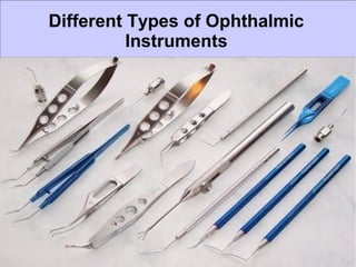
Ophthalmic instruments utilized by means of ophthalmologists as well as eyes doctors to help analyze different kindsconnected with eyes ailments. These kind of machines are quite important as well as important for specific eyes examinations.
Report
Share
Report
Share

Recommended
Autorefractometer
This document discusses the history and principles of autorefractors. It explains that autorefractors use infrared light and meridional refractometry to objectively determine a patient's refractive error without needing subjective feedback. Modern autorefractors are more accurate and efficient than older subjective methods due to advances in electronics. They work by projecting an infrared fixation target and using a Badal optometer and fogging technique to relax accommodation and obtain refractive measurements.
Retinoscopy and its principles
This document provides an overview of retinoscopy, including:
- A brief history and the development of different types of retinoscopes.
- The optical principles behind how retinoscopy works to assess refractive error.
- Different techniques for performing retinoscopy, including static, dynamic, and near retinoscopy.
- Factors that can impact the results or make retinoscopy more difficult in some patients.
- Uses of retinoscopy beyond assessing refractive error, such as detecting other ocular issues.
Tonometry
This document discusses various methods for measuring intraocular pressure (IOP), including direct and indirect techniques. Direct manometry involves inserting a needle into the eye, while indirect methods include indentation tonometry using the Schiotz tonometer, various types of applanation tonometry (Goldmann, Perkins, pneumatic, Tono-Pen), and non-contact tonometry. Factors like ocular rigidity, corneal thickness and curvature can influence tonometry readings. Newer methods like the dynamic contour tonometer and ocular response analyzer aim to provide measurements less affected by corneal properties.
Diplopia charting
Diplopia, or double vision, occurs when more than one image of an object is seen simultaneously. It can be caused by abnormalities in the eyes themselves or issues with eye movement coordination. A diplopia chart is used to evaluate the type and location of double vision by having the patient report the appearance of light sources in different gaze positions. Interpretation of the chart provides clues to which eye muscles may be affected and whether the cause is neurogenic, restrictive, or myogenic in nature. Treatment options include glasses, prisms, eye patching, or strabismus surgery depending on the deviation and goal of eliminating diplopia.
Direct ophthalmoscopy
The direct ophthalmoscope is a handheld instrument used to examine the fundus monocularly within a central 7-10 degree retinal area. It provides 15x magnification. It has illumination and viewing systems, including tungsten bulbs, lenses, filters and apertures to examine the retina, optic disc, vessels, macula and other structures. Key aspects include assessing the optic disc size and color, cup-to-disc ratio, macula, vessels and looking for abnormalities. It allows gross anterior segment examination with additional lenses.
Sturm's Conoid ppt
“sturm Conoid is just a representation of how rays are refracted through two different powered meridians” (eg: a sphero- cylindrical lens). So, instead of one focal point, they form two focal lines.
Sturm’s Conoid/Interval:
Etiology of Sturm’s Conoid :
Focus of Sturm’s Conoid AC/to The Types of Astigmatism:
A scan biometry
A-scan biometry is an ultrasound test used to measure the length of the eye, which is important for determining treatments for sight disorders. It works by emitting a sound beam into the eye and measuring the echoes that bounce back from different structures. The measurements of axial length, corneal curvature, and estimated lens position are used to calculate the ideal intraocular lens power needed after cataract surgery. Accuracy is important as even small errors in measurement can significantly impact the calculated lens power. Different formulas exist to relate the biometry measurements to the appropriate lens power, with newer regression formulas found to be most accurate.
Hypermetropia ppt
This document discusses hyperopia (farsightedness), which occurs when parallel rays of light focus behind the retina at rest. It defines different types of hyperopia such as physiological and pathological. Symptoms include eye strain, headaches, and blurred near vision. Diagnosis involves visual acuity tests, retinoscopy, and subjective refraction. Treatment options include convex lenses, contact lenses, and refractive surgery like LASIK. The prevalence of hyperopia changes with age, from high levels in infants to low levels in adults that may increase again in older age.
Indirect ophthalmoscopy
Indirect ophthalmoscopy allows examination of the peripheral fundus and posterior pole. It should be used when examining patients with symptoms suggesting retinal abnormalities or those with systemic diseases affecting the retina. Indirect ophthalmoscopy involves placing a high-power convex lens in front of the eye to form a real, inverted image of the fundus. There are two methods: monocular uses a handheld lens and provides an erect image, while binocular allows stereoscopic viewing. Proper technique involves dilating the pupil and using a condensing lens held near the eye to view the magnified retinal image.
Astigmatism
Astigmatism is a refractive error where the refraction varies in different meridians. There are two types: regular and irregular. Regular astigmatism has two principal meridians and can be with-the-rule, against-the-rule, oblique, or bi-oblique depending on the axis. Irregular astigmatism has an irregular change in refractive power. Both cause blurred vision and symptoms. Regular astigmatism is treated with cylindrical lenses, contact lenses, or LASIK while irregular astigmatism may require contact lenses, phototherapeutic keratectomy, or surgery.
Optometry instruments
Optometry instruments is a presentation to describe instrument in a beautiful way. use this tool to improve your knowledge. stay blessed. Regards Muhammad Akbar Rashid Qadri.
Trial box
The document describes the components and uses of a trial box, which is a set of lenses, frames, and accessories used to test vision. It contains trial frames that hold spherical, cylindrical, and prismatic lenses in various diopters for refraction testing. Accessories include occluders, filters, charts, and tools like Maddox rods and cross cylinders. The trial box is used for objective and subjective refraction, diagnosing conditions like squint, and assessing binocular vision.
Slit lamp ..
The document discusses slit lamp examinations, which use a high-intensity light source focused as a slit and viewed through a microscope to examine the anterior segment of the eye. It describes the basic components and principles of the slit lamp biomicroscope, various illumination techniques used to examine different ocular structures, and historical developments of the slit lamp.
Trial set
The trial case contains spherical and cylindrical lenses of known power ranging from +0.12D to -20.00D for spherical lenses and +0.25D to -6.00D for cylindrical lenses. It also includes prisms up to 10 prism diopters, trial frames, occluders, pinholes, filters, and other accessories used to perform refraction tests and examinations.
Laser in ophthalmology
The document discusses lasers used in ophthalmology. It begins by defining what a laser is in terms of its acronym parts. It then covers laser physics including absorption, spontaneous emission, and stimulated emission. It describes different types of lasers used in ophthalmology like Nd:YAG, excimer, and diode lasers. Applications covered include treatments for glaucoma, cataracts, retinal diseases, and refractive errors. Mechanisms of laser tissue interaction like photocoagulation and photodisruption are also summarized.
Keratoconus
Keratoconus is a non-inflammatory condition where the cornea progressively thins and changes from a dome shape to a cone shape. It typically develops between ages 8-45. The cornea thins and breaks occur in the Bowman's layer and Descemet's membrane. Diagnosis involves examining for Fleischer's ring, Vogt's striae, and irregular topography. Mild cases are treated with glasses or soft contacts while more severe cases require rigid gas permeable contacts or surgical interventions like intracorneal ring segments, lamellar keratoplasty, or penetrating keratoplasty.
Classification of squint
This document classifies and describes different types of squint (strabismus). It discusses heterophoria vs heterotropia and describes various classifications of squint including by direction of deviation, comitancy, duration, laterality, accommodation, and other categories. The classifications are explained in detail with examples.
Indirect ophthalmoscopy
Indirect ophthalmoscopy has evolved since its introduction in the 1850s to become an indispensable tool for examining the retina. It allows examination of the peripheral retina through the use of a condensing lens held close to the eye. The observer views an enlarged, inverted image of the retina. Several advantages include the ability to compensate for a patient's refractive error, good illumination, and use with scleral indentation to examine the far periphery. Adjustments of the lens diopter and observation distance allow viewing different areas of the retina with varying magnification and field of view. Proper technique involves adjusting the headband-mounted binocular scope and positioning the condensing lens.
Refrective surgery ppt
This document discusses various refractive surgery procedures used to correct refractive errors of the eye, including incisional keratotomy techniques, lamellar procedures, laser ablation procedures, corneal implants, and lens-based procedures. It provides details on common procedures like radial keratotomy, LASIK, PRK, and LASEK. It covers patient evaluation, surgical techniques, potential complications, and advantages of different approaches. Wavefront-guided customized excimer laser surgery is also introduced to correct higher-order aberrations in addition to spherical and cylindrical errors.
Cataract
This document provides an overview of cataracts, including:
- Definitions and types of cataracts such as developmental, acquired, congenital, and secondary.
- Causes of cataracts including age, genetics, trauma, toxins, and medical conditions.
- Classification systems for cataracts based on location, shape, degree of opacity.
- Symptoms such as decreased vision and glare.
- Stages of cortical and nuclear cataracts.
- Secondary cataracts that develop due to underlying ocular diseases.
Vitreous
This document provides information on the anatomy and diseases of the vitreous humor. It discusses that the vitreous humor is a jelly-like structure that fills the back of the eye and provides support. Common diseases include vitreous liquefaction, detachment, hemorrhage, and opacities. Vitreous liquefaction is the most common degenerative change and causes floaters. Posterior vitreous detachment often occurs in older individuals and may lead to retinal tears or breaks. Vitreous opacities can result from inflammatory cells, aggregates, tumors or hemorrhages. Vitreous hemorrhage usually stems from retinal vessels and can cause vision loss.
Pterygium Surgery
The document discusses pterygium, a benign growth of conjunctival tissue that commonly occurs on the nasal side of the eye. It is associated with ultraviolet light exposure. The growth can cause irritation and decreased vision due to induced astigmatism. Treatment options include lubricants, sunglasses, and surgery. For surgery, the growth is removed and a conjunctival autograft is placed to reduce the risk of recurrence compared to primary closure. Post-operative care involves topical antibiotics, steroids, and pain medication. The document also presents a case study of a patient who underwent successful pterygium surgery.
Subjective refraction
The document discusses subjective refraction techniques. It begins by outlining the aims of learning about refraction and subjective refraction techniques. It then defines refraction and discusses the difference between objective and subjective refraction. Several techniques for subjective refraction are described in detail, including Jackson Cross Cylinder, fogging method, duochrome test, Worth Four Dot Test, binocular balancing, and binocular best sphere. The document provides examples and outlines the standard procedure for performing subjective refraction.
Visual acuity testing
Visual acuity is a measure of the eye's ability to see fine detail and discriminate between two points. It is defined as the inverse of the minimum visual angle that can be resolved by the eye. Visual acuity is measured using charts with letters, numbers, or symbols of decreasing size. The Snellen chart is commonly used, with letters sized so that the big "E" subtends 5 minutes of arc at 20 feet. Lower visual acuity is recorded with higher numbers or fractions. Tests measure visual acuity under optimal conditions and help detect vision problems and monitor eye health.
Pseudophakia
This document discusses pseudophakia, which is the condition of having an artificial intraocular lens (IOL) implanted in the eye, usually to treat cataract or aphakia. It describes how IOL power is calculated based on keratometry and axial length measurements to achieve emmetropia. The most common IOL implantation method uses posterior chamber IOLs secured in the ciliary sulcus, but other types include anterior chamber, iris-supported, and scleral fixated IOLs. The refractive status after implantation can be emmetropic, myopic, hyperopic, or induce astigmatism depending on the accuracy of the IOL power calculation. Signs of pseudophak
Instruments
This document describes various instruments used in cataract and lacrimal surgery. It provides details on the characteristics and uses of instruments such as the Barraquer wire speculum, superior rectus holding forceps, conjunctival spring scissor, globe fixation forcep, electrocautery, Bard Parker knife, crescent knife, keratome, cystitome, Kelman-McPherson forceps, lens expressor, irrigation wire vectis, Simcoe's two way irrigation and aspiration canulla, lens holding forcep, Sinskey hook, ring capsule polisher, trephines, Citelli's bone punch, chisel, punctum dilator, and lacrimal probe. These
Contact lens
This document provides information on contact lenses, including their indications, contraindications, types, fitting procedures, parameters, complications, and special considerations. It discusses rigid gas permeable, soft, therapeutic, extended wear, disposable, and cosmetic contact lenses. Key details include the materials used to manufacture different contact lens types, advantages and disadvantages, fitting considerations like base curve and power, and potential post-fitting complications.
VISUALACUITY CHARTS
Visual acuity charts and tests are used to measure the visual acuity or clarity of vision of the eyes. There are two main types of charts - distance vision charts and near vision charts. Common distance vision tests include the Snellen chart, Landolt C chart, and LogMAR chart. The Snellen chart uses letters of decreasing size arranged at a standard test distance of 6 meters or 20 feet to measure visual acuity denoted in fractions like 6/6 or 20/20. Near vision tests include the Jaeger chart which uses printed text of decreasing font size. Visual acuity can be measured for different age groups using specialized pediatric tests that do not require reading letters like preferential looking tests.
Direct Ophthalmoscopy/Fundoscopy
Presentation describing the technique used in direct fundoscopic/opthalmoscopic exam, with some illustration of common pathologies as well as the use of fluorescein to reveal corneal injuries.
Note: there is a typo on slide 7. This slide should instead read "Central Serous Retinopathy (CSR)".
Sources for all imagery and sources listed in references section where possible. I do not claim ownership of any images or graphics. Slides for educational purposes only, and should not replace clinical judgement. No monetary gain was made for this work.
The Eye
This document describes several common diagnostic eye tests and procedures: color vision tests check the ability to distinguish colors, fluorescein angiography looks at blood flow using dye and cameras, keratometry measures corneal curvature, ophthalmoscopy examines the retina and other structures in the back of the eye, refractive error tests check for nearsightedness or farsightedness, slit lamp microscopy uses a high-intensity light to examine the eye, tonometry measures intraocular pressure, and visual acuity tests determine sharpness of vision using a Snellen chart.
More Related Content
What's hot
Indirect ophthalmoscopy
Indirect ophthalmoscopy allows examination of the peripheral fundus and posterior pole. It should be used when examining patients with symptoms suggesting retinal abnormalities or those with systemic diseases affecting the retina. Indirect ophthalmoscopy involves placing a high-power convex lens in front of the eye to form a real, inverted image of the fundus. There are two methods: monocular uses a handheld lens and provides an erect image, while binocular allows stereoscopic viewing. Proper technique involves dilating the pupil and using a condensing lens held near the eye to view the magnified retinal image.
Astigmatism
Astigmatism is a refractive error where the refraction varies in different meridians. There are two types: regular and irregular. Regular astigmatism has two principal meridians and can be with-the-rule, against-the-rule, oblique, or bi-oblique depending on the axis. Irregular astigmatism has an irregular change in refractive power. Both cause blurred vision and symptoms. Regular astigmatism is treated with cylindrical lenses, contact lenses, or LASIK while irregular astigmatism may require contact lenses, phototherapeutic keratectomy, or surgery.
Optometry instruments
Optometry instruments is a presentation to describe instrument in a beautiful way. use this tool to improve your knowledge. stay blessed. Regards Muhammad Akbar Rashid Qadri.
Trial box
The document describes the components and uses of a trial box, which is a set of lenses, frames, and accessories used to test vision. It contains trial frames that hold spherical, cylindrical, and prismatic lenses in various diopters for refraction testing. Accessories include occluders, filters, charts, and tools like Maddox rods and cross cylinders. The trial box is used for objective and subjective refraction, diagnosing conditions like squint, and assessing binocular vision.
Slit lamp ..
The document discusses slit lamp examinations, which use a high-intensity light source focused as a slit and viewed through a microscope to examine the anterior segment of the eye. It describes the basic components and principles of the slit lamp biomicroscope, various illumination techniques used to examine different ocular structures, and historical developments of the slit lamp.
Trial set
The trial case contains spherical and cylindrical lenses of known power ranging from +0.12D to -20.00D for spherical lenses and +0.25D to -6.00D for cylindrical lenses. It also includes prisms up to 10 prism diopters, trial frames, occluders, pinholes, filters, and other accessories used to perform refraction tests and examinations.
Laser in ophthalmology
The document discusses lasers used in ophthalmology. It begins by defining what a laser is in terms of its acronym parts. It then covers laser physics including absorption, spontaneous emission, and stimulated emission. It describes different types of lasers used in ophthalmology like Nd:YAG, excimer, and diode lasers. Applications covered include treatments for glaucoma, cataracts, retinal diseases, and refractive errors. Mechanisms of laser tissue interaction like photocoagulation and photodisruption are also summarized.
Keratoconus
Keratoconus is a non-inflammatory condition where the cornea progressively thins and changes from a dome shape to a cone shape. It typically develops between ages 8-45. The cornea thins and breaks occur in the Bowman's layer and Descemet's membrane. Diagnosis involves examining for Fleischer's ring, Vogt's striae, and irregular topography. Mild cases are treated with glasses or soft contacts while more severe cases require rigid gas permeable contacts or surgical interventions like intracorneal ring segments, lamellar keratoplasty, or penetrating keratoplasty.
Classification of squint
This document classifies and describes different types of squint (strabismus). It discusses heterophoria vs heterotropia and describes various classifications of squint including by direction of deviation, comitancy, duration, laterality, accommodation, and other categories. The classifications are explained in detail with examples.
Indirect ophthalmoscopy
Indirect ophthalmoscopy has evolved since its introduction in the 1850s to become an indispensable tool for examining the retina. It allows examination of the peripheral retina through the use of a condensing lens held close to the eye. The observer views an enlarged, inverted image of the retina. Several advantages include the ability to compensate for a patient's refractive error, good illumination, and use with scleral indentation to examine the far periphery. Adjustments of the lens diopter and observation distance allow viewing different areas of the retina with varying magnification and field of view. Proper technique involves adjusting the headband-mounted binocular scope and positioning the condensing lens.
Refrective surgery ppt
This document discusses various refractive surgery procedures used to correct refractive errors of the eye, including incisional keratotomy techniques, lamellar procedures, laser ablation procedures, corneal implants, and lens-based procedures. It provides details on common procedures like radial keratotomy, LASIK, PRK, and LASEK. It covers patient evaluation, surgical techniques, potential complications, and advantages of different approaches. Wavefront-guided customized excimer laser surgery is also introduced to correct higher-order aberrations in addition to spherical and cylindrical errors.
Cataract
This document provides an overview of cataracts, including:
- Definitions and types of cataracts such as developmental, acquired, congenital, and secondary.
- Causes of cataracts including age, genetics, trauma, toxins, and medical conditions.
- Classification systems for cataracts based on location, shape, degree of opacity.
- Symptoms such as decreased vision and glare.
- Stages of cortical and nuclear cataracts.
- Secondary cataracts that develop due to underlying ocular diseases.
Vitreous
This document provides information on the anatomy and diseases of the vitreous humor. It discusses that the vitreous humor is a jelly-like structure that fills the back of the eye and provides support. Common diseases include vitreous liquefaction, detachment, hemorrhage, and opacities. Vitreous liquefaction is the most common degenerative change and causes floaters. Posterior vitreous detachment often occurs in older individuals and may lead to retinal tears or breaks. Vitreous opacities can result from inflammatory cells, aggregates, tumors or hemorrhages. Vitreous hemorrhage usually stems from retinal vessels and can cause vision loss.
Pterygium Surgery
The document discusses pterygium, a benign growth of conjunctival tissue that commonly occurs on the nasal side of the eye. It is associated with ultraviolet light exposure. The growth can cause irritation and decreased vision due to induced astigmatism. Treatment options include lubricants, sunglasses, and surgery. For surgery, the growth is removed and a conjunctival autograft is placed to reduce the risk of recurrence compared to primary closure. Post-operative care involves topical antibiotics, steroids, and pain medication. The document also presents a case study of a patient who underwent successful pterygium surgery.
Subjective refraction
The document discusses subjective refraction techniques. It begins by outlining the aims of learning about refraction and subjective refraction techniques. It then defines refraction and discusses the difference between objective and subjective refraction. Several techniques for subjective refraction are described in detail, including Jackson Cross Cylinder, fogging method, duochrome test, Worth Four Dot Test, binocular balancing, and binocular best sphere. The document provides examples and outlines the standard procedure for performing subjective refraction.
Visual acuity testing
Visual acuity is a measure of the eye's ability to see fine detail and discriminate between two points. It is defined as the inverse of the minimum visual angle that can be resolved by the eye. Visual acuity is measured using charts with letters, numbers, or symbols of decreasing size. The Snellen chart is commonly used, with letters sized so that the big "E" subtends 5 minutes of arc at 20 feet. Lower visual acuity is recorded with higher numbers or fractions. Tests measure visual acuity under optimal conditions and help detect vision problems and monitor eye health.
Pseudophakia
This document discusses pseudophakia, which is the condition of having an artificial intraocular lens (IOL) implanted in the eye, usually to treat cataract or aphakia. It describes how IOL power is calculated based on keratometry and axial length measurements to achieve emmetropia. The most common IOL implantation method uses posterior chamber IOLs secured in the ciliary sulcus, but other types include anterior chamber, iris-supported, and scleral fixated IOLs. The refractive status after implantation can be emmetropic, myopic, hyperopic, or induce astigmatism depending on the accuracy of the IOL power calculation. Signs of pseudophak
Instruments
This document describes various instruments used in cataract and lacrimal surgery. It provides details on the characteristics and uses of instruments such as the Barraquer wire speculum, superior rectus holding forceps, conjunctival spring scissor, globe fixation forcep, electrocautery, Bard Parker knife, crescent knife, keratome, cystitome, Kelman-McPherson forceps, lens expressor, irrigation wire vectis, Simcoe's two way irrigation and aspiration canulla, lens holding forcep, Sinskey hook, ring capsule polisher, trephines, Citelli's bone punch, chisel, punctum dilator, and lacrimal probe. These
Contact lens
This document provides information on contact lenses, including their indications, contraindications, types, fitting procedures, parameters, complications, and special considerations. It discusses rigid gas permeable, soft, therapeutic, extended wear, disposable, and cosmetic contact lenses. Key details include the materials used to manufacture different contact lens types, advantages and disadvantages, fitting considerations like base curve and power, and potential post-fitting complications.
VISUALACUITY CHARTS
Visual acuity charts and tests are used to measure the visual acuity or clarity of vision of the eyes. There are two main types of charts - distance vision charts and near vision charts. Common distance vision tests include the Snellen chart, Landolt C chart, and LogMAR chart. The Snellen chart uses letters of decreasing size arranged at a standard test distance of 6 meters or 20 feet to measure visual acuity denoted in fractions like 6/6 or 20/20. Near vision tests include the Jaeger chart which uses printed text of decreasing font size. Visual acuity can be measured for different age groups using specialized pediatric tests that do not require reading letters like preferential looking tests.
What's hot (20)
Similar to Different types of ophthalmic instruments
Direct Ophthalmoscopy/Fundoscopy
Presentation describing the technique used in direct fundoscopic/opthalmoscopic exam, with some illustration of common pathologies as well as the use of fluorescein to reveal corneal injuries.
Note: there is a typo on slide 7. This slide should instead read "Central Serous Retinopathy (CSR)".
Sources for all imagery and sources listed in references section where possible. I do not claim ownership of any images or graphics. Slides for educational purposes only, and should not replace clinical judgement. No monetary gain was made for this work.
The Eye
This document describes several common diagnostic eye tests and procedures: color vision tests check the ability to distinguish colors, fluorescein angiography looks at blood flow using dye and cameras, keratometry measures corneal curvature, ophthalmoscopy examines the retina and other structures in the back of the eye, refractive error tests check for nearsightedness or farsightedness, slit lamp microscopy uses a high-intensity light to examine the eye, tonometry measures intraocular pressure, and visual acuity tests determine sharpness of vision using a Snellen chart.
assessment of refractive errors
Refractive error occurs when light is not correctly focused on the retina due to alterations in the shape of the eye, leading to blurred vision. There are several types of refractive error, including myopia, hyperopia, astigmatism, and presbyopia. Doctors use various tests to evaluate refractive error, such as visual acuity tests to check detail vision, retinoscopy to examine the retina's reflection and determine if someone is nearsighted or farsighted, and phoropters to measure refractive error by changing lenses while the patient provides feedback. Corneal topography provides a detailed 3D map of the cornea's shape to help plan for astigmatism surgery.
Behind The Scenes
The document provides an overview of optometry and eye exams. It discusses the history of optometry, what a standard eye exam involves, including case history, refractive tests, binocular and accommodation tests, and ocular health evaluation. It then describes how some optometrists are modernizing exams with new technologies like optical coherence tomography, fundus photography, and automated testing to provide more detailed analysis of the eye.
Eye Tracking Software model of left right all
Eye tracking technology measures eye movements and gaze using cameras and light sources. It has various applications in research and product design. Eye trackers generally have a light source and camera that track eye movement by detecting reflections off the eye. This data provides information on where a person looks, their visual attention, and other eye metrics. Eye tracking gives insights into human visual behavior and decision making.
Optometry
This document provides instructions for measuring eye refraction using an autorefractor. The autorefractor uses infrared light and photoelements to evaluate and determine a patient's eye refraction by measuring the speed, quality, and direction of the reflected light. After positioning and focusing on the eye, the autorefractor emits a light ray that reflects off the retina and is detected by a CCD camera. The measured values for sphere, cylinder, and axis are then printed out.
Ophthalmoscopy
This document discusses ophthalmoscopy, which allows health professionals to examine the inside of the eye. It describes how an ophthalmoscope works using a light source and magnifying lenses to view the posterior structures of the eye. The document outlines the different types of ophthalmoscopes and lenses used, as well as the procedures for direct and indirect ophthalmoscopy examinations. It also notes some potential risks and limitations of direct ophthalmoscopy examinations.
EYE TRACKING TECHNOLOGY
Eye tracking technology measures eye movements and gaze using cameras and light sources. It has various applications in research and product design. Eye trackers generally use infrared light and cameras to track the reflection of light sources off the eye to determine where a person is looking. Researchers can use eye tracking data to observe what stimuli capture people's attention and where their visual focus lies. The technology provides an unobtrusive way to understand how people interact with and process their environments.
Microscopy (Principle, structure, types and abilities of microscope)
Microscopy is the technical field of using microscopes to view objects and areas of objects that cannot be seen with the naked eye (objects that are not within the resolution range of the normal eye).
In this file principle, structure, types and abilities of microscope is discussed.
Microscopy all types of microscope
In this file all the types of microscope along with their resolution are present.
Review the slide for better understanding.
Like and comment below either its useful or not?
retinoscope & retinoscopy.pptx
Retinoscopy is an objective refraction technique that uses a retinoscope to determine a person's refractive error by examining the movement of the light reflex in their eyes. There are different types of retinoscopes including plane mirror and self-illuminated varieties. The exam involves illuminating the retina and observing the light reflex to determine if plus or minus lenses are needed to neutralize movement and find the proper prescription. Cycloplegic eye drops may be used to relax accommodation before performing the exam, especially in children.
Corneal topographer - Medical Equipment
Corneal topography, also known as photokeratoscopy or videokeratography, is a non-invasive medical imaging technique for mapping the surface curvature of the cornea, the outer structure of the eye. Corneal topography is a computer assisted diagnostic tool that creates a three-dimensional map of the surface curvature of the cornea. ... The greatest advantage of corneal topography is its ability to detect irregular conditions invisible to most conventional testing.
A Comprehensive Guide to Eye Tests Conducted During an Eye
If you are too experiencing issues with your eyes or the eyesight, an eye exam will come in handy. Here are some basic types of eye tests to detect issues.
Microscopy
The document discusses different types of microscopes, including compound microscopes and stereomicroscopes. It describes the key parts and principles of operation. Compound microscopes use multiple lenses to magnify specimens and provide a two-dimensional image. Stereomicroscopes use two optical paths to provide a three-dimensional view of surface details. Examples of uses include biology studies, forensics, manufacturing quality control, and more. The document also discusses who may have invented the compound microscope and provides references for further reading.
Direct ophthalmoscope.pptx
you will get information and knowledge about the direct ophthalmic instrument known as ophthalmoscope.
its principle, parts, types, its different filters, techniques, uses, and its method is explained in these slides.
Exophthalmometer
The document discusses the use of an exophthalmometer to measure exophthalmos. It describes how an exophthalmometer works by measuring the distance between the lateral orbital rim and the cornea. It outlines the normal measurement range and discusses different types of exophthalmometers, including the Hertel, Naugle, and Luedde versions. The document also provides guidance on examining a patient for exophthalmos and determining if further investigation is needed.
Autorefractor - Medical Equipment
An autorefractor or automated refractor is a computer-controlled machine used during an eye examination to provide an objective measurement of a person's refractive error and a prescription for glasses or contact lenses. This is achieved by measuring how light is changed as it enters a person's eye.
REPORT OF SOME EQUIPMENTS / INSTRUMENTS USED IN PATHOLOGICAL LABORATORIES
This document discusses various laboratory equipment used in medical testing. It describes microscopes that use light, fluorescence, electrons, and x-rays to view small objects. It also explains hematology analyzers that count and characterize blood cells, haemocytometers that manually count cells, centrifuges that separate blood components, coagulometers that measure clotting, micropipettes that transport small volumes, Sahli haemoglobinometers for hemoglobin estimation, and spirometers that measure lung ventilation. References are provided for additional information on these topics.
Forge OrthoKeratology
This document provides information about orthokeratology (ortho-k), a non-surgical procedure to reshape the cornea using custom contact lenses worn overnight. It corrects refractive errors like nearsightedness, farsightedness, and astigmatism. Ortho-k lenses gently reshape the cornea while worn at night to improve unaided vision during the day. The document describes the ortho-k procedure, lens design and fitting process, potential candidates who may benefit, and follow-up appointment schedule.
Similar to Different types of ophthalmic instruments (20)
Microscopy (Principle, structure, types and abilities of microscope)
Microscopy (Principle, structure, types and abilities of microscope)
A Comprehensive Guide to Eye Tests Conducted During an Eye
A Comprehensive Guide to Eye Tests Conducted During an Eye
REPORT OF SOME EQUIPMENTS / INSTRUMENTS USED IN PATHOLOGICAL LABORATORIES
REPORT OF SOME EQUIPMENTS / INSTRUMENTS USED IN PATHOLOGICAL LABORATORIES
More from Akvan Foren
The Reason Why Buy Private Jets
Private jets offer more customization, security, and convenience than commercial flights. Owners can customize the interior of their private jet. They do not need to go through security checkpoints or metal detectors, and have more reliable travel than commercial flights which are subject to delays. Private jets provide a more secure, customizable, and convenient travel experience than flying commercially.
What are the advantages of laser driver diodes
The laser diode is a one kind of semiconductor device that develop a visible, coherent radiation and infrared spectrum when the current passing through laser diode. It generates less noise, high stability and high energy. If you want to know more advantages of laser drivers, lave a look this presentation, where analog technologies describe it briefly.
Guideline for laser diode driver
Guidelines are provided for driving laser diodes, including using special driver circuits to produce either a constant regulated voltage or constant current. Two main types of laser diodes are described: double heterostructure lasers and vertical cavity surface emitting lasers. Laser diodes find wide use in telecommunications as light sources for fiber optic communication, as well as in barcode readers. Many applications utilize the directed energy property of the optical beam produced by laser diodes.
Top 10 ways to boost your energy
Healthy life is the best key point of our happy life and it started with good energey.So its more important to boost our body enegy. In this PPT we describe some points which are helps to boost energy.
How to get a software project started
Software project management is the one type of art and science of planning and leading software projects. But its most important to start it in a proper way. In this PPT, we are giving some steps to start it, have a look.
Know the advantages and disadvantages of cnc machines
CNC Machinning is one kind of process it mostly used in manufacturing industries, that use to control the machining tools easily. In CNC Machining tools are controlled in this manner include lathers, mills, routers and grinders.
Guideline for buying ophthalmic instruments
There are many online distributors available who provide ophthalmic Instruments. But it's most important to consider some important points before buying it. In this PPT, we have described some important point that has to be taken care of.
What is a digital signature
A digital signature generally relies upon your a variety of encryption. Officially speaking, encryption is actually explained to be a practice by which files that has been delivered collected from one of computer system completely to another could be encoded straight into a questionaire that an additional computer system can easily be able to decode.
What is a digital signature
Digital signatures allow users to securely sign documents electronically without being physically present. They provide flexibility for transactions that can't be completed in person and prevent alterations to signed documents. Several organizations benefit from digital signatures, including mortgage lenders, insurance companies, and law firms, as they enable simple and remote signing of documents without a notary.
What is a digital signature
A digital signature generally relies upon your a variety of encryption. Officially speaking, encryption is actually explained to be a practice by which files that has been delivered collected from one of computer system completely to another could be encoded straight into a questionaire that an additional computer system can easily be able to decode.
What are the responsibilities of criminal lawyer
A criminal lawyer role is most important to represent their defendant in the criminal court system. This entails arraignments, pretrial hearings, relief management meetings, trials and sentence hearings.
Buyers guide to banner advertising
A banner advertising is a highly effective way for your business to announce new products, build your brand, and drive sales. Professional banner ad design that will suit your budget.Get more detail visit at: http://www.bigbanner.com.au
More from Akvan Foren (12)
Know the advantages and disadvantages of cnc machines
Know the advantages and disadvantages of cnc machines
Recently uploaded
Unlocking the Secrets to Safe Patient Handling.pdf
Furthermore, the time constraints and workload in healthcare settings can make it challenging for caregivers to prioritise safe patient handling Australia practices, leading to shortcuts and increased risks.
Pediatric Emergency Care for Children | Apollo Hospital
Pediatric Emergency Care for Children | Apollo HospitalApollo 24/7 Adult & Paediatric Emergency Services
At Apollo Hospital, Lucknow, U.P., we provide specialized care for children experiencing dehydration and other symptoms. We also offer NICU & PICU Ambulance Facility Services. Consult our expert today for the best pediatric emergency care.
For More Details:
Map: https://cutt.ly/BwCeflYo
Name: Apollo Hospital
Address: Singar Nagar, LDA Colony, Lucknow, Uttar Pradesh 226012
Phone: 08429021957
Opening Hours: 24X7Psychedelic Retreat Portugal - Escape to Lighthouse Retreats for an unforgett...
Our aim is to organise conscious gatherings and retreats for open and inquisitive minds and souls, with and without the assistance of sacred plants.
Tips for Pet Care in winters How to take care of pets.
Tips for Pet Care in winters How to take care of pets.
MBC Support Group for Black Women – Insights in Genetic Testing.pdf
Christina Spears, breast cancer genetic counselor at the Ohio State University Comprehensive Cancer Center, joined us for the MBC Support Group for Black Women to discuss the importance of genetic testing in communities of color and answer pressing questions.
一比一原版(EUR毕业证)鹿特丹伊拉斯姆斯大学毕业证如何办理
EUR毕业证offer【微信95270640】《鹿特丹伊拉斯姆斯大学毕业证书原版制作EUR成绩单》【Q微信95270640】《仿制EUR毕业证成绩单鹿特丹伊拉斯姆斯大学学位证书pdf电子图》,A.为什么留学生需要操作留信认证?
留信认证全称全国留学生信息服务网认证,隶属于北京中科院。①留信认证门槛条件更低,费用更美丽,并且包过,完单周期短,效率高②留信认证虽然不能去国企,但是一般的公司都没有问题,因为国内很多公司连基本的留学生学历认证都不了解。这对于留学生来说,这就比自己光拿一个证书更有说服力,因为留学学历可以在留信网站上进行查询!
B.为什么我们提供的毕业证成绩单具有使用价值?
查询留服认证是国内鉴别留学生海外学历的唯一途径,但认证只是个体行为,不是所有留学生都操作,所以没有办理认证的留学生的学历在国内也是查询不到的,他们也仅仅只有一张文凭。所以这时候我们提供的和学校颁发的一模一样的毕业证成绩单,就有了使用价值。
实体公司,专业可靠,办理加拿大毕业证|办美国成绩单|做德国文凭学历认证|办新西兰学位证,办澳洲文凭认证,办留信网认证(网上可查,实体公司,专业可靠)铸就十年品质!信誉!实体公司!
办理国外鹿特丹伊拉斯姆斯大学毕业证书 #成绩单改成绩 #教育部学历学位认证 #毕业证认证 #留服认证 #使馆认证(留学回国人员证明) #(证)等
真实教育部认证教育部存档中国教育部留学服务中心认证(即教育部留服认证)网站100%可查.
真实使馆认证(即留学人员回国证明)使馆存档可通过大使馆查询确认.
留信网认证国家专业人才认证中心颁发入库证书留信网永久存档可查.
鹿特丹伊拉斯姆斯大学鹿特丹伊拉斯姆斯大学毕业证假文凭毕业证 #成绩单等全套材料从防伪到印刷从水印到钢印烫金跟学校原版100%相同.
国际留学归国服务中心:实体公司注册经营行业标杆精益求精!
国外毕业证学位证成绩单办理流程:
1客户提供办理鹿特丹伊拉斯姆斯大学鹿特丹伊拉斯姆斯大学毕业证假文凭信息:姓名生日专业学位毕业时间等(如信息不确定可以咨询顾问:我们有专业老师帮你查询);
2开始安排制作鹿特丹伊拉斯姆斯大学毕业证成绩单电子图;
3鹿特丹伊拉斯姆斯大学毕业证成绩单电子版做好以后发送给您确认;
4鹿特丹伊拉斯姆斯大学毕业证成绩单电子版您确认信息无误之后安排制作成品;
5鹿特丹伊拉斯姆斯大学成品做好拍照或者视频给您确认;
6快递给客户(国内顺丰国外DHLUPS等快递邮寄鹿特丹伊拉斯姆斯大学鹿特丹伊拉斯姆斯大学毕业证假文凭)。地跟着父亲昼夜兼程第二天凌晨就辗转到了父亲的城哇父亲的城真的好大好美哟走出广州火车东站的那一刻山娃感受到了一种从未有过的激动和震撼太美了可爱的广州父亲的城山娃惊喜得几乎叫出声来山娃觉得父亲太伟大了居然能单匹马地跑到这么远这么大这么美的地方赚大钱高楼大厦鳞次栉比大街小巷人潮涌动山娃一路张望一路惊叹他发现城里的桥居然层层叠叠扭来扭去桥下没水却有着水一般的车水马龙山娃惊诧于城里的公交车那么大那么美不的
Can Allopathy and Homeopathy Be Used Together in India.pdf
This article explores the potential for combining allopathy and homeopathy in India, examining the benefits, challenges, and the emerging field of integrative medicine.
DRAFT Ventilator Rapid Reference version 2.4.pdf
Ventilator rapid reference for the Zoll Z cvent. Presented for educational purposes only.
R3 Stem Cell Therapy: A New Hope for Women with Ovarian Failure
Discover the groundbreaking advancements in stem cell therapy by R3 Stem Cell, offering new hope for women with ovarian failure. This innovative treatment aims to restore ovarian function, improve fertility, and enhance overall well-being, revolutionizing reproductive health for women worldwide.
LGBTQ+ Adults: Unique Opportunities and Inclusive Approaches to Care
This webinar helps clinicians understand the unique healthcare needs of the LGBTQ+ community, primarily in relation to end-of-life care. Topics include social and cultural background and challenges, healthcare disparities, advanced care planning, and strategies for reaching the community and improving quality of care.
The Power of Superfoods and Exercise.pdf
Healthy Eating Habits:
Understanding Nutrition Labels: Teaches how to read and interpret food labels, focusing on serving sizes, calorie intake, and nutrients to limit or include.
Tips for Healthy Eating: Offers practical advice such as incorporating a variety of foods, practicing moderation, staying hydrated, and eating mindfully.
Benefits of Regular Exercise:
Physical Benefits: Discusses how exercise aids in weight management, muscle and bone health, cardiovascular health, and flexibility.
Mental Benefits: Explains the psychological advantages, including stress reduction, improved mood, and better sleep.
Tips for Staying Active:
Encourages consistency, variety in exercises, setting realistic goals, and finding enjoyable activities to maintain motivation.
Maintaining a Balanced Lifestyle:
Integrating Nutrition and Exercise: Suggests meal planning and incorporating physical activity into daily routines.
Monitoring Progress: Recommends tracking food intake and exercise, regular health check-ups, and provides tips for achieving balance, such as getting sufficient sleep, managing stress, and staying socially active.
FACIAL NERVE
The facial nerve, also known as cranial nerve VII, is one of the 12 cranial nerves originating from the brain. It's a mixed nerve, meaning it contains both sensory and motor fibres, and it plays a crucial role in controlling various facial muscles, as well as conveying sensory information from the taste buds on the anterior two-thirds of the tongue.
2024 HIPAA Compliance Training Guide to the Compliance Officers
Join us for a comprehensive 90-minute lesson designed specifically for Compliance Officers and Practice/Business Managers. This 2024 HIPAA Training session will guide you through the critical steps needed to ensure your practice is fully prepared for upcoming audits. Key updates and significant changes under the Omnibus Rule will be covered, along with the latest applicable updates for 2024.
Key Areas Covered:
Texting and Email Communication: Understand the compliance requirements for electronic communication.
Encryption Standards: Learn what is necessary and what is overhyped.
Medical Messaging and Voice Data: Ensure secure handling of sensitive information.
IT Risk Factors: Identify and mitigate risks related to your IT infrastructure.
Why Attend:
Expert Instructor: Brian Tuttle, with over 20 years in Health IT and Compliance Consulting, brings invaluable experience and knowledge, including insights from over 1000 risk assessments and direct dealings with Office of Civil Rights HIPAA auditors.
Actionable Insights: Receive practical advice on preparing for audits and avoiding common mistakes.
Clarity on Compliance: Clear up misconceptions and understand the reality of HIPAA regulations.
Ensure your compliance strategy is up-to-date and effective. Enroll now and be prepared for the 2024 HIPAA audits.
Enroll Now to secure your spot in this crucial training session and ensure your HIPAA compliance is robust and audit-ready.
https://conferencepanel.com/conference/hipaa-training-for-the-compliance-officer-2024-updates
TEST BANK For Accounting Information Systems, 3rd Edition by Vernon Richardso...
TEST BANK For Accounting Information Systems, 3rd Edition by Vernon Richardso...rightmanforbloodline
TEST BANK For Accounting Information Systems, 3rd Edition by Vernon Richardson, Verified Chapters 1 - 18, Complete Newest Version
TEST BANK For Accounting Information Systems, 3rd Edition by Vernon Richardson, Verified Chapters 1 - 18, Complete Newest Version
TEST BANK For Accounting Information Systems, 3rd Edition by Vernon Richardson, Verified Chapters 1 - 18, Complete Newest VersionCan coffee help me lose weight? Yes, 25,422 users in the USA use it for that ...
The South Beach Coffee Java Diet is a variation of the popular South Beach Diet, which was developed by cardiologist Dr. Arthur Agatston. The original South Beach Diet focuses on consuming lean proteins, healthy fats, and low-glycemic index carbohydrates. The South Beach Coffee Java Diet adds the element of coffee, specifically caffeine, to enhance weight loss and improve energy levels.
Hypertension and it's role of physiotherapy in it.
This particular slides consist of- what is hypertension,what are it's causes and it's effect on body, risk factors, symptoms,complications, diagnosis and role of physiotherapy in it.
This slide is very helpful for physiotherapy students and also for other medical and healthcare students.
Here is summary of hypertension -
Hypertension, also known as high blood pressure, is a serious medical condition that occurs when blood pressure in the body's arteries is consistently too high. Blood pressure is the force of blood pushing against the walls of blood vessels as the heart pumps it. Hypertension can increase the risk of heart disease, brain disease, kidney disease, and premature death.
Common Challenges in Dermatology Billing and How to Overcome.pptx
Discover the common challenges in dermatology billing and coding. Learn how expert assistance can help improve your practice's productivity.
Recently uploaded (20)
Unlocking the Secrets to Safe Patient Handling.pdf
Unlocking the Secrets to Safe Patient Handling.pdf
Pediatric Emergency Care for Children | Apollo Hospital
Pediatric Emergency Care for Children | Apollo Hospital
Bringing AI into a Mid-Sized Company: A structured Approach
Bringing AI into a Mid-Sized Company: A structured Approach
Psychedelic Retreat Portugal - Escape to Lighthouse Retreats for an unforgett...
Psychedelic Retreat Portugal - Escape to Lighthouse Retreats for an unforgett...
Tips for Pet Care in winters How to take care of pets.
Tips for Pet Care in winters How to take care of pets.
MBC Support Group for Black Women – Insights in Genetic Testing.pdf
MBC Support Group for Black Women – Insights in Genetic Testing.pdf
Can Allopathy and Homeopathy Be Used Together in India.pdf
Can Allopathy and Homeopathy Be Used Together in India.pdf
R3 Stem Cell Therapy: A New Hope for Women with Ovarian Failure
R3 Stem Cell Therapy: A New Hope for Women with Ovarian Failure
LGBTQ+ Adults: Unique Opportunities and Inclusive Approaches to Care
LGBTQ+ Adults: Unique Opportunities and Inclusive Approaches to Care
2024 HIPAA Compliance Training Guide to the Compliance Officers
2024 HIPAA Compliance Training Guide to the Compliance Officers
TEST BANK For Accounting Information Systems, 3rd Edition by Vernon Richardso...
TEST BANK For Accounting Information Systems, 3rd Edition by Vernon Richardso...
Can coffee help me lose weight? Yes, 25,422 users in the USA use it for that ...
Can coffee help me lose weight? Yes, 25,422 users in the USA use it for that ...
Hypertension and it's role of physiotherapy in it.
Hypertension and it's role of physiotherapy in it.
Common Challenges in Dermatology Billing and How to Overcome.pptx
Common Challenges in Dermatology Billing and How to Overcome.pptx
Different types of ophthalmic instruments
- 1. Different Types of Ophthalmic Instruments
- 2. Various types of ophthalmic equipments are used by ophthalmologists to test vision and to determine the health of the eye.
- 3. Some of the most important Ophthalmic instruments: Ophthalmoscope The refractor instrument or the phoropter Keratometer Retinoscope Topographer Slit lamp bio microscope Tonometer Chart projector
- 4. Ophthalmoscope Used to view the retina of the eye
- 5. The refractor instrument or the phoropter Used to measure the glasses prescription.
- 6. Keratometer It is used for measuring the shape of the cornea or the curvature of the eye.
- 7. Retinoscope The equipment being a handheld instrument can evaluate prescription strengths.
- 8. Topographer It is used for creating a cornea map and is used along with the Keratometer.
- 9. Slit lamp biomicroscope Equipment used to examine the structures in the front of the eye.
- 10. Chart projector chart projectors which displays letters and signs on a screen to test the eye sight of the patient.
- 11. Tonometer Equipment which is used for diagnosing glaucoma by measuring the pressure in the eye.
- 12. Buy Online Ophthalmic Instruments at http://surgicalinstruments.com