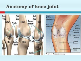
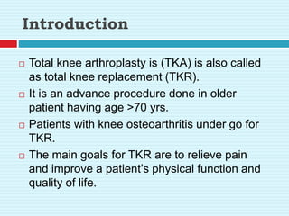
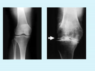
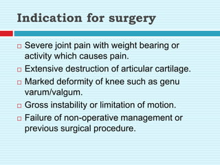
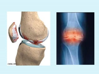
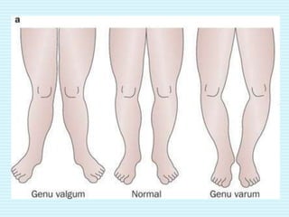
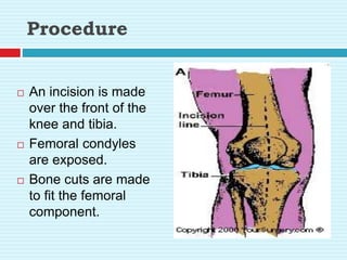
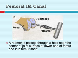
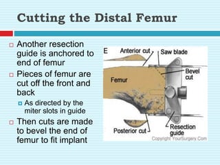
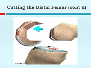
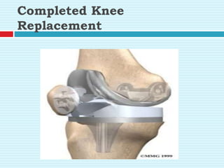
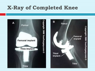
TOTAL KNEE
ARTHROPLASTY
Anatomy of knee joint
 Made up of three bones:
 Femur (thigh bone)
 Tibia (lower leg bone)
 Patella (kneecap)
Anatomy of knee joint
Introduction
 Total knee arthroplasty is (TKA) is also called
as total knee replacement (TKR).
 It is an advance procedure done in older
patient having age >70 yrs.
 Patients with knee osteoarthritis under go for
TKR.
 The main goals for TKR are to relieve pain
and improve a patient’s physical function and
quality of life.
Indication for surgery
 Severe joint pain with weight bearing or
activity which causes pain.
 Extensive destruction of articular cartilage.
 Marked deformity of knee such as genu
varum/valgum.
 Gross instability or limitation of motion.
 Failure of non-operative management or
previous surgical procedure.
Contraindication
 Poor general health
 Severe osteoporosis
 Recurrent or current joint sepsis
 Neuropathic arthropathies
 Quadriceps insufficiency
 Flexion deformity of more than 60 degree
combined with varus & valgum
 Hyperextension more than 20-30 degree
Classification of knee implants
Uni-compartmental
Bi-Compartmental
Tri-Compartmental
1. unconstrained
2. semiconstrained
3.fully constrained
Unicompartmental implants
 These are used to replace the opposing articular
surfaces of the femur & tibia of either the medial or
lateral compartment of knee.
 The opposite compartment & the patello femoral
compartment remain into it.
 This implant are all of unconstrained type.
Bi-compartmental implants.
 These are used to replace the opposing articular
surfaces of the femur & tibia of both the medial or
lateral compartment of knee.
 resurfacing the patello femoral joint.
 This deficit & mechanical loosening of the implant
have resulted in rejection of this approach.
Tri-compartmental implant
 These implant not only replace the opposing surfaces of
the femur & tibia both of the medial & lateral
compartment but also provide for resurfacing the patello-
femoral articulation.
 Implants can be divided conveniently into 3 groups
according to the degree of mechanical constraint
provided.
1.Unconstrained prosthesis
 They are primary resurfacing implants.
 Few implants in this category may provide
minimal restrain to rotation or translation.
 Prosthesis greatly depends on the integrity
of the soft tissue to provide jt stability.
 Soft tissue balancing is necessary to
achieve it stability while restoring proper
limb alignment.
Eg:- Toconley & PCA
2.Semiconstrained prosthesis
 Largest no of prosthesis fall in this category.
 The degree of constrain possed by a given prosthetic
design varies from almost minimal to an extent only
somewhat less than in a fully constrained design.
 Combination of soft tissue release a proper prosthetic
selection may achieve a stable joint, muscle limb
alignment & correction of deformities like flexion
contracture of upto 45 degree & angular deformities of
20-25 degrees.
 Angular deformities associated with ligamentous laxity
are more easily corrected than fixed angular
deformities.
3. Fully constrained prosthesis
 They permit flexion & extension in sggital
plane, but prevent adducting & abducting in
the coronal plane.
 They are
1. hinged constrained implants
-true hinges
-rotating hinges
Procedure
 An incision is made
over the front of the
knee and tibia.
 Femoral condyles
are exposed.
 Bone cuts are made
to fit the femoral
component.
Femoral IM Canal
 A reamer is passed through a hole near the
center of joint surface of lower end of femur
and into femur shaft
Cutting the Distal Femur
 Another resection
guide is anchored to
end of femur
 Pieces of femur are
cut off the front and
back
 As directed by the
miter slots in guide
 Then cuts are made
to bevel the end of
femur to fit implant
Cutting the Distal Femur (cont’d)
Cutting the Tibial Bone
 A resection guide is
attached to front of
tibia
 Direction of the
saw cuts in 3D
 AP tilt
 LM tilt
 Upper end of tibia
is resected
Placing the Femoral
Component
 Metal
component is
held in place by
friction
 In the
cemented
variety
 An epoxy
cement is
used.
Cutting the Tibial Bone
(cont’d)
Placing the Tibial Component
 Metal tray that
will hold plastic
spacer is
attached to the
top of the tibia
Placing the Plastic spacer
 Attached to the metal tray of tibial component
Preparing the Patella
 The
undersurface of
the patella is
removed.
Placing the Patella
Component
 The patella button is
usually cemented
into place behind
the patella
Completed Knee
Replacement
X-Ray of Completed Knee
Immobilization
 Knee is immobilized in bulky compression
dressing for a day or 2 post operatively.
 After the bulky dressing is removed a post knee
splint is often worn but is removed for daily
exercises.
 Cementless arthroplasty may require a longer
period of immobilization than a cemented
procedure to allow ingrowth of bone into the
prosthesis.
 A post knee splint may be indicated for use at
night for as long as 12 weeks post operatively.
Complications
 Intercondylar fracture
 Damage to peripheral nerve (eg. Peroneal nerve)
 Infection
 Joint instability subluxation
 Component loosening
 Risk of wound healing
 DVT
 Deep periprosthetic infection
 Patellar instability or tracking
 Limited knee flexion
 Impaired function of the extensor
mechanism(extensor lag)
Physiotherapy management
 Pre operative physiotherapy
1) breathing exercises.
2) strengthening exercises.
-teach isometric ex for hamstring, glutei &
quadriceps.
-Resistive ex for ankle & foot on the affected
side are taught.
3) ankle-pump exercises.
4)mobilization exercises.
Post operative
 The rehabilitation protocol for a patient with
TKR is dependent on following 3 phases.
1) Maximum protection phase.
2) Moderate protection phase.
3) Minimum protection phase.
Maximum protection phase
 To prevent pulmonary complication
-deep breathing & coughing exercises
-localized and generalized expansion ex to
maintain healthy respiratory system
-if there are secretions in the lungs as aresult
of G.A postural drainage is given.
 To prevent vascular complications
-ATM
 To promote health and reduce post operative
odema & pain.
-strong ankle pumping ex begins immediately
after surgery.
-elevation of foot end to assist venous return to
reduce swelling.
-to relieve pain, TENS may be given at site of
pain.
 To improve strength of quadriceps
-immediately after sx or as soon as patient can
tolerate, SQE are taught to the patient is
encouraged to practice them frequently in a
day.
-last 15 degree extension can be given
keeping pillow under the knee for quads lag.
-supported SLR is taught as early as possible.
-active knee extension in high sitting should be
started and gradually increase repetation,
which should be started on 5th day after
operation.
 To maintain the strength of hip muscles
-static & isometric ex for hip muscles and
hamstring muscles.
 To improve joint mobility for knee
-cpm is given
-active and active assisted ex for knee
-hold and relax should be started(70 degree
5-12 days)
-knee range must not exceed 40 in first 3 P.O
days as a transcutaneous oxygen tension of the
skin near the incision decreases., if it knee flexion
is >40 it may delay wound healing.
 To give proper gait training
-the extent to weight bearing is available
dependent upon the type of prosthesis
implanted.
-full weight bearing (12 weeks) & ambulation
without assistive device may not be
permissible for upto 12 weeks P.O
-with cement fixation weight bearing as
tolerated is permissible immediately after
surgery & increased to full weight bearing over
6 weeks.
yet patient should continue to use a cane
through
moderate & minimum protection phase of
rehabilitation until adequate strength & stability
are achieved.
isometric ex, rythmical stabilization & weight
transference may be practiced in stand but the
splint as retained for walking until 70-90 of flexion
is achieved.
gait training is commenced initially in parallel
bars & in front of mirror, so that the patient can
see & feel the correct pattern of walking.
gait training can then be progressed with a
walker or crutches.
 Managing uneven ground & slopes
 managing stairs
-within 5-12 days stairs case ascend and
descend maximum to be taught
-ability to climb stairs with alternate steps may
not be possible for some weeks.
 To make patient functionally independent
-patient is advised not to flex the knee more
than 90 degree. Hence, sitting on floor & cross
leg sitting is avoided.
-patient is advised to use western toilets & can
be made use the toilet as early as he can walk
with an aid.
Moderate protection phase
 To improve muscular strength
-as healing progresses, multiple angle
isometric resistance ex for quads and hams
can be added.
-adequate strength of knee extensor is most
important for stability of knee during wt.
bearing activities.
-resisted SLR in various position should be
included to increase the strength of hip
muscles.
-as weight bearing permits closed chain, mini
squats, lunges can be added to improve
stability.
 To increase ROM
-by 3-6 weeks knee flexion ex can be given
depending upon type of implant used
-gentle self stretching or contract relax ex are
also added
-stationary bicycle can be used.
-the cycle first may have the sit position as
high as possible to increase knee flexion, and
the seat can be gradually lowered.
 To give gait training
-single crutch walking & well assisted stair
activities is encouraged.
-Gait training with which emphasis on feet
knee swinging can be started with a cane and
in progression to full weight bearing by 6
weeks if cemented is done.
-yet patient should continue using single cane
til 12 weeks.
Minimum protection phase
 By 12th week after surgery, emphasis on
rehabilitation is done on muscle conditioning
so that the patient will have the strength 7
endurance to return to a full level of functional
activities.
-ambulation, stair climbing, & so on are
gradually increased.
Expected results
 Almost all patient report a significant relief of
pain with knee motion and weight bearing.
 Improvement in ROM may continue upto 12-24
months P.O but only minimal changes occur
compared to pre-op status.
 It may take 3 months p.o for a patient to regain
strength in quads & hamstring muscles.

Total knee arthroplasty.pptx

  • 1.
  • 2.
    Anatomy of kneejoint  Made up of three bones:  Femur (thigh bone)  Tibia (lower leg bone)  Patella (kneecap)
  • 3.
  • 4.
    Introduction  Total kneearthroplasty is (TKA) is also called as total knee replacement (TKR).  It is an advance procedure done in older patient having age >70 yrs.  Patients with knee osteoarthritis under go for TKR.  The main goals for TKR are to relieve pain and improve a patient’s physical function and quality of life.
  • 6.
    Indication for surgery Severe joint pain with weight bearing or activity which causes pain.  Extensive destruction of articular cartilage.  Marked deformity of knee such as genu varum/valgum.  Gross instability or limitation of motion.  Failure of non-operative management or previous surgical procedure.
  • 9.
    Contraindication  Poor generalhealth  Severe osteoporosis  Recurrent or current joint sepsis  Neuropathic arthropathies  Quadriceps insufficiency  Flexion deformity of more than 60 degree combined with varus & valgum  Hyperextension more than 20-30 degree
  • 10.
    Classification of kneeimplants Uni-compartmental Bi-Compartmental Tri-Compartmental 1. unconstrained 2. semiconstrained 3.fully constrained
  • 11.
    Unicompartmental implants  Theseare used to replace the opposing articular surfaces of the femur & tibia of either the medial or lateral compartment of knee.  The opposite compartment & the patello femoral compartment remain into it.  This implant are all of unconstrained type.
  • 12.
    Bi-compartmental implants.  Theseare used to replace the opposing articular surfaces of the femur & tibia of both the medial or lateral compartment of knee.  resurfacing the patello femoral joint.  This deficit & mechanical loosening of the implant have resulted in rejection of this approach.
  • 13.
    Tri-compartmental implant  Theseimplant not only replace the opposing surfaces of the femur & tibia both of the medial & lateral compartment but also provide for resurfacing the patello- femoral articulation.  Implants can be divided conveniently into 3 groups according to the degree of mechanical constraint provided.
  • 14.
    1.Unconstrained prosthesis  Theyare primary resurfacing implants.  Few implants in this category may provide minimal restrain to rotation or translation.  Prosthesis greatly depends on the integrity of the soft tissue to provide jt stability.  Soft tissue balancing is necessary to achieve it stability while restoring proper limb alignment. Eg:- Toconley & PCA
  • 15.
    2.Semiconstrained prosthesis  Largestno of prosthesis fall in this category.  The degree of constrain possed by a given prosthetic design varies from almost minimal to an extent only somewhat less than in a fully constrained design.  Combination of soft tissue release a proper prosthetic selection may achieve a stable joint, muscle limb alignment & correction of deformities like flexion contracture of upto 45 degree & angular deformities of 20-25 degrees.  Angular deformities associated with ligamentous laxity are more easily corrected than fixed angular deformities.
  • 16.
    3. Fully constrainedprosthesis  They permit flexion & extension in sggital plane, but prevent adducting & abducting in the coronal plane.  They are 1. hinged constrained implants -true hinges -rotating hinges
  • 17.
    Procedure  An incisionis made over the front of the knee and tibia.  Femoral condyles are exposed.  Bone cuts are made to fit the femoral component.
  • 18.
    Femoral IM Canal A reamer is passed through a hole near the center of joint surface of lower end of femur and into femur shaft
  • 19.
    Cutting the DistalFemur  Another resection guide is anchored to end of femur  Pieces of femur are cut off the front and back  As directed by the miter slots in guide  Then cuts are made to bevel the end of femur to fit implant
  • 20.
    Cutting the DistalFemur (cont’d)
  • 21.
    Cutting the TibialBone  A resection guide is attached to front of tibia  Direction of the saw cuts in 3D  AP tilt  LM tilt  Upper end of tibia is resected
  • 22.
    Placing the Femoral Component Metal component is held in place by friction  In the cemented variety  An epoxy cement is used.
  • 23.
    Cutting the TibialBone (cont’d)
  • 24.
    Placing the TibialComponent  Metal tray that will hold plastic spacer is attached to the top of the tibia
  • 25.
    Placing the Plasticspacer  Attached to the metal tray of tibial component
  • 26.
    Preparing the Patella The undersurface of the patella is removed.
  • 27.
    Placing the Patella Component The patella button is usually cemented into place behind the patella
  • 28.
  • 29.
  • 30.
    Immobilization  Knee isimmobilized in bulky compression dressing for a day or 2 post operatively.  After the bulky dressing is removed a post knee splint is often worn but is removed for daily exercises.  Cementless arthroplasty may require a longer period of immobilization than a cemented procedure to allow ingrowth of bone into the prosthesis.  A post knee splint may be indicated for use at night for as long as 12 weeks post operatively.
  • 31.
    Complications  Intercondylar fracture Damage to peripheral nerve (eg. Peroneal nerve)  Infection  Joint instability subluxation  Component loosening  Risk of wound healing  DVT  Deep periprosthetic infection  Patellar instability or tracking  Limited knee flexion  Impaired function of the extensor mechanism(extensor lag)
  • 32.
    Physiotherapy management  Preoperative physiotherapy 1) breathing exercises. 2) strengthening exercises. -teach isometric ex for hamstring, glutei & quadriceps. -Resistive ex for ankle & foot on the affected side are taught. 3) ankle-pump exercises. 4)mobilization exercises.
  • 33.
    Post operative  Therehabilitation protocol for a patient with TKR is dependent on following 3 phases. 1) Maximum protection phase. 2) Moderate protection phase. 3) Minimum protection phase.
  • 34.
    Maximum protection phase To prevent pulmonary complication -deep breathing & coughing exercises -localized and generalized expansion ex to maintain healthy respiratory system -if there are secretions in the lungs as aresult of G.A postural drainage is given.  To prevent vascular complications -ATM
  • 35.
     To promotehealth and reduce post operative odema & pain. -strong ankle pumping ex begins immediately after surgery. -elevation of foot end to assist venous return to reduce swelling. -to relieve pain, TENS may be given at site of pain.
  • 36.
     To improvestrength of quadriceps -immediately after sx or as soon as patient can tolerate, SQE are taught to the patient is encouraged to practice them frequently in a day. -last 15 degree extension can be given keeping pillow under the knee for quads lag. -supported SLR is taught as early as possible. -active knee extension in high sitting should be started and gradually increase repetation, which should be started on 5th day after operation.
  • 37.
     To maintainthe strength of hip muscles -static & isometric ex for hip muscles and hamstring muscles.  To improve joint mobility for knee -cpm is given -active and active assisted ex for knee -hold and relax should be started(70 degree 5-12 days) -knee range must not exceed 40 in first 3 P.O days as a transcutaneous oxygen tension of the skin near the incision decreases., if it knee flexion is >40 it may delay wound healing.
  • 38.
     To giveproper gait training -the extent to weight bearing is available dependent upon the type of prosthesis implanted. -full weight bearing (12 weeks) & ambulation without assistive device may not be permissible for upto 12 weeks P.O -with cement fixation weight bearing as tolerated is permissible immediately after surgery & increased to full weight bearing over 6 weeks.
  • 39.
    yet patient shouldcontinue to use a cane through moderate & minimum protection phase of rehabilitation until adequate strength & stability are achieved. isometric ex, rythmical stabilization & weight transference may be practiced in stand but the splint as retained for walking until 70-90 of flexion is achieved. gait training is commenced initially in parallel bars & in front of mirror, so that the patient can see & feel the correct pattern of walking. gait training can then be progressed with a walker or crutches.
  • 40.
     Managing unevenground & slopes  managing stairs -within 5-12 days stairs case ascend and descend maximum to be taught -ability to climb stairs with alternate steps may not be possible for some weeks.
  • 41.
     To makepatient functionally independent -patient is advised not to flex the knee more than 90 degree. Hence, sitting on floor & cross leg sitting is avoided. -patient is advised to use western toilets & can be made use the toilet as early as he can walk with an aid.
  • 42.
    Moderate protection phase To improve muscular strength -as healing progresses, multiple angle isometric resistance ex for quads and hams can be added. -adequate strength of knee extensor is most important for stability of knee during wt. bearing activities. -resisted SLR in various position should be included to increase the strength of hip muscles. -as weight bearing permits closed chain, mini squats, lunges can be added to improve stability.
  • 43.
     To increaseROM -by 3-6 weeks knee flexion ex can be given depending upon type of implant used -gentle self stretching or contract relax ex are also added -stationary bicycle can be used. -the cycle first may have the sit position as high as possible to increase knee flexion, and the seat can be gradually lowered.
  • 44.
     To givegait training -single crutch walking & well assisted stair activities is encouraged. -Gait training with which emphasis on feet knee swinging can be started with a cane and in progression to full weight bearing by 6 weeks if cemented is done. -yet patient should continue using single cane til 12 weeks.
  • 45.
    Minimum protection phase By 12th week after surgery, emphasis on rehabilitation is done on muscle conditioning so that the patient will have the strength 7 endurance to return to a full level of functional activities. -ambulation, stair climbing, & so on are gradually increased.
  • 46.
    Expected results  Almostall patient report a significant relief of pain with knee motion and weight bearing.  Improvement in ROM may continue upto 12-24 months P.O but only minimal changes occur compared to pre-op status.  It may take 3 months p.o for a patient to regain strength in quads & hamstring muscles.