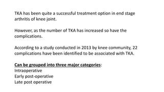
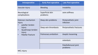
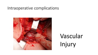
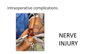
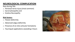
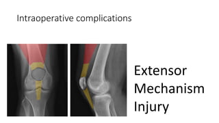
Complications in Total Knee
Arthroplasty
Dr. Vishal
PG Resident
Department of Orthopaedics (ABVIMS & Dr. RML Hospital)
TKA has been quite a successful treatment option in end stage
arthritis of knee joint.
However, as the number of TKA has increased so have the
complications.
According to a study conducted in 2013 by knee community, 22
complications have been identified to be associated with TKA.
Can be grouped into three major categories:
Intraoperative
Early post-operative
Late post operative
Intraoperative Early Post-operative Late Post-operative
Vascular injury Bleeding Instability
Neurological
complications
Superficial skin
problems
Joint stiffness
Extensor mechanism
injury
• patellar tendon
injury
• Quadriceps tendon
injury
• Patellar fracture
Deep skin problems Periprosthetic joint
infection
Deep vein thrombosis Periprosthetic fractures
Pulmonary embolism Aseptic loosening
osteolysis
Patellofemoral joint
problems
MCL injury
Intraoperative complications
Vascular
Injury
Vascular injury
Rare but serious and fatal complication during TKA
Forms:
o Thromboembolism
o Direct vascular laceration
o Pseudoaneurysm
o Arteriovenous fistula
Causes:
Direct damage
Thermal effect of cement polymerization
Joint manipulations
Dislocations
Excessive manipulation
Preventive measures:
Preoperatively
Identify high risk group by proper History and physical examination
Co-morbidities like DM, Htn, Smoking, Vascular claudication
Proper attention to look for-
Coldness of the extremities to be operated
skin atrophy and thinning
prominent vascular structures
ulcerative wound
distal arterial pulse weakness
ankle-brachial index below 0.9
Intra-operatively
o Tourniquet causes intimal damage. Avoid in high risk pt.
o Proper placement of retractors
(posterior retractors to be placed away from 1 cm lateral to midline)
o Careful release of PCL and posterior capsule during distal femoral cut
Management
Suspected vascular injury
Deflate the tourniquet
Perform bleeding control/ revascularisation
(Vascular surgeon consultation)
Close the incision
Acute ischemia cases with delayed diagnosis of
4–6 hours cause irreversible damage.
Prophylactic fasciotomy is performed after
revascularization.
Pseudoaneurysm
Pulsatile swelling in the popliteal fossa.
Cause: due to direct damage to the popliteal
artery during surgery.
Diagnosis: USG
Treatment
Excision of the lesion and repair with vascular
graft is applied after embolization.
Arteriovenous fistula
Less common.
Cause: Injury to the medial and lateral geniculate arteries and its
branches.
Presents as
Pulsatile swelling in the popliteal region that gives “trill.”
Hemarthrosis or pseudoaneurysm may develop.
Diagnosis: Ultrasound and Angiography
Treatment
Should be evaluated together with cardiovascular team
Embolization
Lesion excision
Graft repair
Intraoperative complications
NERVE
INJURY
NEUROLOGICAL COMPLICATION
Rare during TKA.
o Peroneal nerve injury (most common)
o Sacral plexopathy and
o Sciatica Neuropathy
Risk factors:
o Flexion deformity
o Advanced valgus deformity
o Presence of an intra-articular hematoma
o Tourniquet applications exceeding 2 hours
Difficult to detect intraoperatively.
Post operative neurological assessment must be done in each patient.
Management
o Immediate physical therapy should be started.
o EMG examination is to be done after 3 months.
o If no improvement is observed, nerve exploration may be planned
in the future.
Intraoperative complications
Extensor
Mechanism
Injury
Extensor mechanism
consists of quadriceps muscle group, quadriceps tendon,
patella, patellar retinaculum, patellar tendon, and Tibial
tuberosity.
Injury post-op> intra-op
Includes:
Patellar tendon rupture
Quadriceps tendon rupture
Patellar fracture
Patellar tendon rupture
-Usually occurs at the site of insertion to the
tibial tuberosity.
-Intra-tendinous and infrapatellar tendon
rupture may also occur.)
Risk: less than 1%.
Risk increases when patellar tendon mobility
decreases.
-Patella Baja
-Previous surgery
-Severe limitation of movement in the knee
Intra-operative causes:
Forced manipulation in stiff knee.
Tibial bone cutting.
Post-operatively:
falling onto the knee while knee is flexed (m/c)
Patellar tendon injury without trauma is seen by weakening the
tendon after
Non-traumatic: repeated contact of the polyethylene insert
weakens patellar tendon
Presentation:
Swelling
Loss of extension
Palpable defect at the infrapatellar side.
Treatment:
Age, functional status, tendon rupture localization, and soft tissue
status are to be considered.
Treatment:
 Low functional expectation/ unsuitable for surgery: Splinting
and bracing
 Acute patellar tendon rupture intraoperative primarily
requires repair using staple and suture anchors.
 Poor soft tissue quality : reconstruction using biological
materials (hamstring tendon autograft, achilles, peroneal
tendon autograft, and extensor mechanism allograft) or
synthetic materials.
Quadriceps tendon rupture
It is very rare.
Rupture from the insertion side to the patella.
Risk factor:
• Excessive patella cutting
• Previous quadriceps snip
• V-Y tipping
Clinical finding is similar to patellar tendon rupture.
Treatment
Partial tears: Plaster cast
Complete tear: extensor loss greater than 20°.
Requires surgical repair
Unsatisfactory results due to high complication rates
and tendency to re-rupture depending on tendon
quality and soft tissue condition.
Patella fractures
- most common injury among the extensor mechanism injuries.
Causes
o Excessive bone cutting while preparing for patellar component.
o Vascular compromise secondary to lateral release
o Patellar maltracking due to component malposition
o Excessive joint line elevation
o Knee flexion more than 115 degrees
o Thermal necrosis from PMMA
o Direct trauma to the anterior knee.
o Avulsion due to the pull of the quadriceps muscle.
Classification:
Based on integrity of extensor mechanism and stability of
implant.
Type 1: Stable implant and intact extensor mechanism
Type 2: stable implant but a discontinuous extensor mechanism
Type 3: unstable implant and discontinuous extensor
mechanism.
Patellar bone stock is classified as 3A if good and 3B if poor.
Management:
Type I:
knee immobiliser/ cylindrical cast for 6 weeks
Type II:
Transverse middle-third: Tension band wiring and retinacular repair.
Proximal and distal pole fracture.Partial patellectomy
Type III
3A: Fracture fixation
3B: Patellectomy and extensor mechanism repair
Medial collateral ligament injury
Medial collateral ligament (MCL) is important for soft tissue stabilization
and coronal plan stability.
Incidence of iatrogenic MCL injury is 2.2–2.7%.
Factors that increase the risk of MCL injury:
o Using a larger saw blade than femoral condyle
o Delayed excision of medial side osteophytes
o Performing challenging manipulations of varus-valgus
o Patients with flexion contractures
o Patient-related risk factors include obesity and severe deformities
Unrecognized MCL injuries cause early instability and implant wear and thus the
need for early revision.
Intra-op
Sudden instability in the valgus stress test during knee stabilization indicates MCL
injury
T/t options:
i) Repair
varies according to injury level.
 From femoral insertion site: Fixation with screw.
 Through tendon: repairing with insoluble suture technique is recommended.
 From tibial insertion site:insoluble suture anchor technique and staple fixation
ii) Constrained prosthesis use
iii) Revision at the same session
Early post-op
complications
THROMBOEMBOLISM
Prevalence of DVT: 1-84% after TKA (without prophylaxis)
DVT ——> PE
Thrombi in Popliteal vein is more likely to cause PE than that in calf
veins.
Various risk factors have been associated with development of DVT
and TE
Prophylaxis measures have significantly reduced the incidence.
Age: >40yrs
Oesterogen use
H/oStroke
Nephrotic syndrome
Cancer
Prolonged immobility
Previous Thromboembolism
CHF
IBD
Obesity
Varicose vein
Smoking
Hypertension
DM
H/O MI
Risk Factors
Virchow triad
CAPRINI SCORING FOR RISK CATEGORY
https://ortho
toolkit.com/c
aprini/
PROPHYLAXIS
In 2017,Dua et. Al. Reported significant decrease in DVT and VTE cases due to increase in prophylactic measures.
Low risk: Clexane 0.6 mg s.c OD for 3 days f/b oral rivaroxaban 10 mg od for 10
day AND/OR IPCD (if drugs C/I);
To start 6 hr. after removal of drain/epidural catheter.
High risk:
Clexane 0.6mg s.c OD for 5 days f/b oral Rivaroxaban 10 mg od for 10 days
AND IPCD during hospital stay. Dose to be adjusted if creat clearance less than
30 ml/min
USG doppler on 4th day post op in high risk.
C/I to drug therapy
• Intracranial hemorrhage within last yr
• Craniotomy within 2 week
• Intraocular surgery within 2 weeks
• Gastrointestinal OR genitourinary
• Haemorrhage within last month
• Active intracranial lesions/neoplasms
• Hypertensive emergencyEnd stage liver disease (INR > 1.5)
• Bleeding disorder/ coagulopathy
Management of DVT
• Clinical examination: Unreliable
• Radiography:
Venography: Gold std., has R/o Anaphylaxis and DVT also.
Duplex USG: Reliable method to diagnose DVT after TKA
Treatment options
• Therapeutic dose of anticoagulant
• Fibrinolytic drugs (Alteplase, STK)
• IVC filters
• Surgery
INFECTION
INFECTION
Approx. 1.5% of patients develop peri-prosthetic infection in first 2 yrs after TKA.
Risk factors:
Rheumatoid arthritis (esp seropositve men)
Skin ulceration
Previous knee surgery
Use of hinged prosthesis
Obesit
UTI
Steroid use
Renal failure
DM
Poor nutrition
Malignancy
Psoriasis
Prevention
Proper preoperative part preparation
Strict adherence to aseptic technique in OT room
Filtered vertical laminar flow with limited ingress and egress
of OT personelle
Prophylactic antibiotics
Management
Diagnosis
History and Physical examination.
Consistent painful TKA/ Acute onset pain in a previously pain free TKA
O/E
Active Pus discharge
Swelling
Tenderness
Erythema
Painful ROM
Low risk case:
Supacef 1.5 gm 60 min prior to incision/tourniquet application, 2
nd dose(of supacef) 60 min after incision.
High risk case:
Supacef 1.5 gm and teicoplanin 400 mg 60 min prior to
incision/tourniquet application, second dose(of supacef) 60 min
after incision
post op supacef 1.5 gm bd for 3 days
teicoplanin 400 bd for 3 days
ceftum 500 bd for 7 days
DIAGNOSIS
Clinical evaluation
RADIOGRAPH
X-ray:
Bone resorption at bone cement interface.
Cyst formation
Periosteal new bone formation.
ASPIRATION
Standard for diagnosis
Repeated aspiration at gap of two weeks.
Treatment options
o Suppression with antibiotics
o Joint debridement with poly exchange
o Resection arthroplasty
o Knee arthrodesis
o Exchange arthroplasty
Suppression with Antibiotics
Rarely indicated
Indication:
o prosthesis removal is not feasible
o Low virulence organism
Risks:
o Development of resistant strains
o Progressive loosening
o Extensive infection
o Septicaemia
Joint debridement with prosthesis retention:
Indications:
Early (<4 weeks) post op infection
Acute infection in a well fixed prosthesis (>4 weeks after SX with acute
symptom onset)
C/I
Chronic infection
Infection with S.aureus
Several points have been recommended that could lead to
higher success rates for debridement:
o Infectious disease consultation and antibiotic monitoring
o Diagnosis and treatment of hematogenous sources of Infection
o Newer antibiotics
o Six-week duration of postoperative intravenous antibiotics
o Repeat cultures within 2 weeks of the initial debridement and repeat
debridement if these cultures were positive
o Polyethylene exchange at the time of debridement; exchange of gown,
gloves, and instruments; and redraping at the time of wound closure
Resection Arthroplasty
Resection of infected prosthesis and cement and
debridement of synovium
Appose bone ends with heavy sutures or pins
Cast application for 6weeks
Knee Arthrodesis
Provides stable, painless limb with some expected shortening.
Indications:
o High functional demand
o Single joint involvement
o Young age
o Deficient extensor mechanism
o Poor soft tissue coverage
o Immunocompromised pt
o High virulent organism
Knee arthrodesis
Can be performed using:
External fixator
IM nail
External fixator:
Advantage:
o Minimal soft tissue stripping
o Adequate wound access
o Compression at arthrodesis site
Disadvantages:
o Pin tract infection
o Possible NV damage
o Limited stability
o Stress riser at pin removal site
o Remove all the prosthesis
o Cement, hypertrophic synovium and as much scar tissue as possible
o Use EM total knee guides to make bone cuts
o Knee in 10 degree flexion and 5 degree of valgus
o Apply biplanes fixator and cross pins.
o Partial wt bearing is encouraged
o Remove fixator if clinical signs of union present
(usually at 3months)
o Use cylindrical cast, long leg cast/ knee brace
until radiological signs of union present
o Weight bearing as tolerated
Arthrodesis with IM nail
Advantage:
Immediate partial wt bearing allowed
Greater fusion rate
Two stage techniques is recommended
o Complete debridement and component removal
o 4-6 weeks of IV antibiotics
o Repeat joint aspiration after two weeks of IV antibiotic completed
o IM arthrodesis
Reopen the old knee incision and make an incision in the buttock for the
proximal entry portal.
Remove scar tissue from the knee joint to allow apposition of the bone
surfaces.
Ream the tibia antegrade from the knee joint to 1 mm larger than the
diameter of the nail
Ream the femur retrograde or antegrade 1 to 1.5 mm larger than the
diameter of the nail
Drive the nail from the piriformis fossa across the knee to the
level of the distal tibial metaphysis.
Make final corrections in the distal femoral and proximal tibial
surfaces before final driving of the nail and impaction of the
arthrodesis site.
Compression at the fusion site can be done with a femoral
distractor in the compression mode before distal interlocking.
Do not notch the surface of the nail with any power
instruments.
Lock the nail proximally and distally and apply bone graft in
areas of suboptimal apposition
Exchange arthroplasty
Great chance of functional recovery after infected TKA
Two stage arthroplasty is recommended
Prosthesis removal
Debridement
Antibiotic spacer placement
IV antibiotic for 6 weeks
Reimplantation
Use of antibiotic impregnated PMMA
Advantage:
Maintains soft tissue tension
High level local antibiotic delivery
Improved exposure at time of reimplantation
Ability to wt bearing at interval
Above knee amputation
Indication:
Life threatening infection
Persistent local infection with massive bone loss
Late post operative complications
o Instability
o Joint stiffness
o Peri-prosthetic fractures
o Aseptic loosening
o Patello-femoral joint problems
Instability
Third most common cause of revision (17%) after aseptic loosening
and infection.
Presentation:
Pain and swelling with loss of movement.
Abnormal friction and rattling noise in some range of motion. During
walking, varus or valgus orientation and recurvatum can be seen over
knee
Anterior knee pain during sitting up is typical in flexion instabilities.
Risk factors:
Asymmetric gap: most common causes of instability.
Rectangular gap in both flexion and extension after bone excisions and soft tissue
release is required for stability of TKA.
Excess tibial cut: loose extension and flexion.
Excess distal femoral cut: loose extension.
Excess posterior femur cut: loose in flexion.
if the distal femoral incision is excessive, the use of distal femoral augment should
be preferred instead of the use of a thick insert
Joint stiffness
Amount of knee ROM after TKA greatly affect patient satisfaction
after TKA.
Flexion range of at least 90° is required.
A flexion range of less than 90° for 6 weeks after TKA surgery is
defined as a rigid knee.
Sixty-five degrees of flexion is required during walking; 106° of
flexion is required when sitting on a chair and tying shoes.
Risk factors:
Surgical technique:
o Inadequate posterior femoral incision
o Inadequate medial collateral ligament release
o Severe varus deformity
Other factors:
o Hip osteoarthritis
o Heterotropic ossification
o Reflex symptomatic dystrophy
o Arthrofibrosis: most treatment-resistant cause of joint stiffness. It
develops due to excessive increase of fibrous tissue in the joint
Treatment
Physiotherapy
o Prophylactic role, limited role in joint stiffness after TKA.
o Use of continuous passive motion device (CPM) in the early
postoperative period reduces bleeding and is beneficial in preventing
joint stiffness by reducing the formation of fibrosis
Manipulation under anaesthesia:
o Should be performed in cases where knee flexion is below 90°
between 2 weeks and 3 months.
o An average gain of 30–47° was reported
o Revision rates are lower in patients with early manipulation
Surgical procedures
o Arthroscopic release/open release
o limited revision knee arthroplasty/total
revision knee arthroplasty
PERI-PROSTHETIC FRACTURES
oSupracondylar femur fracture
oTibial fracture
Periprosthetic fractures:
Fractures that occur during or after surgery within 15 cm of the knee
joint or within 5 cm of the intramedullary part of the prosthesis, if any
Can occur during or after surgery.
It includes:
Supracondylar femur fracture
Tibia fracture
Supracondylar femur fracture
Risk factors:
o Anterior femoral notching (m/c, if notched use a stemmed femoral
component)
o Osteoporosis
o Female gender
o Revision arthroplasty
o Neurological disorder
Classification: (Rorabeck, Angliss and Lewis)
Type I: Undisplaced fracture with stable prosthesis
Type II: Displaced fracture with stable prosthesis
Type III: Unstable prosthesis with/without fracture displacement
Tibial Fracture:
• Very rare
Classification (felix)
o Type I: Tibial plateau fracture
o Type II: adjacent to stem
o Type III: Shaft fracture distal to stem
o Type IV: Tibial tubercle fracture
Type I
Revision is recommended because tibial component will be in varus alignment.
The medial defect should be closed with bone graft or metal support
Type II
Nonsurgical treatment if the component is stable and there is minimal
displacement
Internal fixation: displaced type 2 fractures
Unstable component: Revision using a long tibial stem to cross the fracture line
Type III/IV
Internal fixation should be performed
Management:
Small lesion and the prosthesis is stable:
o Observation+/- Bisphosphonates and calcium supplementation
If the prosthesis is unstable
o Debridement, polyethylene replacement, and curettage, followed by impac
of the defect with bone graft.
o Revision
Aseptic loosening
The deterioration of the relationship between prosthesis and bone.
Causes
Patient related
Overuse
Osteopenia
Implant design
Cementless prosthesis
Constrained prosthesis
Surgery related:
Malalignment.
In the early period, a radiolucent line is seen between the component and bone
on radiography, and a collapse occurs as the loosening progresses.
Loosening is more common around the tibial component .
In the presence of loosening around the whole component, septic loosening
should be considered
Revision surgery for pain with instability
Osteolysis
o Osteolysis usually occurs due to inflammatory reactions caused by worn
polyethylene particles or in the presence of infection.
o Metal particles can also cause osteolysis.
o Titanium causes more osteolysis than cobalt and chromium. Giant cells that
develop against abrasive particles act by forming a membrane.
Particle size is important for this mechanism.
Sizes range from 1 to 100 micrometers cause osteolysis.
Incidence of osteolysis increases when pres-fit prosthesis is applied; screw
fixation without cement is used or cement breaks. (Contact with cancellous
bone)
Presentation:
Pain, joint effusion, and synovitis due to joint instability.
Radiograph: Focal bone destruction. It can be seen that there is non-
continuity of trabeculae and bone cortex in cancellous bone.
CT and MRI can be used for osteolysis that cannot be detected on
direct radiography.
Patellofemoral joint problems
Pt presents with anterior knee pain.
In a study, Patella and malrotation were among the eight most common
causes of failed TKA
Causes:
Advanced valgus alignment
Previous high tibial osteotomy
Tibial tuberosity osteotomy
Surgical technique to prevent patellofemoral joint problems.
 Component placement: If the femoral component is placed medially,
anteriorly, or flexed, or if there is internal rotation and if the component is
excessive in size, patellofemoral problems may occur finally. Likewise, the
medialization and internal rotation of the tibial component increases the risk.
 Surgical approach type: Midvastus and subvastus interventions that protect the
extensor mechanism more can reduce PF joint problems.
 Lateral release: The need for lateral retinacular release increases PF joint
problems.
 Patella resection amount: When patellar component is used, resection of the
patella with anterior–posterior reduction of 12 mm increases the risk of PF
joint problems
THANK YOU

TKA complications.pptx

  • 1.
    Complications in TotalKnee Arthroplasty Dr. Vishal PG Resident Department of Orthopaedics (ABVIMS & Dr. RML Hospital)
  • 2.
    TKA has beenquite a successful treatment option in end stage arthritis of knee joint. However, as the number of TKA has increased so have the complications. According to a study conducted in 2013 by knee community, 22 complications have been identified to be associated with TKA. Can be grouped into three major categories: Intraoperative Early post-operative Late post operative
  • 3.
    Intraoperative Early Post-operativeLate Post-operative Vascular injury Bleeding Instability Neurological complications Superficial skin problems Joint stiffness Extensor mechanism injury • patellar tendon injury • Quadriceps tendon injury • Patellar fracture Deep skin problems Periprosthetic joint infection Deep vein thrombosis Periprosthetic fractures Pulmonary embolism Aseptic loosening osteolysis Patellofemoral joint problems MCL injury
  • 4.
  • 5.
    Vascular injury Rare butserious and fatal complication during TKA Forms: o Thromboembolism o Direct vascular laceration o Pseudoaneurysm o Arteriovenous fistula Causes: Direct damage Thermal effect of cement polymerization Joint manipulations Dislocations Excessive manipulation
  • 6.
    Preventive measures: Preoperatively Identify highrisk group by proper History and physical examination Co-morbidities like DM, Htn, Smoking, Vascular claudication Proper attention to look for- Coldness of the extremities to be operated skin atrophy and thinning prominent vascular structures ulcerative wound distal arterial pulse weakness ankle-brachial index below 0.9
  • 7.
    Intra-operatively o Tourniquet causesintimal damage. Avoid in high risk pt. o Proper placement of retractors (posterior retractors to be placed away from 1 cm lateral to midline) o Careful release of PCL and posterior capsule during distal femoral cut Management Suspected vascular injury Deflate the tourniquet Perform bleeding control/ revascularisation (Vascular surgeon consultation) Close the incision Acute ischemia cases with delayed diagnosis of 4–6 hours cause irreversible damage. Prophylactic fasciotomy is performed after revascularization.
  • 8.
    Pseudoaneurysm Pulsatile swelling inthe popliteal fossa. Cause: due to direct damage to the popliteal artery during surgery. Diagnosis: USG Treatment Excision of the lesion and repair with vascular graft is applied after embolization.
  • 9.
    Arteriovenous fistula Less common. Cause:Injury to the medial and lateral geniculate arteries and its branches. Presents as Pulsatile swelling in the popliteal region that gives “trill.” Hemarthrosis or pseudoaneurysm may develop.
  • 10.
    Diagnosis: Ultrasound andAngiography Treatment Should be evaluated together with cardiovascular team Embolization Lesion excision Graft repair
  • 11.
  • 12.
    NEUROLOGICAL COMPLICATION Rare duringTKA. o Peroneal nerve injury (most common) o Sacral plexopathy and o Sciatica Neuropathy Risk factors: o Flexion deformity o Advanced valgus deformity o Presence of an intra-articular hematoma o Tourniquet applications exceeding 2 hours
  • 13.
    Difficult to detectintraoperatively. Post operative neurological assessment must be done in each patient. Management o Immediate physical therapy should be started. o EMG examination is to be done after 3 months. o If no improvement is observed, nerve exploration may be planned in the future.
  • 14.
  • 15.
    Extensor mechanism consists ofquadriceps muscle group, quadriceps tendon, patella, patellar retinaculum, patellar tendon, and Tibial tuberosity. Injury post-op> intra-op Includes: Patellar tendon rupture Quadriceps tendon rupture Patellar fracture
  • 16.
    Patellar tendon rupture -Usuallyoccurs at the site of insertion to the tibial tuberosity. -Intra-tendinous and infrapatellar tendon rupture may also occur.) Risk: less than 1%. Risk increases when patellar tendon mobility decreases. -Patella Baja -Previous surgery -Severe limitation of movement in the knee
  • 17.
    Intra-operative causes: Forced manipulationin stiff knee. Tibial bone cutting. Post-operatively: falling onto the knee while knee is flexed (m/c) Patellar tendon injury without trauma is seen by weakening the tendon after Non-traumatic: repeated contact of the polyethylene insert weakens patellar tendon
  • 18.
    Presentation: Swelling Loss of extension Palpabledefect at the infrapatellar side. Treatment: Age, functional status, tendon rupture localization, and soft tissue status are to be considered.
  • 19.
    Treatment:  Low functionalexpectation/ unsuitable for surgery: Splinting and bracing  Acute patellar tendon rupture intraoperative primarily requires repair using staple and suture anchors.  Poor soft tissue quality : reconstruction using biological materials (hamstring tendon autograft, achilles, peroneal tendon autograft, and extensor mechanism allograft) or synthetic materials.
  • 20.
    Quadriceps tendon rupture Itis very rare. Rupture from the insertion side to the patella. Risk factor: • Excessive patella cutting • Previous quadriceps snip • V-Y tipping Clinical finding is similar to patellar tendon rupture.
  • 21.
    Treatment Partial tears: Plastercast Complete tear: extensor loss greater than 20°. Requires surgical repair Unsatisfactory results due to high complication rates and tendency to re-rupture depending on tendon quality and soft tissue condition.
  • 22.
    Patella fractures - mostcommon injury among the extensor mechanism injuries. Causes o Excessive bone cutting while preparing for patellar component. o Vascular compromise secondary to lateral release o Patellar maltracking due to component malposition o Excessive joint line elevation o Knee flexion more than 115 degrees o Thermal necrosis from PMMA o Direct trauma to the anterior knee. o Avulsion due to the pull of the quadriceps muscle.
  • 23.
    Classification: Based on integrityof extensor mechanism and stability of implant. Type 1: Stable implant and intact extensor mechanism Type 2: stable implant but a discontinuous extensor mechanism Type 3: unstable implant and discontinuous extensor mechanism. Patellar bone stock is classified as 3A if good and 3B if poor.
  • 24.
    Management: Type I: knee immobiliser/cylindrical cast for 6 weeks Type II: Transverse middle-third: Tension band wiring and retinacular repair. Proximal and distal pole fracture.Partial patellectomy Type III 3A: Fracture fixation 3B: Patellectomy and extensor mechanism repair
  • 25.
    Medial collateral ligamentinjury Medial collateral ligament (MCL) is important for soft tissue stabilization and coronal plan stability. Incidence of iatrogenic MCL injury is 2.2–2.7%. Factors that increase the risk of MCL injury: o Using a larger saw blade than femoral condyle o Delayed excision of medial side osteophytes o Performing challenging manipulations of varus-valgus o Patients with flexion contractures o Patient-related risk factors include obesity and severe deformities
  • 26.
    Unrecognized MCL injuriescause early instability and implant wear and thus the need for early revision. Intra-op Sudden instability in the valgus stress test during knee stabilization indicates MCL injury T/t options: i) Repair varies according to injury level.  From femoral insertion site: Fixation with screw.  Through tendon: repairing with insoluble suture technique is recommended.  From tibial insertion site:insoluble suture anchor technique and staple fixation ii) Constrained prosthesis use iii) Revision at the same session
  • 27.
  • 28.
    THROMBOEMBOLISM Prevalence of DVT:1-84% after TKA (without prophylaxis) DVT ——> PE Thrombi in Popliteal vein is more likely to cause PE than that in calf veins. Various risk factors have been associated with development of DVT and TE Prophylaxis measures have significantly reduced the incidence.
  • 29.
    Age: >40yrs Oesterogen use H/oStroke Nephroticsyndrome Cancer Prolonged immobility Previous Thromboembolism CHF IBD Obesity Varicose vein Smoking Hypertension DM H/O MI Risk Factors
  • 30.
  • 31.
    CAPRINI SCORING FORRISK CATEGORY
  • 32.
  • 34.
    PROPHYLAXIS In 2017,Dua et.Al. Reported significant decrease in DVT and VTE cases due to increase in prophylactic measures.
  • 35.
    Low risk: Clexane0.6 mg s.c OD for 3 days f/b oral rivaroxaban 10 mg od for 10 day AND/OR IPCD (if drugs C/I); To start 6 hr. after removal of drain/epidural catheter. High risk: Clexane 0.6mg s.c OD for 5 days f/b oral Rivaroxaban 10 mg od for 10 days AND IPCD during hospital stay. Dose to be adjusted if creat clearance less than 30 ml/min USG doppler on 4th day post op in high risk.
  • 36.
    C/I to drugtherapy • Intracranial hemorrhage within last yr • Craniotomy within 2 week • Intraocular surgery within 2 weeks • Gastrointestinal OR genitourinary • Haemorrhage within last month • Active intracranial lesions/neoplasms • Hypertensive emergencyEnd stage liver disease (INR > 1.5) • Bleeding disorder/ coagulopathy
  • 37.
    Management of DVT •Clinical examination: Unreliable • Radiography: Venography: Gold std., has R/o Anaphylaxis and DVT also. Duplex USG: Reliable method to diagnose DVT after TKA
  • 41.
    Treatment options • Therapeuticdose of anticoagulant • Fibrinolytic drugs (Alteplase, STK) • IVC filters • Surgery
  • 42.
  • 43.
    INFECTION Approx. 1.5% ofpatients develop peri-prosthetic infection in first 2 yrs after TKA. Risk factors: Rheumatoid arthritis (esp seropositve men) Skin ulceration Previous knee surgery Use of hinged prosthesis Obesit UTI Steroid use Renal failure DM Poor nutrition Malignancy Psoriasis
  • 44.
    Prevention Proper preoperative partpreparation Strict adherence to aseptic technique in OT room Filtered vertical laminar flow with limited ingress and egress of OT personelle Prophylactic antibiotics
  • 45.
    Management Diagnosis History and Physicalexamination. Consistent painful TKA/ Acute onset pain in a previously pain free TKA O/E Active Pus discharge Swelling Tenderness Erythema Painful ROM
  • 46.
    Low risk case: Supacef1.5 gm 60 min prior to incision/tourniquet application, 2 nd dose(of supacef) 60 min after incision. High risk case: Supacef 1.5 gm and teicoplanin 400 mg 60 min prior to incision/tourniquet application, second dose(of supacef) 60 min after incision post op supacef 1.5 gm bd for 3 days teicoplanin 400 bd for 3 days ceftum 500 bd for 7 days
  • 47.
    DIAGNOSIS Clinical evaluation RADIOGRAPH X-ray: Bone resorptionat bone cement interface. Cyst formation Periosteal new bone formation. ASPIRATION Standard for diagnosis Repeated aspiration at gap of two weeks.
  • 51.
    Treatment options o Suppressionwith antibiotics o Joint debridement with poly exchange o Resection arthroplasty o Knee arthrodesis o Exchange arthroplasty
  • 52.
    Suppression with Antibiotics Rarelyindicated Indication: o prosthesis removal is not feasible o Low virulence organism Risks: o Development of resistant strains o Progressive loosening o Extensive infection o Septicaemia
  • 53.
    Joint debridement withprosthesis retention: Indications: Early (<4 weeks) post op infection Acute infection in a well fixed prosthesis (>4 weeks after SX with acute symptom onset) C/I Chronic infection Infection with S.aureus
  • 54.
    Several points havebeen recommended that could lead to higher success rates for debridement: o Infectious disease consultation and antibiotic monitoring o Diagnosis and treatment of hematogenous sources of Infection o Newer antibiotics o Six-week duration of postoperative intravenous antibiotics o Repeat cultures within 2 weeks of the initial debridement and repeat debridement if these cultures were positive o Polyethylene exchange at the time of debridement; exchange of gown, gloves, and instruments; and redraping at the time of wound closure
  • 55.
    Resection Arthroplasty Resection ofinfected prosthesis and cement and debridement of synovium Appose bone ends with heavy sutures or pins Cast application for 6weeks
  • 56.
    Knee Arthrodesis Provides stable,painless limb with some expected shortening. Indications: o High functional demand o Single joint involvement o Young age o Deficient extensor mechanism o Poor soft tissue coverage o Immunocompromised pt o High virulent organism
  • 57.
    Knee arthrodesis Can beperformed using: External fixator IM nail
  • 58.
    External fixator: Advantage: o Minimalsoft tissue stripping o Adequate wound access o Compression at arthrodesis site Disadvantages: o Pin tract infection o Possible NV damage o Limited stability o Stress riser at pin removal site
  • 59.
    o Remove allthe prosthesis o Cement, hypertrophic synovium and as much scar tissue as possible o Use EM total knee guides to make bone cuts o Knee in 10 degree flexion and 5 degree of valgus o Apply biplanes fixator and cross pins.
  • 60.
    o Partial wtbearing is encouraged o Remove fixator if clinical signs of union present (usually at 3months) o Use cylindrical cast, long leg cast/ knee brace until radiological signs of union present o Weight bearing as tolerated
  • 61.
    Arthrodesis with IMnail Advantage: Immediate partial wt bearing allowed Greater fusion rate Two stage techniques is recommended o Complete debridement and component removal o 4-6 weeks of IV antibiotics o Repeat joint aspiration after two weeks of IV antibiotic completed o IM arthrodesis
  • 62.
    Reopen the oldknee incision and make an incision in the buttock for the proximal entry portal. Remove scar tissue from the knee joint to allow apposition of the bone surfaces. Ream the tibia antegrade from the knee joint to 1 mm larger than the diameter of the nail Ream the femur retrograde or antegrade 1 to 1.5 mm larger than the diameter of the nail
  • 63.
    Drive the nailfrom the piriformis fossa across the knee to the level of the distal tibial metaphysis. Make final corrections in the distal femoral and proximal tibial surfaces before final driving of the nail and impaction of the arthrodesis site. Compression at the fusion site can be done with a femoral distractor in the compression mode before distal interlocking. Do not notch the surface of the nail with any power instruments. Lock the nail proximally and distally and apply bone graft in areas of suboptimal apposition
  • 64.
    Exchange arthroplasty Great chanceof functional recovery after infected TKA Two stage arthroplasty is recommended Prosthesis removal Debridement Antibiotic spacer placement IV antibiotic for 6 weeks Reimplantation
  • 65.
    Use of antibioticimpregnated PMMA Advantage: Maintains soft tissue tension High level local antibiotic delivery Improved exposure at time of reimplantation Ability to wt bearing at interval
  • 66.
    Above knee amputation Indication: Lifethreatening infection Persistent local infection with massive bone loss
  • 67.
    Late post operativecomplications o Instability o Joint stiffness o Peri-prosthetic fractures o Aseptic loosening o Patello-femoral joint problems
  • 68.
    Instability Third most commoncause of revision (17%) after aseptic loosening and infection. Presentation: Pain and swelling with loss of movement. Abnormal friction and rattling noise in some range of motion. During walking, varus or valgus orientation and recurvatum can be seen over knee Anterior knee pain during sitting up is typical in flexion instabilities.
  • 69.
    Risk factors: Asymmetric gap:most common causes of instability. Rectangular gap in both flexion and extension after bone excisions and soft tissue release is required for stability of TKA. Excess tibial cut: loose extension and flexion. Excess distal femoral cut: loose extension. Excess posterior femur cut: loose in flexion. if the distal femoral incision is excessive, the use of distal femoral augment should be preferred instead of the use of a thick insert
  • 70.
    Joint stiffness Amount ofknee ROM after TKA greatly affect patient satisfaction after TKA. Flexion range of at least 90° is required. A flexion range of less than 90° for 6 weeks after TKA surgery is defined as a rigid knee. Sixty-five degrees of flexion is required during walking; 106° of flexion is required when sitting on a chair and tying shoes.
  • 71.
    Risk factors: Surgical technique: oInadequate posterior femoral incision o Inadequate medial collateral ligament release o Severe varus deformity Other factors: o Hip osteoarthritis o Heterotropic ossification o Reflex symptomatic dystrophy o Arthrofibrosis: most treatment-resistant cause of joint stiffness. It develops due to excessive increase of fibrous tissue in the joint
  • 72.
    Treatment Physiotherapy o Prophylactic role,limited role in joint stiffness after TKA. o Use of continuous passive motion device (CPM) in the early postoperative period reduces bleeding and is beneficial in preventing joint stiffness by reducing the formation of fibrosis Manipulation under anaesthesia: o Should be performed in cases where knee flexion is below 90° between 2 weeks and 3 months. o An average gain of 30–47° was reported o Revision rates are lower in patients with early manipulation
  • 73.
    Surgical procedures o Arthroscopicrelease/open release o limited revision knee arthroplasty/total revision knee arthroplasty
  • 74.
  • 75.
    Periprosthetic fractures: Fractures thatoccur during or after surgery within 15 cm of the knee joint or within 5 cm of the intramedullary part of the prosthesis, if any Can occur during or after surgery. It includes: Supracondylar femur fracture Tibia fracture
  • 76.
    Supracondylar femur fracture Riskfactors: o Anterior femoral notching (m/c, if notched use a stemmed femoral component) o Osteoporosis o Female gender o Revision arthroplasty o Neurological disorder
  • 77.
    Classification: (Rorabeck, Anglissand Lewis) Type I: Undisplaced fracture with stable prosthesis Type II: Displaced fracture with stable prosthesis Type III: Unstable prosthesis with/without fracture displacement
  • 80.
    Tibial Fracture: • Veryrare Classification (felix) o Type I: Tibial plateau fracture o Type II: adjacent to stem o Type III: Shaft fracture distal to stem o Type IV: Tibial tubercle fracture
  • 81.
    Type I Revision isrecommended because tibial component will be in varus alignment. The medial defect should be closed with bone graft or metal support Type II Nonsurgical treatment if the component is stable and there is minimal displacement Internal fixation: displaced type 2 fractures Unstable component: Revision using a long tibial stem to cross the fracture line Type III/IV Internal fixation should be performed
  • 82.
    Management: Small lesion andthe prosthesis is stable: o Observation+/- Bisphosphonates and calcium supplementation If the prosthesis is unstable o Debridement, polyethylene replacement, and curettage, followed by impac of the defect with bone graft. o Revision
  • 83.
    Aseptic loosening The deteriorationof the relationship between prosthesis and bone. Causes Patient related Overuse Osteopenia Implant design Cementless prosthesis Constrained prosthesis Surgery related: Malalignment.
  • 84.
    In the earlyperiod, a radiolucent line is seen between the component and bone on radiography, and a collapse occurs as the loosening progresses. Loosening is more common around the tibial component . In the presence of loosening around the whole component, septic loosening should be considered Revision surgery for pain with instability
  • 85.
    Osteolysis o Osteolysis usuallyoccurs due to inflammatory reactions caused by worn polyethylene particles or in the presence of infection. o Metal particles can also cause osteolysis. o Titanium causes more osteolysis than cobalt and chromium. Giant cells that develop against abrasive particles act by forming a membrane. Particle size is important for this mechanism. Sizes range from 1 to 100 micrometers cause osteolysis. Incidence of osteolysis increases when pres-fit prosthesis is applied; screw fixation without cement is used or cement breaks. (Contact with cancellous bone)
  • 86.
    Presentation: Pain, joint effusion,and synovitis due to joint instability. Radiograph: Focal bone destruction. It can be seen that there is non- continuity of trabeculae and bone cortex in cancellous bone. CT and MRI can be used for osteolysis that cannot be detected on direct radiography.
  • 87.
    Patellofemoral joint problems Ptpresents with anterior knee pain. In a study, Patella and malrotation were among the eight most common causes of failed TKA Causes: Advanced valgus alignment Previous high tibial osteotomy Tibial tuberosity osteotomy
  • 88.
    Surgical technique toprevent patellofemoral joint problems.  Component placement: If the femoral component is placed medially, anteriorly, or flexed, or if there is internal rotation and if the component is excessive in size, patellofemoral problems may occur finally. Likewise, the medialization and internal rotation of the tibial component increases the risk.  Surgical approach type: Midvastus and subvastus interventions that protect the extensor mechanism more can reduce PF joint problems.  Lateral release: The need for lateral retinacular release increases PF joint problems.  Patella resection amount: When patellar component is used, resection of the patella with anterior–posterior reduction of 12 mm increases the risk of PF joint problems
  • 89.