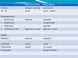
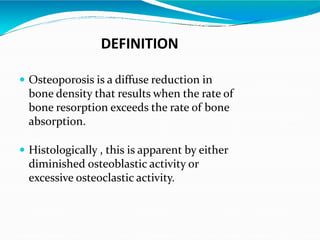
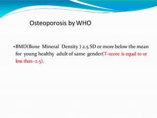
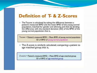
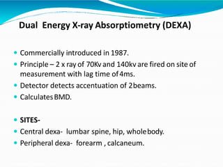
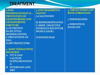
This document provides information on osteoporosis, including its definition, classification, epidemiology, clinical features, investigations used for diagnosis, and management. Osteoporosis is defined as a reduction in bone density leading to an increased risk of fractures. It is classified based on whether it is primary/secondary and type. Diagnosis involves tests like DEXA scans, biomarkers, and imaging. Management includes lifestyle modifications, calcium/vitamin D supplementation, and medications like bisphosphonates, teriparatide, and denosumab that reduce resorption or stimulate bone formation.
This document discusses osteoporosis and bone health. It begins by noting the impact of osteoporosis on older patients, including increased hospital admissions and length of stay. It then discusses definitions of osteoporosis from WHO and treatment gaps. The document covers bone structure, cells, mineralization, remodeling cycles, and factors influencing bone health like hormones and lifestyle. Diagnostic methods like DXA are summarized. Treatment options focused on prevention of fractures through calcium, vitamin D, bisphosphonates, PTH, and fall prevention are outlined.
Osteoporosis is a disease where bones become brittle and weak, increasing the risk of fractures. It is caused by low bone density due to lack of calcium and other minerals in bones. Symptoms include back pain and loss of height. Risk factors include age, gender, family history, and medications like corticosteroids. It is diagnosed through tests like DXA scans and treated through lifestyle changes, medications, and hormone therapy.
Osteoporosis is a progressive bone disease characterized by low bone mass and deterioration of bone tissue, leading to fragile bones that are prone to fractures. It is diagnosed based on bone mineral density measurements. Risk factors include older age, female sex, smoking, excessive alcohol use, low body weight, vitamin D and calcium deficiency, lack of exercise, and certain medical conditions or medications. Treatment focuses on lifestyle modifications to reduce risk factors as well as pharmacological therapies to prevent bone loss and reduce fracture risk. Common medications include bisphosphonates, calcitonin, estrogen therapy, SERMs, teriparatide, and denosumab.
Osteoarthritis (OA) is a progressive degenerative joint disease resulting from the erosion of articular cartilage. It typically affects those over 50 years old and is more common in women. OA can be primary and develop without obvious cause, or secondary due to factors like previous joint injury or deformity. Clinically, OA presents with joint pain, stiffness, and swelling that worsens with use. X-rays show narrowed joint space, osteophyte formation, and subchondral sclerosis. Treatment involves conservative measures like analgesics, exercise, and weight loss initially. Surgery such as arthroscopy, osteotomy, or joint replacement may be considered if conservative treatment fails.
Osteoporosis is a bone disorder characterized by low bone density and mass, leading to an increased risk of fractures. It is a growing epidemic, with over 75 million cases in the US, Europe and Japan expected to increase further. Risk factors include age, gender, family history, lifestyle factors like smoking, sedentary lifestyle, and certain medical conditions or medications. Diagnosis involves clinical evaluation, bone density scans, and biochemical markers. Treatment focuses on lifestyle changes like exercise, calcium intake, and vitamin D; medications include hormones, biphosphonates, and teriparatide injections in severe cases. Preventing osteoporosis requires modifiable lifestyle changes and responsible self-care.
Osteoporosis is a disease characterized by low bone mass and deterioration of bone tissue, leading to fragile bones and increased risk of fractures. It is defined by the World Health Organization as a bone mineral density score of -2.5 or below. Those at highest risk include older adults, especially post-menopausal women, and those with low calcium/vitamin D intake or other risk factors such as smoking. Symptoms may include back pain or loss of height from vertebral fractures. Treatment focuses on calcium and vitamin D supplementation, as well as bisphosphonate medications to decrease bone resorption and reduce fracture risk. Prevention emphasizes building strong bones through diet, exercise and lifestyle habits during childhood and adolescence.
This document provides an overview of osteoarthritis (OA), including its definition as a disease affecting synovial joints marked by cartilage loss and bone changes, leading to pain and impaired movement. It discusses the epidemiology of OA as the most common form of arthritis. The document outlines various causes and clinical subsets of OA and describes the pathological process involving breakdown of collagen and proteoglycans in cartilage. Diagnosis involves history, physical exam, and imaging tests. Treatment options discussed include lifestyle changes, medications, and surgeries such as joint replacement or resurfacing.
Hinduja hospital conducts regular webinars and tweetinars for online users where they can seek advice from expert doctors of hinduja hospital for free. Above is the webinar conducted by hinduja hospital on Osteoporosis where issues like osteoporosis symptoms, osteoporosis prevention, osteoporosis treatment were discussed successfully by Spine Consultant, Dr. Uday Pawar.
To know more about such upcoming webinars and tweetinars from hinduja hospital, visit http://www.hindujahospital.com/communityportal/
Bones provide structure, movement, and protection. Osteoporosis is a bone disorder where density decreases, weakening bones and increasing fracture risk, especially in the spine, hips, wrists. It is usually age-related but can be caused by other factors like medications, diseases, smoking. Diagnosis involves bone density tests and x-rays. Treatment focuses on lifestyle changes and medications like bisphosphonates, calcitonin, raloxifene which slow bone loss and increase density.
Osteoporosis is a systemic skeletal disease characterized by low bone mass and deterioration of bone tissue, leading to increased bone fragility and susceptibility to fractures of the hip, spine, and other bones. It is a major health problem due to consequences like pain, disability, and even death. Diagnosis involves bone mineral density tests like DEXA scans, and management focuses on lifestyle changes and medications to reduce fracture risk such as bisphosphonates, calcitonin, PTH, and SERMs. Effective prevention and treatment can help improve quality of life for those affected by osteoporosis.
Osteoporosis is a progressive bone disease characterized by decreased bone mass and density, leading to an increased risk of fracture. It is caused by an imbalance between bone resorption by osteoclasts and bone formation by osteoblasts. Risk factors include a family history, lifestyle factors like smoking and excessive alcohol, certain medications, and hormonal changes in women after menopause. Symptoms may include fractures, loss of height, and back pain. Diagnosis involves bone mineral density testing. Treatment focuses on lifestyle changes to prevent bone loss, calcium and vitamin D supplements, medications to reduce bone resorption, and surgery for fractures. Nursing interventions include education, medication management, fall prevention, and exercise promotion.
This document provides an overview of osteoporosis including its definition, epidemiology, pathophysiology, causes, clinical features, diagnosis, and treatment. Some key points include:
- Osteoporosis is a skeletal disorder characterized by compromised bone strength and increased risk of fracture. It is defined by the WHO as a bone density 2.5 standard deviations below the mean.
- It is a major global health problem, particularly affecting post-menopausal women and the elderly. Lifetime risk of osteoporotic fractures is 30-50% in females and 15-30% in males.
- Causes include failure to achieve peak bone mass, increased bone resorption, and inadequate bone formation
This document defines osteoporosis and discusses its epidemiology, pathophysiology, risk factors, clinical features, diagnosis and treatment. Osteoporosis is defined as a systemic skeletal disease characterized by low bone density and deterioration of bone tissue, leading to increased bone fragility and fracture risk. It most commonly affects post-menopausal women and the elderly. Diagnosis involves assessing bone mineral density via DEXA scan and evaluating risk factors. Treatment focuses on lifestyle modifications and medications to prevent bone loss and fractures.
Osteoporosis is a disease where bones become brittle and weak, increasing the risk of fractures. It occurs when the body loses more bone than it forms, reducing bone density. Common symptoms include back pain, loss of height, and fractures of the spine, wrists and hips. Risk factors include age, gender, family history, smoking, excessive alcohol, low calcium intake, and medical conditions or medications that reduce bone density. Diagnosis involves tests like DXA scans to measure bone mineral density. Treatment focuses on lifestyle changes, medications, and fall prevention to reduce fractures and complications.
This document provides an introduction and overview of osteoporosis, including its definition, risk factors, diagnosis, and treatment. It begins with defining osteoporosis as a systemic skeletal disease characterized by low bone mass and deterioration of bone tissue, leading to increased bone fragility and fracture risk. It then discusses the main and other risk factors, methods of diagnosis including laboratory tests and DEXA scans, pharmacological treatment options including bisphosphonates, denosumab, teriparatide, and hormone therapies, as well as prevention strategies. The document concludes with several case studies examples to demonstrate treatment approaches.
This document discusses osteoporosis, including its definitions, epidemiology, risk factors, pathophysiology, clinical manifestations, diagnosis, and treatment options. Osteoporosis is a disease characterized by low bone mass and deterioration of bone tissue, leading to fragile bones and increased risk of fractures. It affects millions of people worldwide, especially postmenopausal women, and can be caused by aging, genetics, lifestyle factors, and certain medical conditions or medications. Treatment involves lifestyle modifications like diet, exercise and fall prevention as well as pharmacologic options like calcium, vitamin D, bisphosphonates, and drugs that modify bone metabolism.
Osteoporosis is a metabolic bone disorder characterized by decreased bone mass and density, leading to porous and brittle bones that are prone to fracture. It is classified as either primary or secondary osteoporosis. Primary osteoporosis includes type I, which usually affects postmenopausal women, and type II, which occurs most commonly in those ages 70-85. Symptoms may include fractures after minor trauma. Diagnosis involves imaging tests to measure bone mineral density. Treatment focuses on controlling bone loss and preventing fractures through exercise, medication, supplements, and lifestyle changes. Nursing care emphasizes safety, mobility, nutrition, pain management, and education.
Metabolic bone disease (MBD) encompasses disorders of calcium and phosphorus homeostasis that are often silent until fractures occur. The document discusses several MBDs including osteoporosis, osteomalacia, rickets, and hyperparathyroidism. It provides details on osteoporosis including definitions, classifications, etiology, clinical manifestations, diagnosis through bone densitometry and radiography, assessment methods, and prevention through exercise, nutrition, and drugs.
Osteoporosis is a skeletal disorder characterized by compromised bone strength and increased risk of fracture. It is defined by low bone mineral density (BMD) as measured by dual energy x-ray absorptiometry (DXA) scan. Common sites of osteoporotic fractures include the spine, hips, ribs and wrists. The document discusses the mechanisms of bone loss and risk factors for osteoporosis like aging, menopause, low calcium/vitamin D, smoking and corticosteroid use. It outlines approaches to diagnosis, investigation and management including lifestyle modifications, calcium/vitamin D supplementation, bisphosphonates and hormonal therapy.
Everything you should know about Osteoporosis?
What is Osteoporosis?
Osteoporosis is a disorder of bones characterized by low bone density and a deterioration of bone micro- architecture that enhances bone fragility and increases the risk of fracture
Osteoporosis becomes a serious health threat for aging men & postmenopausal women by predisposing them to an increased risk of fracture
Do you know that?
Osteoporosis is responsible for >1.5 million vertebral and non-vertebral fractures per year
Spine, hip, and wrist fractures are most common.
This document summarizes three metabolic bone disorders: osteoporosis, osteomalacia, and Paget's disease. Osteoporosis is characterized by fragile bones and is caused by reduced bone mass, often occurring after menopause or in aging men. Management includes calcium, vitamin D, and exercise. Osteomalacia is a vitamin D deficiency causing bone softening, with management focusing on calcium, vitamin D, and sun exposure. Paget's disease rapidly destroys bone and can affect the skull and long bones, with an unknown cause. Management includes calcium, vitamin D, weight control, and pain medication.
Osteoporosis is a disease characterized by low bone mass and deterioration of bone structure. It increases the risk of fractures. The World Health Organization defines osteoporosis as a bone density 2.5 standard deviations or more below the mean bone density of healthy young adults. Dual-energy x-ray absorptiometry (DXA) is the gold standard test used to diagnose osteoporosis by measuring bone mineral density at sites like the hip and spine. Lifestyle factors like diet, exercise, smoking and alcohol as well as certain medical conditions and medications can impact bone health and increase the risk of osteoporosis.
This document discusses metabolic bone diseases, including their composition, calcium and phosphate metabolism, and specific diseases. It provides details on osteoporosis, rickets/osteomalacia, Paget's disease, and renal osteodystrophy. The composition of bone includes collagen, proteoglycans, and hydroxyapatite. Calcium and vitamin D intake recommendations are outlined for different populations. PTH, calcitonin, vitamin D, and alkaline phosphatase roles in calcium regulation are summarized. Osteoporosis risk factors and management strategies are highlighted.
Osteoarthritis is a non-inflammatory degenerative condition of the joints characterized by degeneration of articular cartilage and formation of new bone (osteophytes). It commonly affects the hands, knees, hips and spine. Risk factors include age, gender, obesity, injury and genetics. Symptoms include pain, stiffness, swelling and loss of flexibility. Diagnosis is made based on symptoms and confirmed with x-rays or MRI. Treatment involves medications like acetaminophen, NSAIDs, opioids and diacerein as well as complementary therapies and surgery for advanced cases.
Rickets and osteomalacia are diseases caused by a lack of calcium and phosphorus that prevents bone mineralization. Rickets occurs in children before growth plate closure, while osteomalacia occurs in adults. The most common causes are vitamin D deficiency from inadequate intake or absorption and phosphate deficiency. Treatment involves replacing the deficient nutrient, usually with vitamin D, calcium, and phosphate supplements. Monitoring treatment involves checking serum and urine calcium levels until they normalize and x-rays show bone healing.
Osteoporosis is a disease characterized by reduced bone mass and deterioration of bone tissue, leading to an increased risk of bone fractures. It is defined as a bone mineral density (BMD) score of more than 2.5 standard deviations below the average for a young healthy adult. Osteoporosis affects many post-menopausal women and older adults due to age-related bone loss. It has no symptoms until a fracture occurs, with vertebral compression fractures and wrist fractures being common. Diagnosis involves assessing risk factors and performing BMD tests and imaging to evaluate for fractures.
Osteoporosis is a systemic skeletal disease characterized by low bone mass and deterioration of bone microarchitecture, leading to increased bone fragility and risk of fracture. It is diagnosed based on a combination of clinical history, risk factors, physical examination, imaging findings, and bone mineral density (BMD) measurement via dual-energy X-ray absorptiometry. BMD T-scores are used to classify individuals as having normal bone density, osteopenia, or osteoporosis according to World Health Organization criteria. Other imaging techniques like quantitative computed tomography and peripheral BMD measurement can provide additional information.
Metabolic bone diseases by Dr. Bharat Kumar Goud. CBharath Kumar
This document discusses metabolic bone diseases and osteoporosis. It provides details on bone structure, metabolism, modeling and remodeling. Key cell types involved are described like osteoclasts which resorb bone and osteoblasts which form bone matrix. Diagnosis involves measuring bone mineral density by dual-energy x-ray absorptiometry (DXA) and comparing to young adult standards. Treatment focuses on lifestyle and nutritional factors as well as pharmacologic therapies like bisphosphonates, estrogen, and parathyroid hormone to increase bone formation and reduce resorption to prevent fractures.
Calcium homeostasis and Genral OSTEOPOROSIS of human.pptxBirajkc5
Osteoporosis is a disease characterized by low bone mass and structural defects, making bones fragile and prone to fracture. Key risk factors include older age, female sex, family history of fractures, smoking, excessive alcohol, low body weight, and use of corticosteroids. Diagnosis involves assessing bone mineral density via DXA scan and fracture risk factors. Treatment aims to reduce fracture risk and includes bisphosphonates, denosumab, parathyroid hormone, and lifestyle modifications. Secondary causes of osteoporosis include glucocorticoid use, gonadal hormone deficiency, hyperthyroidism, immobilization, and diabetes.
This document provides an overview of osteoporosis, including its definition, bone structure, epidemiology, pathophysiology, risk factors, clinical features, investigations, treatment, and management. It defines osteoporosis as a reduction in bone strength that increases fracture risk. Key points include that it occurs more in women after menopause due to estrogen loss, common risk factors, the roles of osteoblasts and osteoclasts in bone remodeling, biochemical markers used in diagnosis, DXA scans to measure bone mineral density, and first-line pharmaceutical treatments including bisphosphonates.
This document discusses the diagnosis and assessment of osteoporosis. It defines osteoporosis as a systemic skeletal disease characterized by low bone mass and deterioration of bone tissue, leading to bone fragility and susceptibility to fractures. It describes who is at risk and the different types of osteoporosis. Physical exams and various imaging techniques can be used to diagnose osteoporosis such as dual-energy x-ray absorptiometry (DXA) scans, quantitative ultrasound, CT scans, and plain radiography. Factors like BMD T-scores, clinical risk factors, and markers of bone turnover help assess fracture risk in patients.
This document provides an overview of osteoporosis including its definition, characteristic features, classification, contributing factors, clinical manifestations, investigations, diagnosis, and treatment. Some key points:
- Osteoporosis is a skeletal disorder characterized by low bone mass and deterioration of bone tissue, resulting in fragile bones and increased fracture risk. It is defined by the WHO as a bone density 2.5 standard deviations below the mean for young healthy adults.
- It is asymptomatic until a fracture occurs. Common fractures are of the distal radius, vertebral bodies, and hip. Risk factors include age, sex, family history, smoking, alcohol, low body weight, and estrogen deficiency.
- Diagnosis involves investigations
This document provides an overview of osteoporosis including its definition, characteristic features, classification, contributing factors, clinical manifestations, investigations, diagnosis, and management. Some key points:
- Osteoporosis is a skeletal disorder characterized by low bone mass and deterioration of bone tissue, resulting in fragile bones and increased fracture risk. It is defined by the WHO as a bone density 2.5 standard deviations below the mean for young healthy adults.
- It can be primary (postmenopausal or senile) or secondary (caused by other conditions). Risk factors include age, sex, race, family history, smoking, alcohol, estrogen deficiency, low body weight, and lack of activity.
Osteoporosis is a disease characterized by low bone mass and deterioration of bone tissue. It increases bone fragility and risk of fracture. While not a natural part of aging, risk is higher for post-menopausal women over 65 and all races and sexes can be affected. Bone density testing is recommended for women over 65, men over 70, and younger adults with clinical risk factors to diagnose osteoporosis. Dual energy x-ray absorptiometry (DEXA) is the gold standard test which measures bone mineral density at the hip and spine.
Osteoporosis is a systemic skeletal disease characterized by decreased bone density and deterioration in bone quality, leading to compromised bone strength and increased risk of fractures from minor trauma. It is diagnosed based on bone mineral density (BMD) T-scores. Risk factors include age, family history, smoking, alcohol, low body weight, chronic illnesses, and medications like glucocorticoids. Management involves lifestyle modifications, calcium and vitamin D supplementation, and pharmacological interventions like bisphosphonates.
Osteoporosis is a chronic, progressive skeletal disease characterized by low bone mass, microarchitecture deterioration of bone tissue, bone fragility, and a consequent increase in fracture risk.
Multiple atraumatic osteoporotic vertebral fractures: Unusual cause of pain i...Apollo Hospitals
Secondary osteoporosis may not be detected early, and thus the condition remains clinically silent until the patient presents with multiple atraumatic compression fractures. It is devastating for a young patient to develop multiple vertebral fractures in view of the associated morbidity and mortality. To decrease the risk of additional fractures and preserve the quality of life in these patients, interventions should be initiated early. Hence, it is important to consider multiple osteoporotic vertebral fractures as a complication in any patient on prolonged steroid therapy.
This document provides an overview of osteoporosis, including its definition, demographics, risk factors, diagnosis, treatment options and guidelines for follow-up. It defines osteoporosis as a skeletal disorder characterized by compromised bone strength and increased risk of fracture. Key points include that osteoporosis is diagnosed based on fractures or low bone mineral density (BMD), affects millions of Americans especially postmenopausal women, and treatment focuses on increasing calcium/vitamin D intake and medications to reduce fracture risk.
Recent advances in osteoporosis new copyDr Sourya M
Osteoporosis is characterized by low bone mass and deterioration of bone structure, making bones fragile and prone to fractures. Key drugs used to treat osteoporosis include calcium, vitamin D, bisphosphonates, SERMs, calcitonin, PTH and teriparatide, and denosumab. Newer drugs under development include romosozumab, a sclerostin inhibitor that strongly increases bone mineral density, abaloparatide, and integrin antagonists. Non-drug approaches also show promise such as biomaterials and gut serotonin inhibitors.
Osteoporosis is a progressive bone disease characterized by low bone mass and deterioration of bone tissue, making bones brittle and prone to fracture. It is defined as a T-score of -2.5 or below as measured by dual energy x-ray absorptiometry (DEXA) scanning. Key risk factors include older age, female sex, family history, smoking, low body weight and lack of exercise. Treatment focuses on lifestyle modifications like calcium and vitamin D supplementation, exercise and fall prevention, as well as pharmacologic therapies like bisphosphonates.
This document discusses various metabolic and endocrine diseases that can affect bone. It begins with an introduction to metabolic bone diseases and normal bone remodeling. It then discusses specific diseases in detail, including rickets, osteomalacia, scurvy, osteoporosis, hyperthyroidism, Cushing's disease, acromegaly, and hypothyroidism. For each disease, it describes the pathophysiology, clinical features, and key radiographic findings such as changes to bone density and morphology.
Ckd-MBD & osteoporosis the management dilemma Ayman Seddik
This document discusses the management of chronic kidney disease-mineral and bone disorder (CKD-MBD) and osteoporosis in elderly patients. It outlines that CKD-MBD and osteoporosis are common in elderly populations and impact mortality and morbidity. Management is based on the stage of CKD and involves controlling serum phosphorus and calcium levels, using phosphate binders to treat hyperphosphatemia, and treating abnormal PTH levels. Guidelines recommend treating bone disease with bisphosphonates and other osteoporosis medications according to the condition and stage of CKD. The risks and benefits of different treatment options must be considered based on each patient's situation.
Osteoporosis is a systemic skeletal disease characterized by low bone mass and deterioration of bone tissue, leading to increased bone fragility and risk of fractures. It is most commonly seen in elderly women. Bone mineral density testing is used to diagnose osteoporosis, with scores more than 2.5 standard deviations below normal indicating the disease. Treatment options include calcium, vitamin D, exercise, bisphosphonates, calcitonin, parathyroid hormone, and selective estrogen receptor modulators.
This document discusses metabolic bone diseases and provides details on osteoporosis. It begins with an overview of metabolic bone diseases including osteoporosis, rickets/osteomalacia, Paget's disease, and hyperparathyroidism. It then focuses on osteoporosis, defining it as a disease with low bone mass and structural defects leading to fragile bones. Epidemiology, risk factors, pathophysiology, clinical features, investigations including DEXA and other bone density tests, and treatment options like bisphosphonates, teriparatide, and denosumab are summarized. Rickets and osteomalacia are also briefly discussed.
Functional and radiological assessment of displaced midshaft clavicle fractures treated through open reduction and internal fixation surgery using pre-contoured locking compression plates
This document summarizes an original research paper that studied the radiological and functional outcomes of unstable bimalleolar ankle fractures treated surgically using the Baird-Jackson scoring system. The study involved 26 patients between ages 18-60 who underwent open reduction internal fixation surgery using a lateral locking plate and medial malleolar screw(s). Patients were followed for a minimum of 6 months and up to 12 months to assess union, complications, and functional outcomes using the Baird-Jackson scoring system. The majority of patients had good to excellent functional outcomes, with only a few cases of minor complications like infection or stiffness.
This document summarizes principles and concepts in fixing proximal humerus fractures with a locking plate. It describes the anatomy of the proximal humerus, classification systems for these fractures, considerations for surgical treatment, and techniques for open reduction and internal fixation using a locking plate. Key points covered include applied anatomy of the fracture deforming forces, blood supply to the humeral head, Neer's classification system, indications for operative vs non-operative treatment, surgical approach and techniques for placing a locking plate and securing it with screws including the importance of a calcar screw.
The document discusses considerations for correcting varus deformity during total knee arthroplasty for osteoarthritis. It analyzed 20 patients with over 15 degrees of varus deformity who underwent TKA. The goal of TKA is to restore the mechanical axis of the knee to distribute load evenly and reduce wear. This involves correcting alignment through distal femoral and proximal tibial cuts while balancing the extension and flexion gaps. Restoring proper alignment through the joint line and balancing the soft tissues is important for implant longevity and stability.
This document discusses floating knee injuries, which involve ipsilateral fractures of the femur and tibia. It describes the classification system for floating knee injuries, which includes true floating knee injuries (extra-articular fractures of both bones) and various types involving articular fractures. These injuries often result from high-energy trauma and are associated with injuries to other body parts. Treatment involves stabilizing the patient, addressing any life-threatening injuries, and providing initial stabilization of the fractures often using external fixation before definitive surgical fixation of the fractures.
Kosmoderma Academy, a leading institution in the field of dermatology and aesthetics, offers comprehensive courses in cosmetology and trichology. Our specialized courses on PRP (Hair), DR+Growth Factor, GFC, and Qr678 are designed to equip practitioners with advanced skills and knowledge to excel in hair restoration and growth treatments.
Histololgy of Female Reproductive System.pptxAyeshaZaid1
Dive into an in-depth exploration of the histological structure of female reproductive system with this comprehensive lecture. Presented by Dr. Ayesha Irfan, Assistant Professor of Anatomy, this presentation covers the Gross anatomy and functional histology of the female reproductive organs. Ideal for students, educators, and anyone interested in medical science, this lecture provides clear explanations, detailed diagrams, and valuable insights into female reproductive system. Enhance your knowledge and understanding of this essential aspect of human biology.
10 Benefits an EPCR Software should Bring to EMS Organizations Traumasoft LLC
The benefits of an ePCR solution should extend to the whole EMS organization, not just certain groups of people or certain departments. It should provide more than just a form for entering and a database for storing information. It should also include a workflow of how information is communicated, used and stored across the entire organization.
Adhd Medication Shortage Uk - trinexpharmacy.comreignlana06
The UK is currently facing a Adhd Medication Shortage Uk, which has left many patients and their families grappling with uncertainty and frustration. ADHD, or Attention Deficit Hyperactivity Disorder, is a chronic condition that requires consistent medication to manage effectively. This shortage has highlighted the critical role these medications play in the daily lives of those affected by ADHD. Contact : +1 (747) 209 – 3649 E-mail : sales@trinexpharmacy.com
Travel Clinic Cardiff: Health Advice for International TravelersNX Healthcare
Travel Clinic Cardiff offers comprehensive travel health services, including vaccinations, travel advice, and preventive care for international travelers. Our expert team ensures you are well-prepared and protected for your journey, providing personalized consultations tailored to your destination. Conveniently located in Cardiff, we help you travel with confidence and peace of mind. Visit us: www.nxhealthcare.co.uk
8 Surprising Reasons To Meditate 40 Minutes A Day That Can Change Your Life.pptxHolistified Wellness
We’re talking about Vedic Meditation, a form of meditation that has been around for at least 5,000 years. Back then, the people who lived in the Indus Valley, now known as India and Pakistan, practised meditation as a fundamental part of daily life. This knowledge that has given us yoga and Ayurveda, was known as Veda, hence the name Vedic. And though there are some written records, the practice has been passed down verbally from generation to generation.
Osteoporosis - Definition , Evaluation and Management .pdfJim Jacob Roy
Osteoporosis is an increasing cause of morbidity among the elderly.
In this document , a brief outline of osteoporosis is given , including the risk factors of osteoporosis fractures , the indications for testing bone mineral density and the management of osteoporosis
The skin is the largest organ and its health plays a vital role among the other sense organs. The skin concerns like acne breakout, psoriasis, or anything similar along the lines, finding a qualified and experienced dermatologist becomes paramount.
5-hydroxytryptamine or 5-HT or Serotonin is a neurotransmitter that serves a range of roles in the human body. It is sometimes referred to as the happy chemical since it promotes overall well-being and happiness.
It is mostly found in the brain, intestines, and blood platelets.
5-HT is utilised to transport messages between nerve cells, is known to be involved in smooth muscle contraction, and adds to overall well-being and pleasure, among other benefits. 5-HT regulates the body's sleep-wake cycles and internal clock by acting as a precursor to melatonin.
It is hypothesised to regulate hunger, emotions, motor, cognitive, and autonomic processes.
2. DEFINITION
Osteoporosis is a diffuse reduction in
bone density that results when the rate of
bone resorption exceeds the rate of bone
absorption.
Histologically , this is apparent by either
diminished osteoblastic activity or
excessive osteoclastic activity.
3. Osteoporosis byWHO
BMD(Bone Mineral Density ) 2.5 SD or more below the mean
for young healthy adult of same gender(T-score is equal to or
less than-2.5).
4.
5. WHO CRITERIA FOR OSTEOPOROSIS
BMD compared with
young adult
T score
Normal < 1 SD below >/= -1
Low bone mass ( Osteopenia ) 1-2.5 SD below < -1
> -2.5
Osteoporosis >/= 2.5 SD below </= -2.5
Severe osteoporosis >/= 2.5 SD below
PLUS Fracture
6. CLASSIFICATION
RIGGS AND MELTON CLASSIFICATION :
a.Primary osteoporosis –
Type 1 : Postmenopausal
Type 2 : AgeRelatedOsteoporosis
b.Secondary osteoporosis
7. . POST MENOPAUSAL OSTEOPOROSIS (TYPE 1)
Caused by lack of estrogen
Causes PTH to overstimulate osteoclast
AGE RELATED OSTEOPOROSIS (TYPE 2 )
Bone loss due to increased bone turnover
Malabsorption
Mineral and vitamin deficiency
Patients usuallypresentwith fracturesof the hip ,
spine and forearm.
23
9. Epidemiology
In women it is 3 times morecommon than men due to
1.Low peak bone mass (PBM)
2.Hormonal changes at menopause
1 in 3 women over 50 years suffer from osteoporosis.
1 in 5 men over 50 years suffer from osteoporosis.
15% - 30% men and 30%- 50% women suffer fractures related
to osteoporosis in their life time.
Peak incidence :
western countries – 70 -80
years
india – 50 – 60years
11. Factor Target cells Effect
Parathyroid Hormone
(PTH)
Kidney & Bone Stimulate production of Vit-D &
helps resorption of calcium
Calcitonin Bone osteoclasts Inhibits resorptive action of
osteoclasts: lowerscirculating
Calcium.
Calcitriol
(1.25-dihydroxy vit-D3)
Bone Osteoblasts
Bone Osteoclasts,
Kidney,
Intestine
-Stimulates collagen,osteopontin,
osteocalcin synthesis;
-stimulates cell differentiation;
-Stimulates Calcium retention
-Stimulates calcium absorption
Estrogen Bone Stimulates formation of calcitonin
receptors, inhibiting resorption,;
Stimulate bone formation
Testosterone Muscle, Bone Muscle growth, placing stress on
bone to stimulate boneformation
Prostaglandins Osteoclasts Stimulate resorption and bone
formation
Bone Morphogenic
protein
Mesenchyme Stimulate cartilage protein &bone
matrix formation; replication
12. CLINICAL FEATURES
Aka SILENT DISEASE.
Low back ache- usuallymild.
Loss of height
KYPHOSIS
Fractures- m/c at the level of
Dorso-Lumbar junction ,Hip
and Wrist Joint
14. A. DIAGNOSIS
INVESTIGATIONS :
1.ROUTINE INVESTIGATIONS TO RULE OUT SECONDARY OSTEOPOROSIS
2. Plain Radiography
3. PHYSICAL EXAMINATION
4. DEXA (GOLD STANDARD FOR MEASURING BMD )
5.QUANTITATIVE ULTRASOUND
6. BONE TURN OVER MARKERS (BTM)
15. ROUTINE INVESTIGATIONS :
THE PRIMARY AIM IS TO EXCLUDE SECONDARY CAUSES OF OSTEOPOROSIS WHICH
INCLUDES :
COMPLETE BLOOD COUNT
ERYTHROCYTE SEDIMENTATION RATE (ESR)
SR. CALCIUM , PHOSPHATASE , ALBUMIN
ALKALINE PHOSPHATASE
PARATHYROID HORMONE (PTH) IF SERUM CALCIUM LEVEL IS HIGH THAN 10.5 mg %
16. X-ray
Post menopausal osteoporosis : Trabecular
resorption and cortical resorption
Senile osteoporosis: Endostealresorption
Hyperparathyroidism: Sub periostealresorption
Osteoporosis produces increased radiolucency of
vertebral bone. Approximately 30 to 80 percentof
bone tissue must be lost before a recognizable
abnormalitycan bedetected on spinal radiographs.
The main radiographic features of generalized osteoporosis
are cortical thinning and increased radiolucency.
17. CONVENTIONAL RADIOGRAPHY
LS SPINE-
Generalized osteopenia
Thining and accentuation ofcortex
Accentuationof primary trabeculaeand thinning of
secondary trabaculae.
Vertically striated appearance vertebralbody.
18. KLEER KOPER score
Osteoporosis produces increased radiolucency of vertebral
bone. Approximately 30 to 80 per cent of bone tissue must
be lost before a recognizable abnormality can be detected on
spinal radiographs.
21. Dual Energy X-ray Absorptiometry (DEXA)
Commercially introduced in 1987.
Principle – 2 x ray of 70Kvand 140kv are fired on site of
measurement with lag time of4ms.
Detector detects accentuation of 2beams.
CalculatesBMD.
SITES-
Central dexa- lumbar spine, hip, wholebody.
Peripheral dexa- forearm , calcaneum.
22. Dual Energy X-ray Absorptiometry (DEXA)-
2-dimensional study
BMD = Amount of mineral
Area
Accuracy at hip > 90%
Low radiation exposure
Error in
Osteomalacia
Osteoarthritis
Previous fracture
23. BMD Interpretation
T score: standard deviation of the BMD from
the average sex matched 35-year-old
Z score: standard deviation score compared to
age matched control
For every 1 decrease in T score, double risk of
fracture
Regardless of BMD, patients with prior
osteoporotic fracture have up to 5 times risk of
future fracture.
24. INDICATIONS FOR BMD TESTING
In females 60 yrs + and in men 65 yrs +
In postmenopausal women above age 55 based on risk
factorprofile and symptoms.
In postmenopausal women and men age 50 and older
who have had an adult age fracture, to diagnose and
determine degree ofosteoporosis
At dual-energy X-ray absorptiometry (DXA)facilities
using accepted quality assurancemeasures
28. Calcaneum is the most common skeletal site for
quantitative ultrasound assessment because
A.It has a high percentage of trabecular bone that
is replaced more often than cortical bone,
providing early evidence of metabolic change.
B. The calcaneus is fairly flat and parallel,
reducing repositioning errors.
29. The McCue CUBA Ultrasonometry
Technology That Can Assess Osteoporosis
30. CT scan
True volumetric study
Quantitative Computed
Tomography (QCT) utilizes CT
technology to detect low bone
mass and monitors the effects of
therapy in patients undergoing
treatment.
It is a fast, non-invasive exam
that detects low bone mass
earlier and more accurately than
other bone density exams
31. The trabecular BMD is indicated as the
most important parameter, and
interpreted using the
Felsenberg classification based on the
following cut-off values:
Normal BMD > 120 mg/cc
Osteopenia < 120 mg/cc
Osteoporosis < 80 mg/cc
Very high fracture risk < 50 mg/cc
32. Advantages of CT scan over DEXA:
Ability to separate cortical and trabecular bone
Provides true volumetric density in units of mg/cc
No errors due to spinal degenerative changes or aortic calcification
Clinicians and researchers favor DXA because
-Scanners are readily available and relatively
inexpensive.
-The radiation dose is negligible
-The T-score scale, defined by the WHO specifically for DXA, provides a
standardized classification.
33. BIOCHEMICAL MARKERS OF BONE
TURNOVER
1.PREDICT THE RISK OF FRACTURE INDEPENDENTLY
OF BONE DENSITYIN UNTREATED PATIENTS
2.PREDICT RAPIDITY OF BONE LOSS INUNTREATED PATIENTS
3.PREDICT EXTENT OF FRACTURE RISKREDUCTION WHEN
REPEATED AFTER 3-6 MONTHS OF TREATMENT
4. PREDICT MAGNITUDE OF INCREASE IN BMD
5.HELP DETERMINE DURATION OF DRUG HOLIDAY AND WHEN
AND IF MEDICATION SHOULD BE RESTARTED
37. 1.NON
PHARMACOLOGICAL–
PREVENTION OF
OSTEOPOROSIS AND
OSTEOPOROTIC
FARCTURE.
A.NUTRITION
B.LIFE STYLE
MODIFICATIONS
C.PREVENTION OF
FALL
D.HIP PROTECTORS
2. BASICTHERAUPETIC
MEASURES
A. VIT D AND
CALCIUM
SUPPLEMENTATIO
N
B. ESTEROGEN AND
HRT
3.ANTI RESORBTIVE
AGENTS
A.CALCITONIN
B. BISPHOSHPHANTES
C.SERM (SELECTIVE
ESTROGEN RECEPTOR
MODULATOR )
D.DONESUMAB
4. DRUGS STIMULATE
BONE FORMATION
1.TERIPARATIDE
2.STRONTIUM
RANELATE
TREATMENT
38. LIFESTYLE MODIFICATIONS-
a.Physical activity-weight bearing andmuscle
strengthing exercises.
Exercise improves bone strength by 30%to50%.
Exercise should be lifelong.
b.Cessation of smoking,alcohol,high caffeineintake.
c.Adequate sunexposure
39. HIP PROTECTORS
PREVENTS DIRECT IMPACTON PELVIS.
TYPES :
1.Energy absorptiontype
2.Energy shunting types
3.Crash helmet type
4.Airbag type
40. ca
Men age 50–70 should consume 1000 mg/dayof
calcium.
Womenage 51 and olderand men age 71 and older
consume 1200 mg/day of calcium.
Intakes in excess of 1200 to 1500 mg/day mayincrease
the risk of developing kidney stones, cardiovascular
disease, and stroke.
PHARMACOLOGICAL PREVENTION OF
OSTEOPOROSIS
CALCIUM
41. VIT D3
800 to 1000 international units (IU) of vitamin D perday
for adults age 50 andolder.
Treatment of vitamin Ddeficiency-
Adults should be treated with 60,000 IU once aweek) for
4-6 weeks to achieve a level of approximately 30 ng/ml.
This regimen should be followed by
maintenance therapy of 1500–2000 IU/day.
42. Pharmacologic therapy
All patients being considered for treatment of
osteoporosisshould also be counseled on risk factor
reduction including the importance of calcium,
vitamin D, and exercise as part of any treatment
program forosteoporosis.
Prior to initiating treatment, patients should be
evaluated forsecondarycausesof osteoporosisand
have BMD measurements by central DXA, when
available, and vertebral imaging studies when
appropriate.
Biochemical marker levels should be obtainedif
monitoring of treatment effects isplanned.
43. Who should be considered for
treatment?
Postmenopausal women and men age 50 and older
presenting with the following should beconsidered-
A hip or vertebral fracture (clinically apparent or found on
vertebralimaging).
T-score ≤−2.5 at the femoral neck, hip joint , or lumbar
spine.
Low bone mass (T-score between −1.0 and −2.5 at the
femoral neck or lumbarspine)
a 10-year probability of a hip fracture ≥3 % or a 10-year
probability of a major osteoporosis-relatedfracture
≥20 %.
45. 1.Alendronate-
prevention -5 mg daily and 35 mg weeklytablets.
treatment -10 mg daily tablet, 70 mg weeklytablet.
Alendronate is also used in treatmentof osteoporosis in
men and women takingglucocorticoids.
2.Ibandronate-
2.5 mg daily for 3 years
3.Risedronate-
prevention and treatment -5 mg daily tablet; 35mg
weekly tabletfor 6 months.
46. 4.Zoledronic acid
prevention and treatment -5 mg by intravenous
infusionoverat least 15 min onceyearly for
treatment and onceevery 2 years for prevention.
47. Drug safety
Side effects for all oral bisphosphonates gastrointestinal
problems such as difficulty swallowing and oesophagitis
andgastritis.
All bisphosphonates are contraindicated inpatients
with estimated GFR below 30–35ml/min.
osteonecrosis of the jaw (ONJ) can occurwith long-
term use of bisphosphonates(>5year).
Although rare, low-trauma atypical femurfractures
may be associated with the long-term use of
bisphosphonates (e.g., >5 years ofuse).
48. Teriparatide
Teriparatide is approved for the treatment of
osteoporosis in postmenopausal women and menat
high risk forfracture.
It is also approved for treatment in men and women at
high risk of fracture with osteoporosis associated with
sustained systemic glucocorticoidtherapy.
DOSE-20 μg daily subcutaneousinjection for 18 months.
Regular monitoring of sr. calcium and uric acid at 1,6 and
12 months.
49. Calcitonin
Treatment of osteoporosis in women who are at least
5 years postmenopausal when alternative treatments
are notsuitable.
200 IU delivered as a singledaily intranasal spray.
Intranasal calcitonin can cause rhinitis, epistaxis, and
allergicreactions.
Verysmall increase in the risk of certain cancers.
50. SERM (SELECTIVE ESTROGEN RECEPTOR MODULATOR)
Used for both prevention and treatment of osteoporosis.
RALOXIFENE-60mg/day.
53. PREVENTATION TREATMENT
Calcium 500mg to 1500 mg 1000 to 1500
Vit – D 400IU 400IU – 800IU
Bi phosphonates
1. Alendronate 5mg/day 10mg/day
2. Ibandronate - 150mg/month
3. Rsidronate - 5mg/day
4. Zolendronic acid 5mg once in 2 year 5mg once /1 year
SERMS
Rolaxifen 5mg/day 10mg/day
Calcitonin 200 IU 200IU
Teriparatide 20ug/d 20-40ug/d
Donesumab - 60mg/6 months
54. SURGERY IN VERTEBRAL FRACTURES
Vertebroplasty
To reduce vertebralfracture–associated pain
Kyphoplasty
To restore height or to treat the deformity
associated with osteoporotic vertebralfractures
Pedicle screw fixation
In progressive vertebral collapse or deformity
55.
56. Role of Orthopaedic Surgeons
The goals of surgical treatment ofosteoporotic
fractures include
Rapid mobilization and return to normalfunction and
activities.
Back / Bowel / Bladder care.
Avoid too much manipulations.
Progressive physiotherapy.